Finally, I would like to thank my family and friends for being such an amazing support system through the stress of these past few semesters. Six human breast cancer cell lines (MDA-MB-231, LM, BoM, T47D, MCF7 and MCF7-BoM) were treated with three differently targeted phospholipase inhibitors (bromoenol lactone, tricyclodecan-9-yl-xanthogenate and halopemid) as well as different dietary oils (rapeseed oil, avocado oil, coconut oil, olive oil, sesame oil and soybean oil). Cell viability assays were performed to examine the effects of each inhibitor and oil at various concentrations on the proliferation of each breast cancer cell line.
The goal was to detect changes in cardiolipin concentration within each cell line after exposure to a variety of different phospholipase inhibitors and oils. Liquid chromatography-mass spectrometry (LC-MS) was performed on a cardiolipin standard as well as lipid extract samples from each cell line in order to detect changes in cardiolipin composition before and after the addition of test compounds. Because the MS results for the cell lines did not show a peak for the cardiolipin standard, we cannot use these results as a valid tool for determining differences in cardiolipin composition between samples.
However, BEL, halopemid, D609, canola oil, avocado oil, coconut oil and olive oil have been shown to cause a statistically significant difference in the cell viability of various breast cancer cell lines.
Introduction
- Breast Cancer Background
- Metastasis
- Mitochondria and Cardiolipin Background
- Cardiolipin Remodeling
- Conclusions
However, approximately 90% of deaths associated with breast cancer are still due to metastasis of the tumor cells1. The role of mitochondria in the regulation of cell death signals allows them to definitively control apoptosis. Chemical structure of Cardiolipin16. The four fatty acyl chains in CL, represented by R1, R1¢, R2 and R2¢, can exist in varying lengths and degrees of unsaturation.
Unlike most membrane lipids, which are synthesized in the endoplasmic reticulum, cardiolipin is mainly synthesized in the inner mitochondrial membrane and is involved in various, diverse functions of the mitochondria17. The four fatty acid tails of the cardiolipin along with the negative charges associated with the two phosphate heads. This MLCL can be reacylated by tafazzin, a Taz1 gene product located on the outside of the inner mitochondrial membrane as well as the inside of the outer mitochondrial membrane17.
Tafazzin's key role in the two-step method of CL remodeling is further demonstrated by the fact that tafazzin deficiency terminates the acylation step of the two-step method17. Cardiolipin synthesis in the inner mitochondrial membrane is highly conserved between yeast and mammals. Mass spectrometry serves as the primary method for lipid analysis and works by “detecting ions (as charged lipid molecules) after their separation within.
Efforts have been made to determine what changes within a cancer cell allow its metastatic potential, and the answer may lie within the tumor cell's mitochondrial membrane.

Cardiolipin Profile in Metastatic Breast Cancer Cells
Introduction
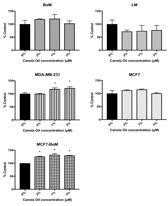
In addition to the phospholipase inhibitors, the cell lines were also treated with various dietary oils to determine the effect of these oil types and concentrations on breast tumor cell proliferation. Canola oil contains a higher ratio of ω-3 fatty acids to ω-6 fatty acids (a 1:2 ratio) compared to corn oil25. Studies have shown that the chemopreventive mechanism of ω-3 fatty acids in canola oil has to do with the.
In contrast, high levels of ω-6 fatty acids are linked to increased prostaglandin production, which increases associated cytokine activity. Research has shown that diets high in ω-6 fatty acids can also increase the growth rate of cancers other than colon cancer, including breast cancer25. Six dietary oils were selected (canola oil, avocado oil, coconut oil, olive oil, sesame oil, and soybean oil).
While canola oil has a relatively low ratio of ω-3 fatty acids to ω-6 fatty acids (1:2), olive oil has a much higher ω-6 fatty acid content, with a ratio of 1:10. In a study by Serge Hardy and colleagues, they examined the effect of saturated and unsaturated free fatty acids (FFA) on the proliferation and apoptosis of the breast cancer cell line MDA-MB-23128. Saturated fatty acids reduce the level of mitochondrial CL, which is required to retain cytochrome c.
The release of cytochrome c, a mitochondrial proapoptotic protein, results in the activation of caspases that cause cell death28. After performing cell viability experiments for each breast cancer cell line with the different phospholipase inhibitors and oils, mitochondria were isolated from the different cell cultures and liquid chromatography-mass spectrometry was performed to analyze their cardiolipin profiles. The aim was to study the working hypothesis for this experiment: that cardiolipin composition plays an important role in the mitochondrial functioning of metastatic breast cancer cells.
The BoM and LM cell lines are bone metastatic and lung metastatic subclones derived from the MDA-MB-231 cell line6,7. MDA-MB-23, T47D and MCF7 were all derived from the pleural effusion of a metastatic breast cancer patient. The T47D and MCF7 cell lines were derived from a luminal gene cluster, whereas MDA-MB-231 was derived from a basal gene cluster.

Results
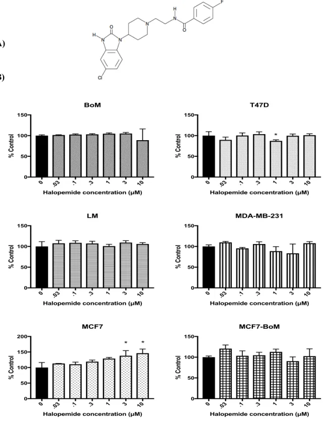
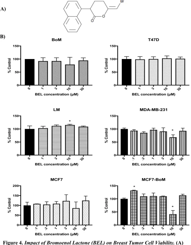
Impact of bromoenol lactone (BEL) on breast tumor viability. B) Percentage control of BEL inhibitor (B1552) on breast cancer cell lines. Liquid chromatography-mass spectrometry was performed on the breast cancer cells before the addition of the three inhibitors. The cardiolipin standard peak can be seen at 3.32 minutes on the MS of the cardiolipin-only sample.
The cardiolipin peak was not shown on the MS results for the cell lines, which does not allow comparison of the cells.
Discussion
The cardiolipin standard peak can finally be observed in the MS results for the sample containing only cardiolipin. However, the MS results for the breast cancer cell lines did not show the peak for the cardiolipin standard. Therefore, we cannot use the MS data we collected as a valid means of comparing cardiolipin composition before and after the addition of the inhibitory compounds.
However, statistically significant differences in cell viability outcomes were observed in the breast tumor cell lines as a result of treatment with each of the three phospholipase inhibitors (BEL, halopemid, D609), as well as canola oil, avocado oil, coconut oil, and olive oil.
Materials and Methods
- Cell viability assay
- Mitochondrial isolation
- Cardiolipin extraction
- Mass spectrometry
- Data
- Statistic analysis
Once the cells were washed, 10 ml of the prepared medium was added back to each culture. Then, the medium was again aspirated and 1 ml of trypsin-EDTA (0.25%, Gibco) was used to detach the cells. Once the cells were liquefied, 10 ml of the prepared medium was added to inactivate the trypsin.
Because BEL is stable at a maximum concentration of 25 g/L, the stock solution was prepared to be 20 g/L. Another 100 µL of the 30 µM sample was added to a third tube along with 900 µL of media to yield a 3 µM sample. The inhibitors in different concentrations were added to the cells seeded on 96-well plates.
Next, to stain the cells, 80 µL of SRB was added to the cells at room temperature for 10 min. After rinsing with 1% acetic acid three times, 100 µL of Tris-base solution was added to the cells. The PIERCE Mitochondria Isolation Kit for Cultured Cells was used to extract intact mitochondria from the cell cultures.
Then, 800 µL of reagent A was added to each sample and the samples were vortexed at medium speed for five seconds. Then 10 µL of reagent B was added and the samples were again vortexed for five seconds, this time at maximum speed. The instrument used to perform the mass spectrometry analysis was Waters Xevo G2-S QTOF. Cardiolipin (1 µg/ml) in isopropyl alcohol (i-PrOH) served as the standard for the mass spectrometry analysis.
The cardiolipin standard was then intended to be used as a means of comparing cardiolipin changes in each sample after the addition of the phospholipase inhibitors. This was done by dividing the average absorbance of the specific condition by the average absorbance for the control samples in each plate. Morbidity of local therapy for locally advanced metastatic breast cancer: an analysis of the Surveillance, Epidemiology, and End Results (SEER)-Medicare registry. Breast Cancer Research and Treatment, 2018, doi:10.1007/s y.