The results showed that the expression of I antigen on the erythrocyte surface is independent of the control mechanism of the biosynthesis of the beta chain of hemoglobin. Incompatibility of the red blood cells resulted in complement-mediated lysis and release of hemoglobin. The recognition of the ABO blood groups was followed by the development of safe blood transfusion.
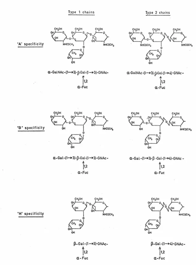
These efforts lead to the establishment of the terminal sugar sequences in polysaccharide chains of glyco-proteins which confer A,B and H specificity (7), in summary. This enzyme transfers D-galactose from uridine diphosphate galactose to the terminal non-reducing end of the H-active structure (9). This locus controls the enzyme that transfers L-fucose to the terminal galactose of the oligosaccharide.
One important aspect of the 'Lewis' locus is the lack of participation in the biosynthesis of blood group substances present in erythrocyte membranes.
The I blood groups in man
This indicates that the presence of substance I in saliva does not reflect the I status of red blood cells. Auto-anti-I agglutinins have been found in the sera of patients with acquired hemolytic anemia of the cold antibody type (26). A high incidence of cold agglutinins directed against the blood group I system has been reported in a Melanesian population living in a climate similar to that of the Orinoco Delta in Venezuela (51).
The development of the ABH antigens of the red cells has been studied by numerous investigators (5). Only in the case of thymic leukemia are there cells outside the thymus with TL+ antigens. The occurrence of embryo-specific molecules in patients with primary cancer of the liver was reported by.
Adenocarcinomas have also been reported to produce “carcinoembryonic” antigens found in the serum of patients with colorectal cancer (65).
9 tsirt h
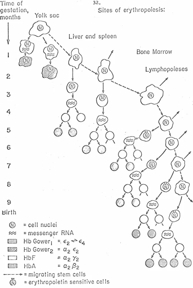
Presumably, the stem cells of the same organ populate the bone marrow until the seventh and eighth months of pregnancy. These results indicate that there is no restriction of the synthesis of a particular type of hemoglobin to a particular hematopoietic site. Nucleated erythrocytes originating from yolk sac blood islands appear between days 8 and 12 of gestation and synthesize predominantly embryonic hemoglobin (69).
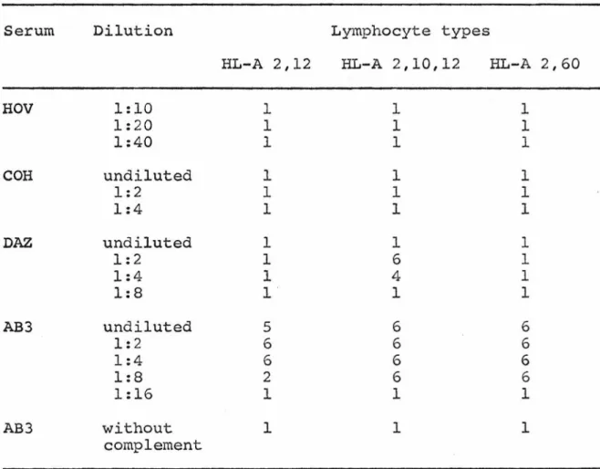
Several hereditary defects have been studied as they affect the biosynthesis of hemoglobin polypeptide chains. Stainless steel loops were used for serial dilution except in the inhibition studies where the surface tension of the solutions tested for inhibition varied considerably. Serum from patient AB3 was also a gift from the Los Angeles Red Cross Blood Bank.
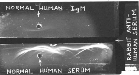
The heavy and light chains of the cold agglutinin molecules were separated on Sephadex G-200 columns in SM guanidine hydrochloride. The amino terminal sequences of the light chains were determined according to Edman (77) using an automatic Protein sequencer. Cells were washed from the clot at 37°c and the samples were studied within 24 hours of their extraction.
These experiments proved that the stromata were feasible material for chemical and enzymatic modifications, and could be used to obtain information about the molecular nature of the I-antigen. The digested membrane particles were centrifuged at 50,000 g for 15 minutes and both the pellet and the supernatant were tested for the presence of the I antigen. Fingerprints were prepared from the trypsin and subtilisin-digested stromata according to Katz et al.
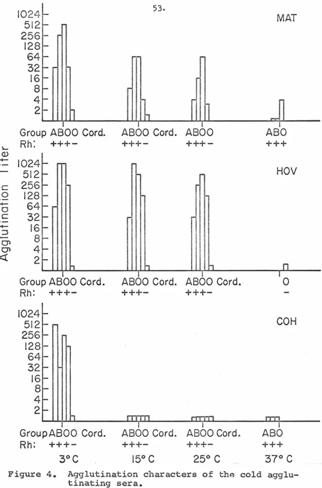
A dilution of an anti-blood type serum with an agglutination strength of 2+ was mixed with an equal volume of the solution being tested for the presence of blood type activity. The results for the three cold agglutinin sera are summarized in Figure 4 • The three sera showed different agglutination.
ABO +++
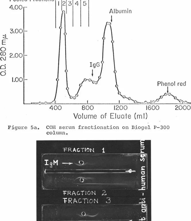
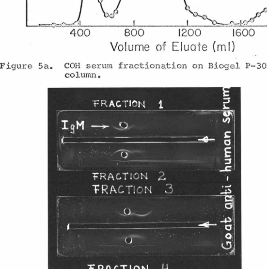
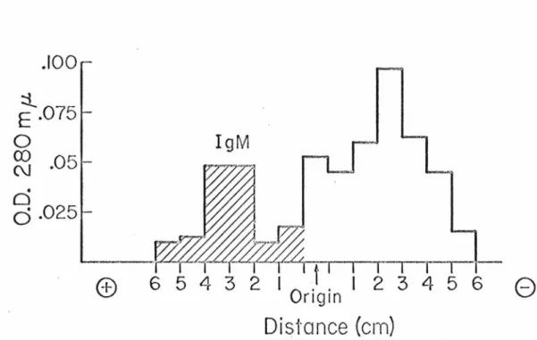
Anti-I normal in one individual of 1, DAZ, I cells agglutinated positive at 37°C and no additional cold-acting antibodies were found in this serum. The titer was very low: a 1:3 dilution gave l+ agglutination. Cold purified agglutinins behaved like the sera from which they were derived in agglutination of adult red blood cells of different ABO groups and their stromata. IgM from cold agglutinin serum after COH pneumonia was purified by disk gel electrophoresis and immunoelectrophoresis as shown in Figure 9a. The heavy and light chains were separated after reduction of disulfide bonds with dithiothreitol and alkylation with ethyleneimine.
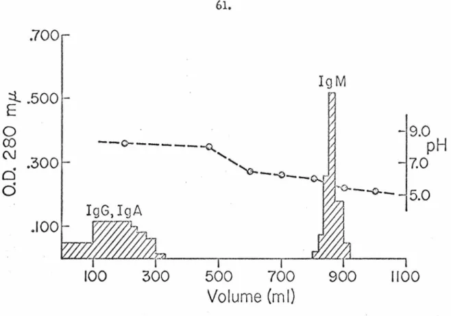
The fractionation on a previously calibrated Sephadex G-200 column in 5 M guanidine hydrochloride is depicted in Figure 8,9. • The light chains did not react with rabbit anti- The results also indicate that over 80% of the IgM globulin, the limit of detection in this system, is I-specific cold agglutinin in the serum of COH. Two major changes in postnatal erythrocyte. development was examined; one was the development of the I antigen and the other was the rate of hemoglobin in adults.
Quantitative differences were observed in I agglutinability of the two subpopulations of cells indicating that fractionation had been achieved. The rate of adult hemoglobin appearance and the rate of disappearance of "I negative" cells as a function of age .. are summarized in Figure 10 • The results show a much greater variation in the appearance of "I positive" cells than in the disappearance of HbFo At an age of 120 days a variation as high as 45% was observed, while the ·greatest variation in HbF was of the order of 20%. The values in this case must fall between the two solid lines, and the straight line of the average values will have a slope of 0.5.
Any dilution of the anti-I sera resulted in an uneven drying of the cells with frequent cell breakage. This would amount to approximately 10% of the population in umbilical cord blood; the rest of the cells will contain varying amounts of HbA and HbF. If there were two clearly distinct populations, one would expect that 32% of the cells would have given optical absorbance of less than 0.160, and the rest would not be affected by the extraction.
None of the samples had any cells containing exclusively HbF, as determined from the optical absorptions of the unfractionated cells after extraction. These results prove that the expression of the I antigen is independent of the hemoglobin content of the cell during postnatal development. It is assumed that the amount of absorbed antibody is a direct indication of the number of available antigenic sites.
The success of the experiment lies in the binding specificity and strength of the antibody used.
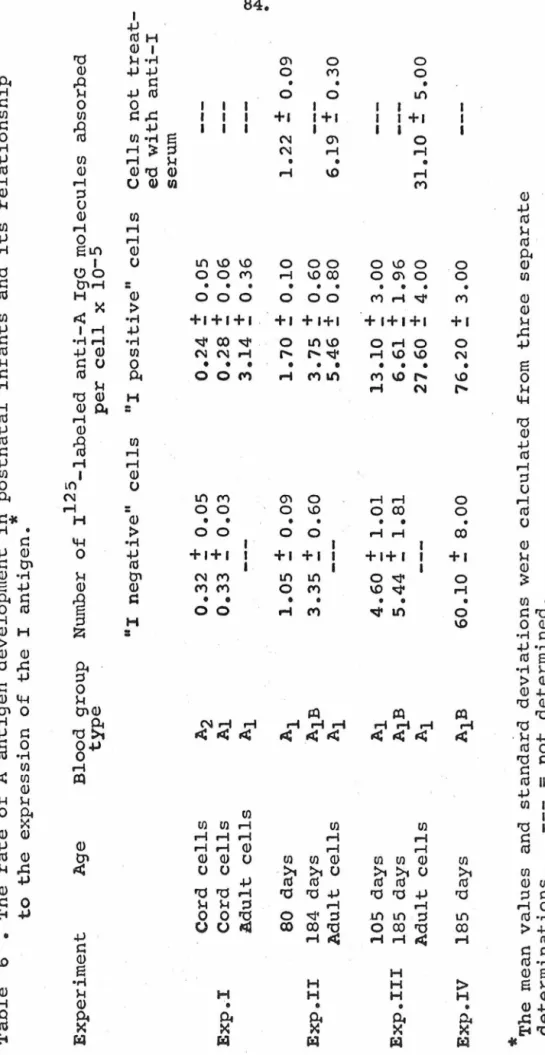
Each labeled antibody preparation was absorbed using type A or type 0 cells to eliminate most of the nonspecific binding. Two different approaches were used to determine the number of antibodies adsorbed to the cells. The data for experiments I, II, and III summarized in Table 6 were obtained by this method_ In the other method used.
Unreasonably high values were obtained in this way, but the difference between the two subpopulations of cells remained comparable to the results of the previous experiments. Separate groups of labeled antibodies were used in these experiments and they accounted for the differences in the values for the mature cells. The values obtained in experiment IV were tenfold higher than in the previous experiments due to the different method used to measure absorbed radioactivity.
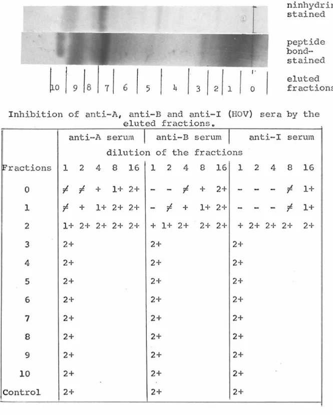
This indicates that there was no artifact due to the elution technique used to evaluate the amounts of labeled antibody molecules absorbed into the two subpopulations of cells. Modifications of I antigen can be detected by eliminating the inhibition capacity of I-positive stromata in the agglutination system of anti-I cold agglutinins and human erythrocytes from adults. Lyophilized stromata treated with these reagents absorbed anti-I IgM molecules to the same extent as the untreated samples.
Partial digestion with trypsin and chymotrypsin failed to eliminate the inhibitory capacity of the stroma. 42% of membrane carbohydrates were oxidized in this procedure, as measured by the Anthrone method. Concentrations of treated and untreated stromal samples were adjusted to the same protein concentrations.
50 microliters of diluted antiserum was mixed with 50 microliters of serially diluted stromata suspension in buffered saline, incubated for two hours at 4°C, and the unabsorbed part of the adult red blood cell membranes and umbilical cord with respect to the localization of the I ANTIGEN.
After chromatography of subtilisin-digested samples, the hydrophilic fraction represented 4-5% and the lipophilic material 35-45% of the stromal dry weight. Pooled fractions 1,2 and 3 of the leading peak from SE-Sephadex column separation contained only proteins with. Thirty percent of the digested sample was recovered after chromatography in the lipid fraction with Rf 0.8-1.0.
Thr, like the sample was prepared from the hydrophilic fraction of the subtilisin-digested whole stroma. The importance of the two-dimensional distribution of molecules on the membrane surface has been recognized by several investigators. This does not mean that molecules with I specificity are not expressed on. the surface, but implies only a low-density distribution.
This conclusion is drawn from the study of the context. between the appearance of adult hemoglobin and the expression of the I agglutinability. Stem cells present in the bone marrow are capable of repopulating the tissues of the immune and hernopoietic systems. This suggests that the major protein components of cells do not change much from birth to adulthood.
Another explanation would be on the basis that the display of the molecules responsible for the specificity is the critical factor in anti-I agglutinin absorption. This would mean that the two-dimensional distribution of the molecules is responsible for the difference in IgM binding capacity on the cell surface. Once this molecular pattern is disrupted, the difference becomes less noticeable and the specific inhibition of the two preparations may reflect only the number of molecules present in the membrane.
Part of the difficulty in evaluating this observation is rooted in the problem of antigen detection. The amino acid composition of the largest hydrophilic molecular fraction is similar to the composition reported for glycopeptides carrying blood group activity M, N. Approximately 10% of total I activity was recovered in this fraction and about 10% of B activity.
Thesis submitted to the School of Hygiene and Public Health at Johns Hopkins University.