EDITORS-IN-CHIEF
B. Morales-Nin, Mediterranean Institute for Advanced Studies, Esporles, Spain Email: [email protected]
A.E. Punt, University of Washington, Seattle, Washington, United States of America E-mail: [email protected]
EDITORS
J. Cao, North Carolina State University, Morehead City, North Carolina, United States. E-mail: [email protected]
J. M. Cope, NOAA Fisheries, Northwest Fisheries Science Center, Seattle, Washington, United States. E-mail: [email protected] I. Figueiredo, Portuguese Institute for Sea and Atmosphere (IPMA), Algés, Portugal, [email protected]
N. Madsen, Aalborg University, Aalborg, Denmark, E-mail: [email protected]
R Millar,University of Auckland, Auckland, New Zealand, Email:[email protected] M. T. Spedicato, COISPA, Bari, Italy, E-mail:[email protected]
C. Speir, NOAA Fisheries, Southwest Fisheries Science Center, Santa Cruz, California, United States of America,E-mail: cameron.
[email protected]
J. Viñas, University of Girona, Girona, Spain. E-mail: [email protected]
EDITORIAL ADVISORY BOARDR. Ahrens, Hawaii, USA J. Alós, Illes Balears, Spain P. Arechavala, Illes Balears, Spain
S. X. Cadrin, Dartmouth, Massachusetts, USA A. Campos, Lisboa, Portugal
L. G. Cardoso, Rio Grande, Brazil S. J. Cooke, Ottawa, Canada M. Cutler, Massachusetts, USA P. Díaz-Jaimes, Mexico City, Mexico N. N. Fabré, Maceió, Brazil
K. Ganias, Thessaloniki, Greece M. H. Griffiths, Wellington, New Zealand E. Grimaldo, Trondheim, Norway J. C. Groeneveld, Durban, South Africa Cemal Gücü, Mersin, Turkey
R. Henriques, Pretoria, South Africa B. Herrmann, Hirtshals, Denmark A. Hesp, Western Australia, Australia K. Kroetz, Arizona, USA
R. Lae, Brest, France
B. J. Langseth, Washington, USA Y. Li, Raleigh, North Carolina, USA
A. Linnane, South Australia, Australia F. Lucena, Recife, Brazil
D. MacLennan, Perth, UK F. Maltagliati, Pisa, Italy
W. Mandelman, Massachusetts, USA E. Massuti, Illes Balears, Spain M. N. Maunder, California, USA R. Millar, Auckland, New Zealand
J. Morgan, Newfoundland and Labrador, Canada F. G. O'Neill, Aberdeen, UK
H. Okamura, Yokohama, Japan H. Özbilgin, Mersin, Turkey C. Pinto, Genova, Italy
I. van Putten, British Columbia, Canada A. D. Rijnsdorp, Ijmuiden, Netherlands G. A. Rose, British Columbia, Canada U. R. Sumaila, British Columbia, Canada S. Tian, Shanghai, China
C.Tibuzio, Seattle, USA
I. Zarraonandia, Vask Country, Spain
Fisheries Research 264 (2023) 106731
Available online 3 May 2023
0165-7836/© 2023 Elsevier B.V. All rights reserved.
A comprehensive description of the exoskeleton of six Lobster species (Genus Panulirus) in Aceh Province, Indonesia
Yusrizal Akmal
a,b, Irfannur Irfannur
a,*, Muliari Muliari
c, Agung Setia Batubara
d, Muchammad Yunus
e, Hani Plumeriastuti
f, Yeni Dhamayanti
gaDepartment of Aquaculture, Faculty of Agriculture, Universitas Almuslim, Bireuen, Indonesia
bSains Veteriner Doctoral Program, Faculty of Veterinary Medicine, Universitas Airlangga, Surabaya, Indonesia
cDepartment of Marine Science, Faculty of Agriculture, Malikussaleh University, Aceh Utara, Indonesia
dDepartment of Biology, Faculty of Mathematics and Natural Sciences, Universitas Negeri Medan, North Sumatra, Indonesia
eDepartment of Veterinary Science, Faculty of Veterinary Medicine, Universitas Airlangga, Surabaya, Indonesia
fDepartment of Veterinary Parasitology, Faculty of Veterinary Medicine, Universitas Airlangga, Surabaya, Indonesia
gDepartment of Veterinary Anatomy, Faculty of Veterinary Medicine, Universitas Airlangga, Surabaya, Indonesia
A R T I C L E I N F O Handled by Jason M. Cope Keywords:
Anatomy Morphology Cephalothorax Abdomen Caudal
A B S T R A C T
Lobster (Panulirus spp.) is a leading commodity on a local and export scale in Aceh Province, Indonesia, where the population has experienced a significant decline in recent years. This study describes the exoskeleton morphology of six lobster species distributed in Aceh Province, Indonesia. The research was carried out in 2022 with stages covering sample preparation, sample shooting, and morphological analysis. The results of the study showed that there were six lobster species in Aceh including Panulirus penicillatus, P. homarus, P. longispes, P. ornatus, P. versicolor, and P. polyphagus. The anatomy of the lobster exoskeleton of six species of P. penicillatus, P. homarus, P. longispes, P. ornatus, P. versicolor and P. phloyphagus has marked differences, where each part of the cephalothorax (cervical sulcus, sternum, epistome, maxillaped, pereiopod), Abdominal (segment, pleura, pleopod) and caudal (telson, uropod, and protopod) have different shapes. In addition, differences were also found in the color and presence of spines on the dorsal body.
1. Introduction
Lobster is a high economic commodity from Aceh Province (Wahju and Riyanto, 2017). This commodity can be a strategic solution for the economic development of fishermen (Irfannur et al., 2017). Currently, more than 20 lobster species have been identified in the world (Lipcius and Eggleston, 2000), some of which are distributed in the waters of Aceh, including Panulirus homarus, P. longipes longipes, P. ornatus, P.
penicillatus, P. polyphagus, and P. versicolor (Damora et al., 2021).
Lobster is a resource that can be recovered (renewable resources) but increased fishing without any restrictions causes population decline (Priyambodo et al., 2020). The high intensity of lobster catching on the south west coast of Aceh is feared to threaten the sustainability of lobster resources in Aceh Province. Damora et al. (2021) also revealed that overfishing has been occurring in the Aceh Province since 2008.
Therefore, information regarding the inventory of lobster species is important as a conservation effort so that future management of this
lobster can be designed. One of the conservation efforts that needs to be done is species identification based on exoskeleton analysis. Lobster management in Indonesia has been regulated by the Minister of Marine Affairs and Fisheries Regulation Number 12 of 2020 (PERMEN, 2020) According to the Cockcroft et al. (2011), currently lobster is declared to be still in the low risk category, but it is predicted that in the next few years it will increase to be threatened with extinction if management is not carried out now.
Several studies that have been reported include studies of lobster morphology (Simpson, 1985; Meeren and Uksnoy, 2000), lobster his- topathology exposed to heavy metals (Maharajan et al., 2012), repro- ductive system histology (Pillai et al., 2014), digestive system (Welden et al., 2015), genetic structure (Irisarri et al., 2018). Meanwhile, studies regarding the identification of lobsters based on exoskeletons are still very limited, causing this research to be feasible to be developed. The exoskeleton is a morphological character key for identifying lobster species (Raabe et al., 2007; Davies et al., 2014) and its implications for
* Corresponding author.
E-mail address: [email protected] (I. Irfannur).
Contents lists available at ScienceDirect
Fisheries Research
journal homepage: www.elsevier.com/locate/fishres
https://doi.org/10.1016/j.fishres.2023.106731
Received 8 November 2022; Received in revised form 3 April 2023; Accepted 21 April 2023
mechanical behavior (Cheng et al., 2008). In addition, identification of lobsters based on exoskeleton characters is easier and cheaper than other analyses. Exoskeleton analysis is also one of the lobster conser- vation efforts by identifying intra-interspecies characters for future management plans.
2. Materials and methods 2.1. Site and time of research
The research was conducted in 2022 with research stages covering preparation, photography, and morphological analysis. Sampling was carried out in the coastal area of Aceh Jaya Regency, Aceh Province, Indonesia (Fig. 1). Lobster morphology preparation and analysis was completed at the Integrated Aquaculture Laboratory, Almuslim Uni- versity, Indonesia.
2.2. Sample preparation
The lobster used in this study was obtained from the direct catch of fishermen in Lhok Ringai, Aceh Jaya Regency, Aceh Province, Indonesia. The sampling distance is about 2–4 miles from the coast. Ten samples per lobsters species were obtained, with the weights around 500 g and lengths ranging from 18 to 25 cm.
Collected samples were kept alive by sprinkling individuals with water, wet sand and sawdust. The goal is to keep the lobster in low metabolic activity (fainting). Before the lobster samples were put into the box, each lobster sample was wrapped in paper (two layers of flip chart paper per lobster sample and flip chart paper size 65×100 cm)
and tightly closed with a total of 10 lobsters per box and then the samples were sent to the laboratory for further analysis (lobster samples were photographed first, then identified by exoskeleton).
2.3. Sample photography
Lobster photos were taken in each dorsal, ventral, lateral, medial, anterior and posterior using a Canon EOS 200D digital camera. Photo- graphs of samples were taken in an upright position, with a green background, and using a measuring instrument (ruler with an accuracy of 0.05cm) as the size of sample. Image editing is done using Adobe Photoshop CS 6 software (Table 1).
2.4. Exoskeleton determination
The results of image editing are illustrated scientifically using manual methods, where each lobster species is drawn in detail using a pencil covering all specific exoskeleton characters (Akmal et al., 2022).
The illustration starts with the whole figure of lobster body, the dorsal and ventral cephalothorax, the dorsal and ventral abdominal somites, the dorsal and ventral caudal sections, the pereiopod, pleopod and maxillaped sections. Determination of anatomical nomenclature using references to studies that have been reported, namely Goy (2010), L˝ow et al. (2016), and Hettiarachchi et al. (2022).
2.5. Data analysis
The data are presented in the tables and scientific illustrations. Data analysis was conducted descriptively by comparing the specific
Fig. 1. Map of lobster fishing areas in Aceh Jaya Regency, Aceh Province, Indonesia.
characters between the collected lobster species.
3. Results
3.1. Morphology of lobster
Lobsters have an exoskeleton which is a hard layer and is equipped with spines. Each lobster species has specific characteristics such as differences in colors. In addition, the development of large and small spines starts from the head to the tail, but on the head the spines are more dominant. Morphologically, lobsters are divided into three major regions consisting of the cephalothorax, abdomen and caudal regions.
The cephalothorax region is the anterior part of the combined head and chest that is wrapped in a slightly cylindrical carapace, swells and fuses with the thorax. Abdomen region is part of the body in the form of segments consisting of 6 segments. Segments 1–3 are called tergums, while segments 4–6 are called pleurons. The caudal region is the tail formed by the urupod and telson.
3.1.1. Cephalothorax
The cepha area consists of the antennae, antennae, cornea, rostrum,
ophthalmic artery, protochepalon, oris, maxilliped, epistome, and mandible. Antennules have a stem shape that is not elongated, at each base of the anterior part of the rostrum branching into two parts resembling a vertical V. Antennae have a round rod shape with a long tapered tip that exceeds the length of the antennae, and has sharp, jagged spines on the posterior antenna. Antennule functions as a tool for touch, taste and smell. In addition, the antenna is also used as a pro- tective device to detect food or objects from a distance, while the antennae function as chemical and mechanical sensors, namely air, temperature, moisture content, vibration, and detect objects at close range. Cornea is a compound eye that is stalked and is located diventral lateral to the rostrum process. Cornea serves to distinguish between dark and light, observe movement, and determine the position of food.
Rostrum is the leading part of the cepha which has a sharp tip and serves as a tool to attack the enemy. The rostrum consists of the rostrum processus and the rostrum spine. Rostrum spine is a sharp-tipped spine and is located anteriorly. Rostrum processus has a curved and sharp shape at the top of the eye. Rostrum processus serves as a tool for the body’s defense in a state of danger. The ophthalmic artery is a branch that supplies blood to the eye. The ophthalmic artery has the function of carrying blood away from the eye and branching into smaller vessels Table 1
Comparison of the exoskeleton structure of six Panulirus species in Aceh Province, Indonesia.
No Region Structure P. penicillatus P. homarus P. ornatus P. polyphagus P. versicolor P. longispes 1 Cephalothorax Form ✓ Width and length
in the thorax ✓ Long and slender
in the thorax ✓ Length and width in the thorax
✓ Long and slender in the thorax
✓ Width and length in the thorax
✓ Long and slender in the thorax
✓ There are more small bumps around the thorax
✓ There are a few small bumps on the thorax
✓ There are a few small bumps on the thorax
✓ The little bumps are barely visible
✓ There are no small bulges on the thorax
✓ There are many small bumps on the thorax Spines ✓ There are more
spines but blunt and irregularly located
✓ There are few spines but sharp and irregularly located
✓ There are many spines but blunt and slightly irregular
✓ There are few spines but sharp and parallel
✓ There are some
sharp spines ✓ There are more spines but sharp and slightly irregular
✓ The front spines are developing well
✓ The front spines are not well developed
✓ The front spines are developing well
✓ The front spines are developing well
✓ The front spines are developing well
✓ The front spines are developing well
✓ The front spines are 4 sharp and parallel spines
✓ The front spines are 3 spines, there are 1 small blunt spine and 2 large sharp spines
✓ The front spines have 2 small blunt and 2 large sharp spines
✓ The front spines are 2 large sharp spines
✓ The front spines have 2 small and 2 large sharp spines
✓ The front spines are 2 large and sharp spines
Cepha
dorsal ✓ Has a complete sulcus to the thorax
✓ Does not have a sulcus in the thorax
✓ Does not have a sulcus in the thorax
✓ Has a complete sulcus to the thorax
✓ Has a complete sulcus to the thorax
✓ Has a complete sulcus to the thorax
✓ Spina on pointed
carapace ✓ Spina on sharp
carapace ✓ Spina on pointed
carapace ✓ Spina on sharp
carapace ✓ Spina on pointed carapace
✓ Spina on sharp carapace Cepha
ventral ✓ There are 6 legs and there is no protrusion at the end of the sixth leg
✓ There are 6 legs and there is no protrusion at the end of the sixth leg
✓ There are 6 legs and there is no protrusion at the end of the sixth leg
✓ There are 6 legs and there is no protrusion at the end of the sixth leg
✓ There are 6 legs and there is a protrusion at the end of the sixth leg
✓ There are 6 legs and there is a protrusion at the end of the sixth leg
✓ There are no curves in the middle of the body
✓ In the middle of the body there is an indentation
✓ In the middle of the body there is an indentation
✓ There are no curves in the middle of the body
✓ There are no curves in the middle of the body
✓ In the middle of the body there is an indentation
✓ There are 3 blunt spines and 3 sharp spines on the underside of the antenna
✓ There are 3 sharp spines on the underside of the antenna
✓ There are 3 sharp spines on the underside of the antenna
✓ There are 3 sharp spines on the underside of the antenna
✓ There are 3 sharp spines on the underside of the antenna
✓ There are 3 sharp spines and 6 blunt spines on the underside of the antenna
2 Abdomen ✓ Pleopod sheet
wide and long ✓ Pleopod sheet
wide ✓ Pleopod sheet
wide and long ✓ Pleopod sheet
long and pointed ✓ Pleopod sheet long and pointed
✓ Pleopod sheet wide and long
✓ The lateral side is pointed and slender
✓ The lateral side is pointed and slender
✓ Wide and pointed lateral side
✓ Wide and pointed lateral side
✓ Wide and pointed lateral side
✓ The lateral side is pointed and slender
3 Caudal ✓ Telson spines are
rough and parallel
✓ Telson spines are smooth and parallel
✓ Telson spines are rough and blunt
✓ Fine telson
spines ✓ Telson spines are rough and parallel
✓ Telson spines are rough and parallel
that open into a network of spaces, as well as a series of sinuses that drain blood back to the heart. The protocephalon is the stalk and the place where the blood vessels unite in the eye. The protocephalon functions as a sensory device as well as to detect food.
Maxillaped is a form of small walking legs that have fine hairs on each part of the foot. Maxillaped has a function as a tool to filter food and bring food to the mouth. The mandible is formed from a pair of segmental appendages that correspond to the pedipalps of Chelicherata.
The mandible functions as a sense, regulating the movement of water around the gills. The epistome is the underside of the head formed by a broad plate below the base of the antenna. The epistome has a function to protect the muscles attached to the epistome wall. The oris is a cavity where food and water enter which is located on the ventral part of the cepha surrounded by the mandible and maxillaped.
The dorsal thorax region consists of the cervical sulcus, branchios- tegite, while the ventral thorax consists of the sternum, and pereiopod.
Pereiopods have 5 pairs of walking legs which have fine hairs on the ends of the feet, and in female pereiopods on the fifth leg there is a small capitan while the male pereiopod does not have a capitate. Pereiopods are 5 pairs of lobster walking legs to move around. The cervical sulcus is a line that forms an indentation on the dorsal of the lobster’s head.
Sulcus cervival has a different form of curve in each species. The ster- num is located on the ventral surface of five pairs of foot bearing seg- ments consisting of left and right, the first to fifth foot bearing segments and has a different indentation shape in each species. The sternum serves as a place of attachment for the pereiopod and protects the organs below the sternum. Branchiostegite is the side of the lobster head that serves to protect the gills. Branchiotegite has an elongated shape located lateral to the lobster head.
3.1.2. Abdominal
The abdominal region consists of the pleopods, pleura and abdom- inal somites. Pleopod is a locomotion tool in the form of 4 pairs of swimming legs, which have different ends such as length, width, and point. Pleopods function to swim with a push backwards, as a counter- weight to swimming when at the bottom of the water, the female pleopod has a biramous and long bear where the eggs are and are cemented. The pleura is an open branch space on each body between the carapace edge and the base of the perciopod that points upward. The pleura has a wide, pointed, slender lateral side and serves as a liaison between the thorax and the abdominal somites. Abdomen somites is part of the lobster body which consists of 6 segments and the abdomen so- mites serves to assist the movement of the lobster.
3.1.3. Caudal
The caudal region consists of the anus, uropod, protopod, and telson.
The anus has a round shape that is wide and flat, splitting into two parts on the lower surface of the telson, the anus is an organ of reproduction and digestion. Uropods are the leaf appendages of the last caudal segment. Uropod has an oval shape and at the end of its base there are fine spines, and serves as a swimming tool for balance. Telson has the same shape as the uropod but the telson is located in the middle of the tail, has a strong propulsion function for swimming. The protopod is the anterior dorsal part which is the basis of the telson and uropod parts associated with the sixth somites. Uropod and telson is the tail that is connected to form a fan and serves as a means of controlling the di- rection back and forth when swimming.
3.2. Lobster exoskeleton anatomy comparison
In the dorsal cephalothorax region (carapace) of P. penicillatus has a greenish-brown color, and the ventral part is yellowish green (Fig. 2A).
Yellow spots also appear on P. penicillatus and P. homarus ventrally. The dorsal part of the cephalothorax of P. homarus has a blue-green color and on the ventral side it is yellowish (Fig. 2B). In the cephalothorax region of P. longispes on the dorsal side it has a dark brown to reddish color, while on the ventral side it is light brown and there are white spots (Fig. 2C).
On the dorsal cephalothorax of P. ornatus has a blue color mixed with a little black, and on the ventral part it is white mixed with orange color and there are orange spots (Fig. 2D). P. versicolor in the dorsal cepha- lothorax region has a black color which is more dominant than tur- quoise, while in the ventral section it has white color and there are black spots (Fig. 2E). The dorsal cephalothorax of P. pholypagus has a light brown color and the ventral side is orange (Fig. 2F) (Figs. 3 –8).
On the dorsal cephalothorax of P. penicillatus has spines that grow irregularly and has two large spines of rostrum spine that develop anteriorly. The anteriodorsal rostrum has four sharp spines, consisting of two medium spines and two small spines, while in the anterior part of the epistome there are three medium-sized blunt spines (Fig. 9A). The spines of the dorsal cephalothorax of P. homarus grow irregularly and have two large spines (rostrum spine) that develop anteriorly. In addi- tion, the anteriodorsal rostrum has three sharp spines, while the anterior epistome has three blunt spines (Fig. 10A).
In the dorsal cephalothorax of P. longispes has spines that grow par- allel and neatly arranged, there are two large spines that develop anteriorly, while the anteriodorsal section has four sharp spines, namely two small spines and two large spines. In addition, in the anterior part of
Fig. 2.Genus Panulirus. (A) Panulirus penicillatus, (B) Panulirus homarus, (C) Panulirus longispes, (D) Panulirus ornatus, (E) Panulirus versicolor, (F) Panulirus pholypagus.
Bar scale: 2 cm.
Fig. 3. The morphology of Panulirus penicillatus appears dorsal (A) and ventral (B). Where AN: antennule, AT: antennae, PR: pereiopod, SC: sulcus cervical, BC:
branchiostegite, PO: pleopod, PL: pleura, C: cornea, PC: processus rostrum, MP: maxilliped, SN: sternum, ANS: anus, ADS: abdomen somites, CT: cephalothorax, CD:
caudal. Bar scale: 2 cm for Fig. A dan B; Bar scale: 1 cm for figure ♂ and ♀.
the epistome there are nine, namely three large and sharp spines and six small blunt spines (Fig. 11A). In the dorsal region of the cephalothorax of P. ornatus has spines that develop regularly, and in the anteriodorsal section there are four spines, namely two blunt spines and two sharp spines. The spina rostrum has two large spines developing anteriorly, while the anterior epistome has three small and blunt spines (Fig. 12A).
Spines on the dorsal cephalothorax of P. versicolor which have spines on the cepha and spina rostrum have two large spines that develop anteriorly. On the anteriodorsal side there are two blunt spines and two sharp spines. Anterior to the epistome are three large, sharp spines (Fig. 13A). On the dorsal cephalothorax of P. pholypagus has spines that
grow parallel and neatly arranged. The rostrum spine has two large spines and develops anteriorly. The anteriodorsal section has two large and sharp spines, while the anterior epistome has two sharp, large and small spines (Fig. 14A).
On the dorsal side of the P. penicillatus abdomen has six segments that are reddish green (Fig. 2A), while on the ventral side of the abdomen there are four pairs of pleopod sheets that are wide and long (Fig. 15A) and have a dark green color (Fig. 2A). Meanwhile, on the lateral side of the abdomen there are white spots surrounding the pleura which are wide and pointed (Figs. 2A and 15B). In the dorsal abdominal region of P. homarus has six greenish-brown segments (Fig. 2B), while in the Fig. 4. The morphology of Panulirus homarus appears dorsal (A) and ventral (B). Where AN: antennule, AT: antennae, PR: pereiopod, SC: sulcus cervical, BC:
branchiostegite, PO: pleopod, PL: pleura, C: cornea, PC: processus rostrum, MP: maxilliped, SN: sternum, ANS: anus, ADS: abdomen somites, CT: cephalothorax, CD:
caudal. Bar scale: 2 cm for Fig. A dan B; Bar scale: 1 cm for figure ♂ and ♀.
ventral abdomen there are four pairs of pleopod sheets that are wide (Fig. 16A) and have an orange color on each sheet (Fig. 2B). On the lateral side of the abdomen there are white spots surrounding the pleura which are slender and pointed (Figs. 2B and 16B). P. ornatus has six dorsal abdominal segments which are blue-black in color (Fig. 2D). On the ventral abdomen there are four pairs of pleopod sheets that are long and wide (Fig. 17A) and each sheet has an orange color (Fig. 2D). On the lateral side of the abdomen there is a pleura that is broad and pointed (Fig. 17B) and has white spots surrounding it (Fig. 2D).
On the dorsal of P. longispes abdomen region has six segments that are dark reddish brown (Fig. 2C) and on the ventral abdomen there are four
pairs of pleopod sheets that are long and wide (Fig. 18A) and reddish brown on each sheet (Fig. 2C). On the lateral side of the abdomen there are white and red spots surrounding the pleura which are slender and tapered (Figs. 2C and 18B). On the dorsal abdomen of P. versicolor has six segments that are light green mixed with black and white (Fig. 2E) and on the ventral abdomen it has four pairs of pleopod sheets that are long and slender (Fig. 19A) and have black and white on the abdomen (Fig. 2E). On the lateral side of the abdomen is the pleura, which is broad and pointed (Fig. 19B), and has white lines surrounding it (Fig. 2E).
On the dorsal of P. pholypagus abdomen region has six segments that are light brown mixed with dark brown (Fig. 2F) while on the ventral Fig. 5. The morphology of Panulirus versicolor appears dorsal (A) and ventral (B). Where AN: antennule, AT: antennae, PR: pereiopod, SC: sulcus cervical, BC:
branchiostegite, PO: pleopod, PL: pleura, C: cornea, PC: processus rostrum, MP: maxilliped, SN: sternum, ANS: anus, ADS: abdomen somites, CT: cephalothorax, CD:
caudal. Bar scale: 2 cm for Fig. A dan B; Bar scale: 1 cm for figure ♂ and ♀.
abdomen there are 4 pairs of pleopod sheets that are wide and slender (Fig. 20A) and pink on each sheet (Fig. 2F). Meanwhile, on the lateral side of the abdomen there is a pleura that is wide and pointed (Fig. 20B) and has white spots around it (Fig. 2F).
In the dorsal of P. penicillatus caudal region has five fin sheets (Fig. 15A) consisting of uropods and telsons with reddish-green
coloration on each sheet (Fig. 2A) accompanied by the anteriodorsal part there are small parallel and sharp spines. The posterior part of the uropod has a broadened shape while the posterior telson is not dilated (Fig. 15A). P. homarus in the dorsal caudal section contained five fin sheets (Fig. 16A), consisting of a uropod and telson with orange color on each sheet (Fig. 2B) accompanied by a few blunt smooth spines on the Fig. 6.The morphology of Panulirus ornatus appears dorsal (A) and ventral (B). Where AN: antennule, AT: antennae, PR: pereiopod, SC: sulcus cervical, BC:
branchiostegite, PO: pleopod, PL: pleura, C: cornea, PC: processus rostrum, MP: maxilliped, SN: sternum, ANS: anus, ADS: abdomen somites, CT: cephalothorax, CD:
caudal. Bar scale: 2 cm for Fig. A dan B; Bar scale: 1 cm for figure ♂ and ♀.
Fig. 7. The morphology of Panulirus longispes appears dorsal (A) and ventral (B). Where AN: antennule, AT: antennae, PR: pereiopod, SC: sulcus cervical, BC:
branchiostegite, PO: pleopod, PL: pleura, C: cornea, PC: processus rostrum, MP: maxilliped, SN: sternum, ANS: anus, ADS: abdomen somites, CT: cephalothorax, CD:
caudal. Bar scale: 2 cm for Fig. A dan B; Bar scale: 1 cm for figure ♂ and ♀.
Fig. 8. The morphology of Panulirus pholypagus appears dorsal (A) and ventral (B). Where AN: antennule, AT: antennae, PR: pereiopod, SC: sulcus cervical, BC:
branchiostegite, PO: pleopod, PL: pleura, C: cornea, PC: processus rostrum, MP: maxilliped, SN: sternum, ANS: anus, ADS: abdomen somites, CT: cephalothorax, CD:
caudal. Bar scale: 2 cm for Fig. A dan B; Bar scale: 1 cm for figure ♂ and ♀.
anteriodorsal side. The posterior part of the uropod has a wide triangular shape while the posterior telson has a wide round shape (Fig. 16A). On the dorsal caudal part of P. ornatus there are five fan-shaped sheets (Fig. 17A) which have an orange color (Fig. 2D), and on the ante- riodorsal there are sharp parallel fine spines on the uropod and telson.
Posteriorly the uropod and telson have a broadened shape (Fig. 17A).
The dorsal of P. longispes caudal region has five fan-shaped sheets (Fig. 18A) with reddish brown color (Fig. 2C), and on the anteriodorsal region there are sharp parallel fine spines on the uropod and telson
sections. The posterior region of the uropod has a broadened shape while the posterior telson is not dilated (Fig. 18A). On the dorsal of P. versicolor caudal region has a light green color (Fig. 2E) of five fin sheets, and on the anteriodorsal it has a few blunt fine spines. The posterior part of the uropod has a broadened shape while the posterior telson is not dilated (Fig. 19A). P. pholypagus caudal dorsally has five fin sheets (Fig. 20A) which have a pink color (Fig. 2F) and on the ante- riodorsal there are few sharp fine spines. Posteriorly the uropod and telson have a broadened shape (Fig. 20A).
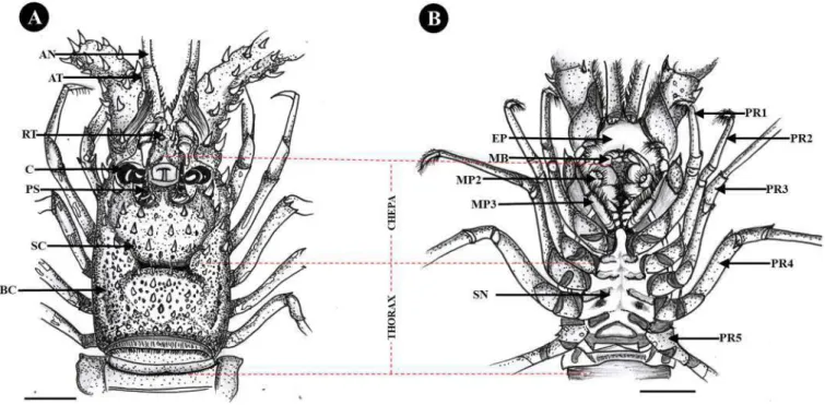
Fig. 9. The cephalothorax morphology of Panulirus penicillatus appears dorsal (A) and ventral (B). Where AN: antennule, AT: antennae, PR: pereiopodes, RT: rostrum, C: cornea, PC: prosesus rostrum, SC: sulcus cervicalis, BC: branchiostegite, SN: sternum, MP: maxilliped, MB: mandible, EP: epistome. Bar scale: 2 cm.
Fig. 10. The cephalothorax morphology of Panulirus homarus appears dorsal (A) and ventral (B). Where AN: antennule, AT: antennae, PR: pereiopodes, RT: rostrum, C: cornea, PC: prosesus rostrum, SC: sulcus cervicalis, BC: branchiostegite, SN: sternum, MP: maxilliped, MB: mandible, EP: epistome. Bar scale: 2 cm.
In the dorsal cephalothorax region of P. penicillatus, there is a cervical sulcus which is wide, long and clearly shaped and reaches the thoracic region. In addition, there are many small and blunt spines (Fig. 9A). The dorsal cephalothorax of P. Homarus is long and slender, and has an indistinct cervical sulcus, and there are small blunt projections (Fig. 10A). In the dorsal cephalothorax of P. longispes has a clear cervical sulcus that reaches the thoracic region, and is long, slender and has
smaller blunt projections (Fig. 11A).
In the dorsal of P. ornatus cephalothorax region has a clear cervical sulcus shape and wide, long, and a few small blunt projections (Fig. 12A). In the dorsal cephalothorax region of P. versicolor has a wide, long size and there is a cervical sulcus shape that is not clearly visible.
There are only a few small blunt projections in the cepha (Fig. 13A).
P. pholypagus in the dorsal cephalothorax region is long, slender and has Fig. 11. The cephalothorax morphology of Panulirus longispes appears dorsal (A) and ventral (B). Where AN: antennule, AT: antennae, PR: pereiopodes, RT: rostrum, C: cornea, PC: prosesus rostrum, SC: sulcus cervicalis, BC: branchiostegite, SN: sternum, MP: maxilliped, MB: mandible, EP: epistome. Bar scale: 2 cm.
Fig. 12. The cephalothorax morphology of Panulirus ornatus appears dorsal (A) and ventral (B). Where AN: antennule, AT: antennae, PR: pereiopodes, RT: rostrum, C: cornea, PC: prosesus rostrum, SC: sulcus cervicalis, BC: branchiostegite, SN: sternum, MP: maxilliped, MB: mandible, EP: epistome. Bar scale: 2 cm.
a clear cervical sulcus shape and reaches up to the thoracic region. There are many small blunt projections (Fig. 14A).
In the ventral cephalothorax region of P. penicillatus has six pereio- pods (walking legs) which have many fine hairs on the tips of the feet. At the base of the sixth leg there is no extension towards the median ster- num (Fig. 9B). In the ventral cephalothorax of P. homarus has six pe- reiopods with a few fine hairs on each toe and a median extension of the sternum at the base of the sixth leg (Fig. 10B). In the ventral
cephalothorax of P. longispes has six pereiopods which have many fine hairs on the tips of the feet. Meanwhile, at the base of the sixth pereiopod there is a medial extension of the sternum (Fig. 11B).
On the ventral cephalothorax of P. ornatus has six pereiopods which are overgrown with fine hairs on the tips of the feet and there is no median extension of the sternum at the base of the sixth leg (Fig. 12B). In the ventral cephalothorax of P. versicolor has six pereiopods which are overgrown with fine hairs at the ends of the legs. There is an indistinct Fig. 13.The cephalothorax morphology of Panulirus versicolor appears dorsal (A) and ventral (B). Where AN: antennule, AT: antennae, PR: pereiopodes, RT: rostrum, C: cornea, PC: prosesus rostrum, SC: sulcus cervicalis, BC: branchiostegite, SN: sternum, MP: maxilliped, MB: mandible, EP: epistome. Bar scale: 2 cm.
Fig. 14.The cephalothorax morphology of Panulirus polyphagus appears dorsal (A) and ventral (B). Where AN: antennule, AT: antennae, PR: pereiopodes, RT:
rostrum, C: cornea, PC: prosesus rostrum, SC: sulcus cervicalis, BC: branchiostegite, SN: sternum, MP: maxilliped, MB: mandible, EP: epistome. Bar scale: 2 cm.
extension towards the median of the sternum at the base of the sixth leg (Fig. 13B). On the ventral cephalothorax of P. pholypagus has six pe- reiopods with a few fine hairs at the ends of their feet. Meanwhile, at the base of the sixth pereiopod, there is a medial extension of the sternum (Fig. 14B).
In the ventral cephalothorax region of P. penicillatus, there is a ster- num that has an indistinct indentation (Fig. 9B). In the ventral cepha- lothorax of P. homarus did not have a clear sternal shape (Fig. 10B). In the ventral cephalothorax of P. longispes, there is a sternum which has a clear indentation (Fig. 11B). In the ventral cephalothorax region of Fig. 15. Abdominal and caudal morphology of Panulirus penicillatus appears dorsal (A) and ventral (B). Where PT: protopod, PO: pleopod, PL: pleura, ADS: abdomen somites, TS: telson, ED: endopod, EP: exopod, ANS: anus, TS: tail fan. Bar scale: 2 cm.
Fig. 16. Abdominal and caudal morphology of Panulirus homarus appears dorsal (A) and ventral (B). Where PT: protopod, PO: pleopod, PL: pleura, ADS: abdomen somites, TS: telson, ED: endopod, EP: exopod, ANS: anus, TS: tail fan. Bar scale: 2 cm.
P. ornatus, there is an indistinct sternal shape (Fig. 12B). In the ventral cephalothorax of P. versicolor did not have a clear sternal shape (Fig. 13B). In the ventral cephalothorax region of P. pholypagus has a
distinct sternal shape (Fig. 14B).
In the ventral cephalothorax region in the mouth, there are two auxiliary legs called maxillipeds. In the third maxillaped of P. penicillatus Fig. 17.Abdominal and caudal morphology of Panulirus ornatus appears dorsal (A) and ventral (B). Where PT: protopod, PO: pleopod, PL: pleura, ADS: abdomen somites, TS: telson, ED: endopod, EP: exopod, ANS: anus, TS: tail fan. Bar scale: 2 cm.
Fig. 18. Abdominal and caudal morphology of Panulirus longispes appears dorsal (A) and ventral (B). Where PT: protopod, PO: pleopod, PL: pleura, ADS: abdomen somites, TS: telson, ED: endopod, EP: exopod, ANS: anus, TS: tail fan. Bar scale: 2 cm.
had many fine hairs around it (Fig. 21B). In the ventral cephalothorax of P. homarus, there is a third maxillaped which has many fine hairs around it (Fig. 22B). In the third dimaxillaped of P. longispes has many fine and
dense hairs in the ventral cephalothorax region (Fig. 23B). On the third maxillaped of P. ornatus, there are few fine hairs (Fig. 24B). In the third maxillaped of P. versicolor had many fine and dense hairs (Fig. 25B). In Fig. 19. Abdominal and caudal morphology of Panulirus versicolor appears dorsal (A) and ventral (B). Where PT: protopod, PO: pleopod, PL: pleura, ADS: abdomen somites, TS: telson, ED: endopod, EP: exopod, ANS: anus, TS: tail fan. Bar scale: 2 cm.
Fig. 20. Abdominal and caudal morphology of Panulirus polyphagus appears dorsal (A) and ventral (B). Where PT: protopod, PO: pleopod, PL: pleura, ADS: abdomen somites, TS: telson, ED: endopod, EP: exopod, ANS: anus, TS: tail fan. Bar scale: 2 cm.
Fig. 21. Morphology of the cephalothorax of Panulirus penicillatus appears anterior (A) and ventral (B). Where OA: ophthalmic artery, PC: Protochepalon, PS: Processus rostum, C: cornea, SR: spina rostrum, O: Oris, MB: mandible, MP: maxillaped, EP: epistome.
Fig. 22. Morphology of the cephalothorax of Panulirus homarus appears anterior (A) and ventral (B). Where OA: ophthalmic artery, PC: Protochepalon, PS: Processus rostum, C: cornea, SR: spina rostrum, O: Oris, MB: mandible, MP: maxillaped, EP: epistome.
Fig. 23. Morphology of the cephalothorax of Panulirus longispes appears anterior (A) and ventral (B). Where OA: ophthalmic artery, PC: Protochepalon, PS: Processus rostum, C: cornea, SR: spina rostrum, O: Oris, MB: mandible, MP: maxillaped, EP: epistome.
the third maxilliped of P. pholypagus has many fine hairs (Fig. 26B).
In the dorsal abdomen region of P. penicillatus has clear and tight grooves on each segment (Fig. 15A). On the dorsal abdomen region of P. homarus has an indistinct and sparse indentation in the segment (Fig. 16A). On the dorsal abdomen region of P. longispes has a well- defined and tight indentation at the segment (Fig. 18A). On the dorsal abdomen region of P. ornatus, there is an indistinct and tight indentation at the segment (Fig. 17A). On the dorsal abdomen region of P. versicolor has indistinct and sparse indentations in its segments (Fig. 19A). On the dorsal abdomen region of P. pholypagus has a well-defined and tight indentation on the segment (Fig. 20A).
The dorsal caudal (tail) region of P. penicillatus has a broad, slender protopod with a slightly pronounced thickness of indentation (Fig. 15A).
In the dorsal caudal region of P. homarus has a protopod shape with a distinct thickness of indentation that is wide and long (Fig. 16A). In the dorsal caudal region of P. longispes has a protopod that is long and slender and has a distinct thickness of indentation (Fig. 18A). In the dorsal caudal region of P. ornatus has a protopod shape with a clear
thickness and has a width and length (Fig. 17A). The dorsal caudal portion of P. versicolor has a long, slender protopod with a distinct thickness of indentation (Fig. 19A). P. pholypagus has a protopod that is wide and slender and has a slightly pronounced thickness of indentation (Fig. 20A).
4. Discussion
Morphologically, lobster have a body that is divided into three re- gions, namely the cephalothorax, abdomen and caudal (Irawan, 2013).
The cephalothorax region is the anterior part of the combined head and chest that is wrapped in a slightly cylindrical carapace, swells and merges with the thorax. The abdominal region consists of 6 segments (Tavares, 2014). Segments 1–3 are called tergums, while segments 4–6 are called pleurons. The caudal region is the tail formed by the urupod and telson.
The lobster’s cylindrical carapace is fused to the thorax (Quinn, 2023). The lobster’s eyes are large enough in the form of compound eyes Fig. 24.Morphology of the cephalothorax of Panulirus ornatus appears anterior (A) and ventral (B). Where OA: ophthalmic artery, PC: Protochepalon, PS: Processus rostum, C: cornea, SR: spina rostrum, O: Oris, MB: mandible, MP: maxillaped, EP: epistome.
Fig. 25. Morphology of the cephalothorax of Panulirus versicolor appears anterior (A) and ventral (B). Where OA: ophthalmic artery, PC: Protochepalon, PS: Processus rostum, C: cornea, SR: spina rostrum, O: Oris, MB: mandible, MP: maxillaped, EP: epistome.
accompanied by eye stalks consisting of thousands of ommatidia to spy on the surroundings. The cornea is dark brown, black or white pig- mented, or unpigmented and opaque. The epistome consists of a narrow anterior portion between the antennae and a posterior portion usually fully armed with spines on the perimeter of the circular margin to which the labrum is attached. Mandibles are so named due to the fact that their main organs for biting and chewing are a pair of jaws, the mandibles, formed from a pair of segmental appendages that correspond to the pedipalps chelicerata.
In crustaceans, the mandible is preceded by two pairs of antennae (Dahms, 2000). The pleomeric pleura in the anterior is usually not dilated. Male lobsters have strong ventral medial spines. In female lobsters, there are 8 pairs of swimming legs and are used for attachment of eggs. Each leg will interlock around the egg collection (Yeap et al., 2022). When carrying eggs, these legs sometimes move like a fanning motion. This movement can provide the oxygen supply needed for the eggs it carries (Ziegler and Forward, 2007).
The characteristics of spiny lobster are almost the same as freshwater crayfish, the difference is that in the cephalothorax of freshwater cray- fish is triangular in shape, wide, and there are spines around the rostrum (Sukmajaya and Suharjo, 2003), while the spiny lobster has a spiny appearance or rostrum that surrounds the entire cephalothorax. There are other specific differences between spiny lobster and freshwater crayfish, where in spiny lobster there are no additional legs and a claw, while in freshwater crayfish they have legs and a claw.
Lobster antennae are on the outside of the peduncular spine and spinose segments. The flagellum antennae are well developed, usually with short plumose setae extending beyond the tip of the telson. Sca- phocerite varies from long and slender to wide and rounded. Usually the lateral margins are serrated and there are one or two longitudinal carinae (Kartika et al., 2022). Some stenopodids also have multiple dorsal and ventral spinules on the scaphocerite. The epistome is mem- branous, suboval to triangular anteriorly with median and submedian spines.
The first maxilliped has endopods of 2 or 3 segments (unsegmented in Macromaxillocaris and Globospongicola), the first two segments with many long plumosa setae, the distal segment when glabrous or with 1–2 plumosaceta (Goy, 2010). The base is large, rounded anteriorly with an outer border supporting a simple solid margin of long setae. The second maxilliped with a very strong 5 segment endopod is reflected medially at the carpo-propoda joint. The dactylus is suboval with a dense margin of short setae along the distodorsal margin. The propodus is wider than the dactylus, densely setose on the dorsal and ventral margins, usually having proximal denticles or solid teeth in Stenopodids and Macro- maxillocarids. Third maxilliped with highly developed, pediform, 5 segment endopod with fused base and coxa. Many long medial setae in
most segments of the exopod are long and slender, with long distal plumose setae.
All pereiopods are uniramous; the first three are chelate; and the third pair is usually the largest, with the second longest pair in Para- spongicola and fifth longest in Macromaxillocaris. Pereiopods are five pairs of lobster walking legs to move around (Tavares, 2014). There is a small claw in female lobster, while in male lobster there is no claw.
The pleopod of the first pair is uniramous, while the posterior pair is biramous (Irawan, 2013). No one bears an internal attachment. The egg-laying female lobster develops ovigerous setae in pleopods and inspongicolids, the second to fifth pleopods rotated at 90◦ from their original position. Similar evidence has been found in other crustacean taxa such as Globospongicola nudibranchus, G. spinulatus, and Porcellio scaber (Komai and Saito, 2006; Wolff, 2009).
Uropods do not have diaeresis. The outer edges of exopods are usu- ally serrated and have two elongated ridges. The telson is subtriangular in the Stenopodidae and Macromaxillocarididae, usually with two rows of longitudinal spines on the back, a lateral spine about midway on each outer margin, and ending in two or three small teeth (Bochini et al., 2020). In the Spongicolidae telson is round or subquadrangular, with two rows of not very pronounced dorsal longitudinal, lateral outer margins and terminal teeth (Saito et al., 2006).
Exoskeleton analysis is related to the measurement of the carapace of lobsters, where this character can be used as an indicator of the size of lobsters worth catching. Lobster at the size of maturity are the ones most valued by the fishery (lobsters have experienced gonad maturity for the first time). Based on previous reports showing P. homarus first matured at carapace size reaching 5.75 cm (Kulmiye et al., 2006), P. longipes 5.5 cm (Gomez et al., 1994), P. ornatus 9.81 cm (Zakaria and Kassim, 1999), P. penicillatus 6.66 cm (Chang et al., 2007), and P. versicolor 7.8 cm (Frisch, 2007). Determining the size of lobsters worth catching is important as an effort to keep them sustainable. In addition, the Ministry of Maritime Affairs and Fisheries of the Republic of Indonesia has also issued regulations regarding the prohibition of catching lobsters (Pan- ulirus spp.) which are laying eggs, weighing <150 g/lobster and cara- pace length <6 cm (PERMEN, 2020). The most important management of lobsters is to determine the size of the catch. Lobsters are categorized as fit to be caught determined by the length of the carapace when the gonads first mature. Therefore, it is necessary to limit the size of catch for each lobster species by fishermen.
Furthermore, lobster management can be carried out by making regulations such as lobster marine protected areas (MPAs). In this area, fishing for lobsters is prohibited, because this area is a habitat for spawning, caring for larvae, and foraging for lobsters. The imple- mentation of MPAs in Norway has had a significant positive impact on lobster populations, including behavior, demographics, density, growth, Fig. 26. Morphology of the cephalothorax of Panulirus polyphagus appears anterior (A) and ventral (B). Where OA: ophthalmic artery, PC: Protochepalon, PS: Processus rostum, C: cornea, SR: spina rostrum, O: Oris, MB: mandible, MP: maxillaped, EP: epistome.
and phenotypic diversity (Knutsen et al., 2022). MPAs also contributed to an increase in catch-per-unit-effort (CPUE) to 245% and increased the average size of lobsters to 13% in the Norwegian Skagerrak coast (Moland et al., 2013).
5. Conclusions
Exoskeleton analysis successfully identified 6 lobster species based on shape, size and color in the cephalothorax (cervical sulcus, sternum, epistome, maxillaped, pereiopod), abdominal (segment, pleura, pleopod), caudal (telson, uropod and protopod) and presence of spines on the dorsal body. The formulation of a lobster management plan in Aceh Province needs to be covered by a regulation so that the lobster population can be controlled. The draft regulation could be in the form of clustering fishing areas, limiting catch size, and prohibiting catching during the spawning season.
CRediT authorship contribution statement
Yusrizal Akmal: Conceptualization, Formal analysis, Investigation, Project administration, Writing – original draft. Irfannur Irfannur:
Data curation, Formal analysis, Investigation, Methodology, Resources, Writing – review & editing. Muliari Muliari: Conceptualization, Investigation, Methodology, Writing – review & editing. Agung Setia Batubara: Conceptualization, Investigation, Methodology, Writing – review & editing. Muchammad Yunus: Conceptualization, Investiga- tion, Writing – review & editing. Hani Plumeriastuti: Conceptualiza- tion, Investigation, Writing – review & editing. Yeni Dhamayanti:
Conceptualization, Investigation, Writing – review & editing.
Declaration of Competing Interest
The authors declare that they have no known competing financial interests or personal relationships that could have appeared to influence the work reported in this paper.
Data availability
Data will be made available on request.
Acknowledgments
We would like to thank the Ministry of Education, Culture, Research, and Technology Indonesia for funding this research through the Pene- litian Desentralisasi (PDUPT)) scheme (Number: 268/E5/PG.02.00. PT/
2022, 058/LL13/AKA/LT/2022, 388/LPPM-Umuslim/KP- PDUPT/
2022).
References
Akmal, Y., Muliari, M., Humairani, R., Zulfahmi, I., Burhanuddin, A.I., Budimawan, B., Batubara, A.S., 2022. Species authentication of Tor spp. (family Cyprinidae) in Indonesia based on osteocranium structure and biometric data. Zool. Anz. 299, 21–30. https://doi.org/10.1016/j.jcz.2022.05.001.
Bochini, G.L., Cunha, A.M., Terossi, M., Almeida, A.O., 2020. A new genus and species from Brazil of the resurrected family Macromaxillocarididae Alvarez, Iliffe &
Villalobos, 2006 and a worldwide list of Stenopodidea (Decapoda). J. Crust. Biol. 40, 704–714. https://doi.org/10.1093/jcbiol/ruaa064.
Chang, J.Y., Sun, L.C., Chen, Y., Yeh, Z.S., Chiang, C.W., 2007. Reproductive biology of the spiny lobster, Panulirus penicillatus, in the southeastern coastel waters off Taiwan.
Mar. Biol. 151, 553–564. https://doi.org/10.1007/s00227-006-0488-9.
Cheng, L., Wang, L., Karlsson, A.M., 2008. Image analyses of two crustacean exoskeletons and implications of the exoskeletal microstructure on the mechanical behavior.
J. Mat. Res. 23, 2854–2872. https://doi.org/10.1557/JMR.2008.0375.
Cockcroft, A., Butler, M., MacDiarmid, A., 2011. Panulirus ornatus. IUCN Red. List Threat. Species 2011 e.T169987A6700058. URL {https://dx.doi.org/10.2305/IUCN.
UK.20111.RLTS.T169987A6700058.en} (Accessed on 21 September 2022).
Dahms, H.U., 2000. Phylogenetic implications of the crustacean nauplius. Hydrobiol 417, 91–99. https://doi.org/10.1023/A:1003886818724.
Damora, A., Fadli, N., Andriyono, S., Suman, A., 2021. The potential of the spiny lobster fishery in Aceh waters: A short review. IOP Conf. Ser. Earth Env. Sci. 869, 012049 https://doi.org/10.1088/1755-1315/869/1/012049.
Davies, C.E., Whitten, M.M., Kim, A., Wootton, E.C., Maffeis, T.G., Tlusty, M., Vogan, C.
L., Rowley, A.F., 2014. A comparison of the structure of American (Homarus americanus) and European (Homarus gammarus) lobster cuticle with particular reference to shell disease susceptibility. J. Inver. Pathol. 117, 33–41. https://doi.
org/10.1016/j.jip.2014.01.001.
Frisch, A.J., 2007. Population biology and fishery ecology of the painted crayfish, Panulirus versicolor, on the Geet Barrier Reef [thesis]. Australia (AU). James Cook University,. URL { https://researchonline.jcu.edu.au/2128/} (Accessed on 27 February 2023).
Gomez, E.D., Juinio, M.A.R., Bermas, N.A., 1994. Reproduction of Panulirus longipes longipes in Calatagan, Batangas, Philippines. The Philippine Scientist Special Issue Proceeding 3rd Natural Symposium. Marine Science,, Philipines.
Goy, J.W., 2010. Infraorder Stenopodidea Claus. 1872. Book of Chapter. Brill,, Leiden.
https://doi.org/10.1163/9789004187801_009.
Hettiarachchi, S.A., Hyeon, J.Y., Mahardini, A., Kim, H.S., Byun, J.H., Kim, H.J., Jeong, J.G., Yeo, J.K., Kim, S.K., Kim, S.J., Heo, Y.S., Sathyadith, J., Kang, D.H., Hur, S.P., 2022. DNA barcoding and morphological identification of spiny lobsters in South Korean waters: a new record of Panulirus longipes and Panulirus homarus homarus. PeerJ 10, e12744. https://doi.org/10.7717/peerj.12744.
Irawan, B., 2013. Karsinologi dengan penjelasan deskriptif dan fungsional. Airlangga University Press,, Surabaya.
Irfannur, I., Wahju, R.I., Riyanto, M., 2017. Komposisi hasil tangkapan dan ukuran lobster dengan jaring insang di perairan Kabupaten Aceh Jaya. J. Albac. 1, 211–223.
https://doi.org/10.29244/core.1.2.211-223.
Irisarri, I., Singh, P., Koblmuller, S., Dowdall, J.T., Henning, F., Franchini, P., Fischer, C., Lemmon, A.R., Lemmon, E.M., Thallinger, G.G., Sturmbauer, C., Meyer, A., Meter, A., 2018. Phylogenomics uncovers early hybridization and adaptive loci shaping the radiation of Lake Tanganyika Cichlid fishes. Nat. Comm. 9, 3159.
https://doi.org/10.1038/s41467-018-05479-9.
Kartika, W.D., Wulandari, T., Siburian, J., Shalehati, F., Oktaviani, N., 2022. Kajian bioekologi Crustacea berbasis teknologi dalam upaya pengembangan edu-ekowisata di Kabupaten Tanjung Jabung Barat. Biospec 15, 80–88. https://doi.org/10.22437/
biospecies.v15i2.15731.
Knutsen, J.A., Kleiven, A.R., Olsen, E.M., Knutsen, H., Espeland, S.H., Sørdalen, T.K., Thorbjørnsen, S.H., Hutchings, J.A., Fern´andez-Chac´on, A., Huserbråten, M., Villegas-Ríos, D., Halvorsen, K.T., Kleiven, P.J.N., Langeland, T.K., Moland, E., 2022.
Lobster reserves as a management tool in coastal waters: Two decades of experience in Norway. Mar. Pol. 136, 104908 https://doi.org/10.1016/j.marpol.2021.104908.
Komai, T., Saito, T., 2006. A new genus and two new species of Spongicolidae (Crustacea, Decapoda, Stenopodidea) from the South-West Pacific. Trop. Deep-Sea Benth. 24, 265–284.
Kulmiye, J.A., Mavuti, M.K., Goeneveld, C.J., 2006. Size at onset of spiny lobsters Panulirus homarus at Mambrui. Kenya Afr. J. Mar. Sci. 28, 121–128. https://doi.org/
10.2989/18142320609504133.
Lipcius, R.N., Eggleston, D.B., 2000. Introduction: Ecology and fishery biology of spiny lobsters. Blackwell Scientific,, Oxford. https://doi.org/10.1002/9780470698808.ch.
L˝ow, P., Moln´ar, K., Kriska, G., 2016. Atlas of Animal Anatomy and Histology. Springer,, Switzerland.
Maharajan, A., Rajalakshmi, S., Vijayakumaran, M., Kumarasamy, 2012. Sublethal effect of copper toxicity against histopathological changes in the spiny lobster, Panulirus homarus (Linnaeus, 1758). Biol. Trac. Elem. Res. 145, 201–210. https://doi.org/
10.1007/s12011-011-9173-z.
Meeren, G.I.V.D., Uksnoy, L.E., 2000. A comparison of claw morphology and dominance between wild and cultivated male European lobster. Aqua. Int 8, 77–94. https://doi.
org/10.1023/A:1009225001318.
Moland, E., Olsen, E.M., Knutsen, H., Garrigou, P., Espeland, S.H., Kleiven, A.R., Andr´e, C., Knutsen, J.A., 2013, Lobster and cod benefit from small-scale northern marine protected areas: inference from an empirical before–after control-impact study. Proc.
Roy. Soc. B Biol. Sci. 280, 20122679. https://doi.org/10.1098/rspb.2012.2679.
PERMEN K.P. (Ministry of Maritime Affairs and Fisheries of the Republic of Indonesia), 2020, Regulation Minister of Marine Affairs and Fisheries of the Republic of Indonesia Number 56/PERMEN-KP/2016 about prohibition of lobster catching and/
or release (Panulirus spp.), crab (Scylla spp.), and portunid crab (Portunus spp.) from the region Republic of Indonesia. Ministry of Maritime Affairs and Fisheries of the Republic of Indonesia, Jakarta.
Pillai, S.L., Nasser, M., Sanil, N.K., 2014. Histology and ultrastructure of male reproductive system of the Indian spiny lobster Panulirus homarus (Decapoda:
Palinuridae). Rev. Biol. Trop. 62, 533–541. https://doi.org/10.15517/rbt.
v62i2.8641.
Priyambodo, B., Jones, C.M., Sammut, J., 2020. Assessment of the lobster puerulus (Panulirus homarus and Panulirus ornatus, Decapoda: Palinuridae) resource of Indonesia and its potential for sustainable harvest for. Aquac. Aqua. 528, 735563 https://doi.org/10.1016/j.aquaculture.2020.735563.
Quinn, B.K., 2023, Palaeontological implications of the precedence of the lobster genus Phyllamphion Reinhardt, 1849 over Palinurellus Von Martens, 1878 (Decapoda, Achelata, Palinuridae), Crust. 96, 97–102. https://doi.org/10.1163/15685403 -bja10260.
Raabe, D., Al-Sawalmih, A., Yi, S.B., Fabritius, H., 2007. Preferred crystallographic texture of α-chitin as a microscopic and macroscopic design principle of the exoskeleton of the lobster Homarus americanus. Act. Biomater. 3, 882–895. https://
doi.org/10.1016/j.actbio.2007.04.006.
Saito, T., Tsuchida, S., Yamamoto, T., 2006. Spongicoloides iheyaensis, a new species of deep-sea sponge-associated shrimp from the Iheya Ridge, Ryukyu Islands, southern
Japan (Decapoda: Stenopodidea: Spongicolidae). J. Crust. Biol. 26, 224–233.
https://doi.org/10.1651/C-2650.1.
Simpson, M.I., 1985. A study of Meyerella Magna (M′Coy) and other Macrurous Crustacean from the lower Cretaceous of England. Thesis, Geol. Dep., Univ. Glasg.
Sukmajaya, I.Y., Suharjo, I., 2003. Lobster air tawar; komoditas perikanan prospektif.
AgroMedia,, Jakarta.
Tavares, M., 2014. Lobsters. In: Carpenter, K.E., De Angelis, N. (Eds.), The Living Marine Resources of the Eastern Central Atlantic. FAO, Rome.
Wahju, R.I., Riyanto, M., 2017. Komposisi hasil tangkapan dan ukuran lobster dengan jaring insang di perairan Kabupaten Aceh Jaya. Albacore J. Pen. Per. Laut. 1, 211–223. https://doi.org/10.29244/core.1.2.211-223.
Welden, N.A., Taylor, A.C., Cowie, P.R., 2015. Growth and gut morphology of the lobster Nephrops norvegicus. J. Crust. Biol. 35, 20–25. https://doi.org/10.1163/1937240X- 00002298.
Wolff, C., 2009. The embryonic development of the malacostracan crustacean Porcellio scaber (Isopoda, Oniscidea). Dev. Gen. Evo 219, 545–564. https://doi.org/10.1007/
s00427-010-0316-6.
Yeap, A.L., de-Souza-Valente, C., Hartnett, F., Conneely, E.A., Bolton-Warberg, M., Davies, S.J., Johnson, M.P., Wan, A.H., 2022. Barriers in European spiny lobster (Palinurus elephas) aquaculture: What we know so far? Rev. Aqua 14, 2099–2121.
https://doi.org/10.1111/raq.12693.
Zakaria, M.Z., Kassim, A., 1999. Site at maturity stages of lobster Panulirus ornatus Fabricus. J. Mar. Biol. Assoc. Ind. 41, 125–129.
Ziegler, T.A., Forward, R.B., 2007. Control of larval release in the Caribbean spiny lobster, Panulirus argus: role of chemical cues. Mar. Biol. 152, 589–597. https://doi.
org/10.1007/s00227-007-0712-2.