Thesis by
Pun To (Douglas) Yung
In Partial Fulfillment of the Requirements
for the Degree of
Doctor of Philosophy
California Institute of Technology
Pasadena, California
2008
(Defended May 9, 2008)
© 2008
Pun To (Douglas) Yung
All Rights Reserved
Acknowledgment
I would like to express my deepest gratitude to my thesis adviser, Dr. Adrian Ponce, for his guidance and support. It is due to his scholastic guidance and encouragement that ultimately made this work possible. I appreciate the time and attention of my committee members: Professor Morteza Gharib, Professor Scott Fraser, and Professor Changhuei Yang. Given their busy schedules, it has been kind of them to play a role in my course of study.
I owe my gratitude to all members of the Ponce Group who have made this dissertation possible, and because of them my graduate experience has been one that I will cherish forever. Elizabeth Lester has kindly introduced me to techniques in microbiology. Alan Fung has taught me principles in mechanical engineering and instrument design. I have enjoyed working with Elizabeth Lester, Alan Fung, Morgan Cable, Hannah Shafaat and Wanwan Yang. I would like express thanks to my summer undergraduate mentees, Wilson Sung, Christine Tarleton, and William Fan, who have contributed important pieces to my thesis work. I would like to acknowledge the other individuals who have contributed to the completion of this thesis: Dr. James Kirby, Dr.
Xenia Amashukeli, Dr. Christine Pelletier, Raymond Lam, Dr. Michael Kempf, Michael Lee, Dr. Fei Chen, Dr. Anita Fisher, Dr. Steve Monacos, Dr. Stephanie Connon, and Dr.
Donald Obenhuber.
I appreciate the support from the many reliable staff members working at JPL and Caltech, including Stacey Klinger, Robert Downer, Icy Ma, Steve Gould, Lillian Kremar, Joe Drew, Cora Carriedo, and Carlos Hernandez. Last but not least, I have to thank Linda Scott for being the most efficient professional in bioengineering. I would like to thank
Ray Pini, Amir Ettehdieh and Universal Detection Technology for helpful discussions. I thank Fei Chen for her help in a preliminary vaporized hydrogen peroxide experiment and access to the spacecraft assembly clean room. I thank Steris Corporation for preparing vaporized hydrogen peroxide treated-endospore strips.
I am also indebted to my friends and family for their continuous support, love and patience. Particularly, I would like to express my heartfelt gratitude to Jerry Ruiz and Karen Chan. I warmly appreciate the encouragement from Dr. Stephen Shum and Dr.
James Tong from high school, college, to graduate school.
“The most beautiful and most profound emotion we can experience is the sensation of the mystical. It is the source of all true science. He to whom the emotion is a stranger, who can no longer wonder and stand rapt in awe, is as good as dead.”
Albert Einstein
Abstract
Bacterial endospores are formed in genera such as Bacillus and Clostridium in times of incipient stresses. Derivative of their remarkable resistance and ubiquity, endospores are delivery vehicles for anthrax attack, biological indicators for checking sterilization efficacy, and candidates for Panspermia and potential extraterrestrial life, thereby underscoring the significance of their rapid detection. In this thesis project, spectroscopy and microscopy methods are studied to measure the release of a unique constituent, dipicolinic acid (DPA), via germination as a proxy for endospore viability. In particular, a luminescence time-gated microscopy technique (called microscopy endospore viability assay, acronym: µEVA) has been developed to enumerate germination-capable aerobic endospores rapidly based on energy transfer from DPA to terbium ions doped on a solid matrix upon UV excitation. The distinctive emission and millisecond lifetime enable time-resolved imaging to achieve a sensitivity of one endospore.
Effective air sampling of endospores is crucial in view of the potential catastrophe caused by the dissemination of airborne anthrax endospores. Based on time-gated spectroscopy of terbium-DPA luminescence, the Anthrax Smoke Detector has been built to provide real-time surveillance of air quality for timely mitigation and decontamination.
This technology also finds application in the monitoring of airborne endospore bioburden as an indicator of total biomass in a closed spacecraft system in order to safeguard the health of astronauts.
Sterilization validation is of prime concern in the medical field and planetary protection to prevent cross-contaminations among patients and planets. µEVA has yielded faster and comparable results compared with the culture-based NASA standard assay in
assessing surface endospore bioburden on spacecraft materials and clean rooms surfaces.
The current analysis time has been expedited from 3 days to within an hour in compliance with planetary protection requirements imposed on landers and probes designed for life detection missions.
From the perspective of astrobiology, endospores are time capsules preserving geological history and may exist as dormant lives in analogous extraterrestrial environments. µEVA has successfully recovered ancient endospores in cold biospheres (Greenland ice core, Antarctic Lake Vida, polar permafrost) and hyper-arid biospheres (Atacama Desert) on Earth as templates for determining life longevity and the search of extinct or extant life on Mars and other icy celestial bodies. Result authenticity has been validated by a comprehensive suite of experiments encompassing culture-based and culture-independent techniques such as epifluorescence microscopy, flow cytometry, fluorometry, bioluminescence and 16s rRNA analysis. In conclusion, µEVA is a sensitive analytical tool that opens a new realm in microbiology to provide insights into air sampling, sterility assessment and exobiology.
Table of contents
ACKNOWLEDGMENT………iii
ABSTRACT ...vi
TABLE OF CONTENTS ... viii
LIST OF FIGURES ... xv
LIST OF TABLES ... xviii
CHAPTER 1: INTRODUCTION ... 1
1.1INTRODUCTION ... 1
1.2LIFE CYCLE OF AN SPORE-FORMING BACTERIA ... 5
1.3DPA-TRIGGERED TERBIUM PHOTOLUMINESCENCE ASSAY ... 7
1.4OUTLINE OF THESIS ... 10
1.5REFERENCES ... 16
CHAPTER 2: REVIEW OF ENDOSPORE DETECTION TECHNOLOGY ... 21
2.1ABSTRACT ... 21
2.2INTRODUCTION ... 21
2.3BIOMARKER METHODS ... 24
2.3.1 Calcium dipicolinate ... 25
2.3.2 Dipicolinic acid ... 27
2.3.3 DNA ... 31
2.4OPTICAL PROPERTY AND STAINABILITY ... 34
2.4.1 Direct epifluorescent filtration technique ... 34
2.4.2 Coulter counter ... 35
2.4.3 Method of Schaeffer and Fulton ... 35
2.4.4 Phase contrast microscopy ... 36
2.4.5 Electron microscopy ... 38
2.4.6 Flow cytometry ... 38
2.5METABOLISM ... 39
2.5.1 Impedance measurement ... 39
2.5.2 Microcalorimetry ... 40
2.5.3 ATP firefly luciferin-luciferase assay ... 40
2.6BIOCHEMICAL TESTS ... 44
2.7COLONY FORMATION ... 46
2.7.1 Culturing (plate count and most probable number) ... 47
2.7.2 Turbidity measurement ... 47
2.8SPORE COAT ... 48
2.8.1 Immunoassay + Flow cytometry ... 49
2.8.2 Immunofluorescence resonance energy transfer ... 49
2.9SPORE DETECTION INSTRUMENTS ... 50
2.10CONCLUSION ... 54
2.11REFERENCES ... 55
CHAPTER 3: AN AUTOMATED FRONT-END MONITOR FOR ANTHRAX SURVEILLANCE SYSTEMS BASED ON THE RAPID DETECTION OF AIRBORNE ENDOSPORES ... 61
3.1ABSTRACT ... 61
3.2INTRODUCTION ... 62
3.3MATERIALS AND METHODS ... 64
3.3.1 Chemicals ... 64
3.3.2 Biological samples ... 65
3.3.3 Anthrax Smoke Detector ... 65
3.3.4 Spectrometry ... 66
3.3.5 Simulated anthrax attack ... 67
3.4RESULTS ... 68
3.4.1 Spectrometer time-gated performance ... 69
3.4.2 Quantification of DPA and spores in water ... 69
3.4.3 ASD response to simulated anthrax attack ... 70
3.5DISCUSSION ... 71
3.6CONCLUSION ... 73
3.7REFERENCES ... 74
CHAPTER 4: AIRBORNE ENDOSPORE BIOBURDEN AS AN INDICATOR OF SPACECRAFT CLEANLINESS ... 82
4.1ABSTRACT ... 82
4.2INTRODUCTION ... 82
4.3METHODS AND PROCEDURE ... 85
4.3.1 Chemicals ... 85
4.3.2 Microbiological samples ... 85
4.3.3Microbial Event Monitor ... 86
4.3.4 Air Sample Collection ... 87
4.3.5 Determination of total biomass ... 88
4.3.6 Correlating airborne and total biomass in a laboratory controlled environment ... 90
4.3.7 Correlating airborne and total biomass in indoor and outdoor environemnts ... 91
4.3.8 Comparison of biofilm-forming environmental isolate with lab-strain B. subtilis ... 92
4.3.9 Data Analysis ... 93
4.4RESULTS ... 94
4.4.1 Comparison test of 3 different air samplers ... 94
4.4.2 Aerosolized biofilm endospore testing in the laboratory ... 94
4.4.3 Correlation of airborne and surface endospores in a closed laboratory environment ... 95
4.4.4 Correlation of AEB and total biomass in indoor environments ... 96
4.4.5 Correlation of AEB and total biomass in outdoor environments ... 97
4.4.6 Comparison of biofilm-forming environmental-strain B. subtilis and lab-strain B. subtilis endospores ... 98
4.5DISCUSSION ... 98
4.6CONCLUSION ... 102
4.7REFERENCES ... 103
CHAPTER 5: RAPID STERILIZATION ASSESSMENT BY MONITORING INACTIVATION OF GERMINABLE BACILLUS ENDOSPORES ... 114
5.1ABSTRACT ... 114
5.2INTRODUCTION ... 114
5.3METHODS ... 117
5.3.1 Chemicals ... 117
5.3.2 Preparation of endospore stock suspension ... 117
5.3.3 Sample Preparation for µEVA experiments ... 118
5.3.4 The µEVA instrument... 119
5.3.5 Endospore germination and germinable endospore assignment ... 119
5.3.6 Phase contrast microscopy for measuring total endospore concentration ... 120
5.3.7 Inactivation experiments ... 120
5.3.8 Statistical analysis ... 121
5.3.9 Spectroscopy ... 122
5.4RESULTS ... 123
5.4.1 Monitoring single endospore germination dynamics ... 123
5.4.2 Sensitivity, dynamic range, and false positive rate ... 124
5.4.3 Monitoring thermal and UV sterilization of Bacillus atrophaeus endospores ... 125
5.5DISCUSSION ... 126
5.6REFERENCES ... 129
CHAPTER 6: TIME-GATED LUMINESCENCE MICROSCOPY OF SURFACE
BACTERIAL SPORES AS A RAPID BIOBURDEN ASSESSMENT OF SPACECRAFT
HARDWARE AND ASSEMBLY ENVIRONMENTS ... 140
6.1ABSTRACT ... 140
6.2INTRODUCTION ... 141
6.3METHODS ... 143
6.3.1 Chemicals ... 143
6.3.2 Preparation of endospore stock suspension ... 144
6.3.3 Coupon cleaning and inoculation ... 144
6.3.4 Swab-rinse method ... 145
6.3.5 NASA Standard Assay ... 147
6.3.6 Sample preparation for µEVA experiments ... 147
6.3.7 The µEVA instrument... 148
6.3.8 Endospore germination and assignment ... 149
6.3.9 Viability ratio ... 150
6.3.10 Surface sampling and NASA standard assay ... 150
6.3.11 Vaporized hydrogen peroxide inactivation ... 151
6.3.12 Oxygen plasma inactivation study ... 152
6.3.13 Clean room sampling ... 152
6.3.14 Statistical analysis ... 152
6.4RESULTS ... 153
6.4.1 Surface sampling of pure spore suspension on metal coupons ... 154
6.4.2 Surface sampling of endospores in a spacecraft assembly cleanroom ... 155
6.4.3 Monitoring of endospore inactivation to vaporized hydrogen peroxide ... 155
6.4.4 Monitoring of endospore inactivation to oxygen plasma ... 156
6.5DISCUSSION AND CONCLUSION ... 156
6.6REFERENCES ... 160
CHAPTER 7: METHOD DEVELOPMENT FOR ASTROBIOLOGIAL EXPLORATION ... 168
7.1ABSTRACT ... 168
7.2SPORULATION OF PSYCHROPHILIC ENDOSPORES ... 168
7.2.1 Introduction ... 169
7.2.2 Methods ... 171
7.2.3 Results ... 177
7.2.4 Discussion ... 180
7.2.5 Conclusion... 183
7.3MICRO- AND MACRO-GERMINATION KINETICS ... 183
7.3.1 Introduction ... 184
7.3.2 Methods ... 187
7.3.3 Results ... 195
7.3.4 Conclusion... 198
7.4METHODS TO INCREASE CULTURABILITY ... 199
7.4.1 Introduction ... 199
7.4.2 Methods ... 201
7.4.3 Results ... 204
7.4.4 Discussion ... 206
7.5REFERENCES ... 208
CHAPTER 8: ENDOSPORES IN COLD BIOSPHERE... 222
8.1ABSTRACT ... 222
8.2INTRODUCTION ... 223
8.3MATERIALS AND METHODS ... 230
8.3.1 Materials ... 230
8.3.2 Validation of ice core decontamination protocol ... 231
8.3.3 GISP2 ice core handling ... 231
8.3.4 Permafrost handling ... 234
8.3.5 Ice core analysis using spectroEVA ... 235
8.3.6 Ice core analysis using µEVA ... 237
8.3.7 Ice core cultivation ... 239
8.3.8 Lake Vida Cultivation ... 241
8.3.9 Permafrost cultivation ... 242
8.3.10 Permafrost metabolic study ... 243
8.3.11 Fluorescence microscopy ... 245
8.3.12 Phase contrast microscopy ... 247
8.3.13 Method of Schaeffer and Fulton ... 248
8.3.14 Flow cytometry ... 249
8.4RESULTS ... 250
8.4.1 Validation of ice core decontamination protocol ... 251
8.4.2 Ice core SpectroEVA ... 251
8.4.3 Ice core µEVA ... 255
8.4.4 Ice core microscopy and flow cytometry ... 256
8.4.5 Ice core cultivation ... 257
8.4.6 Lake Vida analysis ... 258
8.4.7 Permafrost analysis ... 260
8.5DISCUSSION ... 264
8.5.1 Comparison of spectroEVA and µEVA at 295-m depth ... 266
8.5.2 Viability assessment at 295-m depth ... 266
8.5.3 Towards endospore longevity experiments ... 268
8.5.4 Our results in the context of literature reports ... 276
8.5.5 Insights from Lake Vida analysis ... 277
8.5.6 Insights from permafrost analysis ... 278
8.6.CONCLUSION ... 280
8.7REFERENCES ... 282
CHAPTER 9: ENDOSPORES IN HYPER-ARID BIOSPHERE ... 309
9.1ABSTRACT ... 309
9.2INTRODUCTION ... 310
9.3METHODS ... 315
9.3.1 Materials ... 315
9.3.2 Soil sampling ... 315
9.3.3 Air sampling ... 316
9.3.4 Resistance experiment ... 317
9.3.5 Water augmentation experiment ... 317
9.3.6 Measure of soil water activity, pH, EH, eC and temperature in field ... 318
9.3.7 Anion exchange chromatography ... 319
9.3.8 Total organic carbon (TOC) measurement ... 319
9.3.9 Phospholipid fatty acid (PLFA) analysis ... 319
9.3.10 Radioactive carbon dating ... 319
9.3.11 Cell extraction from soils ... 320
9.3.12 Cultivation ... 321
9.3.13 µEVA ... 324
9.3.14 SpectroEVA ... 325
9.3.15 Extraction of DPA ... 327
9.3.16 ATP measurement ... 328
9.3.17 Microscopic enumeration ... 329
9.3.18 Ecology study and strain identification ... 330
9.3.19 Sporulation ... 331
9.3.20 Spore size measurement ... 332
9.3.21 Hydrophobicity ... 332
9.3.22 Germination ... 333
9.3.23 UV inactivation ... 334
9.4RESULTS &DISCUSSION ... 335
9.4.1 Spatial and temporal heterogeneity in Site A and Site E ... 336
9.4.2 Water augmentation ... 340
9.4.3 Air sampling ... 342
9.4.4 Depth profile ... 343
9.4.5 Determination of DPA by terbium dipicolinate fluorescence assay ... 345
9.4.6 Other interesting sampling locations ... 345
9.4.7 Ecological Study ... 347
9.4.8 Germination model ... 348
9.4.9 Germinability and culturability between lab strains and environmental strains ... 350
9.4.10 Role of DPA on spore resistance ... 352
9.4.11 Inheritance of properties in spores ... 353
9.5CONCLUSION ... 354
9.6REFERENCES ... 358
CHAPTER 10: CONCLUSION ... 389
10.1DEVELOPMENT OF AUTOMATED BACILLUS SPORE VIABILITY INSTRUMENTATION FOR STERILIZATION VALIDATION,BIODEFENSE AND ASTROBIOLOGY ... 390
10.1.1 Statement of problem ... 391
10.1.2 Background and relevancy to previous work ... 393
10.1.3 General methodology and procedures to be followed ... 395
10.1.4 Proposed work ... 396
10.1.5 Expected results ... 402
10.1.6 Significance and application ... 402
10.2DEVELOPMENT OF A RAPID ENDOSPORE DETECTOR ... 405
10.2.1 Statement of problem ... 406
10.2.2 Background ... 407
10.2.3 General methodology ... 410
10.2.4 Expected results ... 418
10.2.5 Conclusion ... 421
10.3REFERENCES ... 422
List of figures
Figure 1.1 Images of endospores ... 18
Figure 1.2 Life cycle of an endospore-forming bacterium ... 19
Figure 1.3 Photochemistry of Tb-DPA luminescence assay ... 20
Figure 2.1 Energy transition of the metal-to-ligand charge transfer between DPA and Fe2+ ... 59
Figure 2.2 Comparison of Anthrax Smoke Detector and PCR detection system ... 60
Figure 3.1 Energy transfer and Stokes shift of Tb-DPA luminescence assay ... 76
Figure 3.2 Instrument prototype of the Anthrax Smoke Detector (ASD) ... 77
Figure 3.3 Filter set transmittance spectra of the ASD time-gated spectrometer ... 78
Figure 3.4 Dose response of the ASD spectrometer to pure DPA ... 79
Figure 3.5 Comparison of the ASD spectrometer performance on pure DPA and heat-treated endospores ... 80
Figure 3.6 ASD measurement of a simulated anthrax attack ... 81
Figure 4.1 Operating principles of different air samplers ... 105
Figure 4.2 Bioefficiency comparison of different air samplers ... 106
Figure 4.3 Correlation of airborne and surface endospores in a laboratory-controlled closed system ... 107
Figure 4.4 Correlation of airborne and surface endospores in everyday environments ... 108
Figure 4.5 Air sampling measurement as a timecourse of resident activity tested in the Marshall Space Flight Center ... 109
Figure 4.6 Air sampling in the Atacama Desert, Chile ... 110
Figure 4.7 Phase contrast micrograph of the sporulating culture of a biofilm-forming spore-former isolated from Mojave Desert, CA ... 111
Figure 4.8 A 2-compartmental model describing the dynamics between airborne and total biomass in a closed system ... 112
Figure 5.1 Instrument prototype and principle of µEVA ... 131
Figure 5.2 Germination timecourses monitored by refractility loss and DPA release ... 133
Figure 5.3 Comparison of the detection of low concentration of endospores (< 50 cfu mL-1) using µEVA and a culture-based method ... 134
Figure 5.4 Monitoring of the culturability and germinability loss of during heat inactivation and UV irradiation experiments ... 135
Figure 5.5 Single endospore germination of lab-strain and environmental endospores ... 136
Figure 5.6 Excitation spectrum of pure DPA and germinating spores measured at front face configuration on an agarose substrate ... 137
Figure 5.7 Comparison of µEVA and TSA plating at high endospore concentration regimes ... 138
Figure 5.8 Comparison of spectroEVA and µEVA ... 139
Figure 6.1 Spectral overlap of Tb-DPA luminescence spectra with the experimnetal filter set in the ASD spectrometer ... 162
Figure 6.2 Flowchart illustration of the NASA standard assay procedure ... 163
Figure 6.3 Recovery of endospores from coupons surface ... 164
Figure 7.1 Phase contrast images of psychrophilic endospore-forming bacteria ... 212
Figure 7.2 Recovery of endospores from coupon surface ... 213
Figure 7.3 Determination of optimal heat-shock temperature for screening mesophilic and psychrophilic endospores from a mixed endospore population ... 214
Figure 7.4 Thermal inactivation curves of typical mesophilic and psychrophilic endospores ... 215
Figure 7.5 Single endospore germination of B. subtilis fitted to a model based on a rectangular pulse approximation ... 216
Figure 7.6 Characterization of the microgermination model parameters of lab-strain and environmental-strain endospores ... 217
Figure 7.7 Effectiveness of a stochastic model in modeling macrogermination kinetics ... 218
Figure 7.8 Effect of germinant incubation in the enhancement of endospore culturability ... 219
Figure 8.1 Validation of ice core decontamination protocol using a fabricated ice core ... 287
Figure 8.2 Ice core decontamination and handling procedure ... 288
Figure 8.3 Permafrost decontamination procedure ... 289
Figure 8.4 SpectroEVA results on ice cores (age = 295 yr) ... 290
Figure 8.5 SpectroEVA results on ice cores (age = 20,000 yr) ... 291
Figure 8.6 µEVA image and endospore germination timecourse in an ice core sample (age = 295 yr) ... 292
Figure 8.7 Microscopy and flow cytometry analyses on ice cores ... 293
Figure 8.8 SpectroEVA results on Lake Vida brine water ... 294
Figure 8.9 Phase contrast micrograph of an aerobic subculture of Lake Vida sample – presence of two endospores in one mother cell ... 295
Figure 8.10 Size distribution of endospores observed in Lake Vida ... 296
Figure 8.11 Germination timecourse and µEVA time-lapse images of endospores found in permafrost ... 297
Figure 8.12 Epifluorescence microscopy images of microorganisms found in permafrost ... 298
Figure 8.13 Use of tetrazolium chloride to detect metabolic activities in a permafrost sample ... 299
Figure 8.14 Monitoring of ATP and DPA levels of a permafrost sample over 7 days upon exposure to nutrients ... 300
Figure 8.15 Viability Venn diagram ... 301 Figure 8.16 Viability plots of 12 ice cores along a depth transect (from 295 yr to 110,000 yr) ... 302 Figure 9.1 Comparison of microbial abundance of sites A & E based on heterotrophic cell counts
in the Atacama Desert ... 372 Figure 9.2 Measurement of in situ microbial response to water augmentation in the Atacama
Desert ... 373 Figure 9.3 Measurement of microbial response to water augmentation under simulated laboratory
conditions ... 374 Figure 9.4 Weather monitoring and air sampling in the Atacama Desert ... 375 Figure 9.5 Culture-based, µEVA, TOC and PLFA analyses on 6 samples along a depth transect in
a soil pit in the Atacama Desert ... 376 Figure 9.6 Determination of germination-capable endospores in the Atacama Desert by µEVA ... 377 Figure 9.7 Qualitative determination of DPA from Atacama soil samples using spectroEVA ... 378 Figure 9.8 Comparison of spread plating and µEVA results across 30 different heat shocked
Atacama Desert soil samples ... 379 Figure 9.9 Hydrophobicity of lab-strain and environmental-strain endospores ... 384 Figure 9.10 Kinetics of L-alanine-induced germination on lab-strain and environmental-strain
endospores isolated from the Atacama Desert ... 385 Figure 9.11 DPA density vs. spore volume across thermophilic, mesophilic and psychrophilic lab
strains and environmental strains ... 386 Figure 9.12 UV and heat inactivation plots of endospore isolated from the Atacama Desert ... 387 Figure 9.13 Correlation of DPA density to thermal resistance and UV resistance across
thermophilic, mesophilic and psychrophilic lab strains and environmental strains ... 388 Figure 10.1 Photochemistry of Tb-DPA assay and µEVA instrumentation ... 425 Figure 10.2 Prototype design of a portable µEVA instrument ... 426 Figure 10.3 Germination timecourse of single B. atrophaeus endospores monitored by DPA
release and refractility change ... 427 Figure 10.4 Design of a high throughout biological indicator module ... 428 Figure 10.5 96-well filtration unit ... 429 Figure 10.6 Prototype design of a microfluidic chip encompassing different microbial detection
assays ... 430 Figure 10.7 Microfluidic implementation of the ATP luciferin-luciferase assay... 431
List of tables
Table 3.1 Bioefficiency comparison between different air samplers ... 113
Table 6.1 µEVA and NASA standard assay sampling results in a class 100 clean room facility ... 167
Table 7.1 Properties of laboratory-strain and environmental-strain psychrophilic endospores ... 220
Table 7.2 Germination parameters of laboratory-strain and environmental-strain psychrophilic endospores ... 221
Table 8.1 Summary of permafrost microbiological analysis data ... 304
Table 8.2 Summary of GISP2 ice core, Lake Vida and permafrost analysis data ... 305
Table 8.3 Summary of the viability ratios of GISP2, Lake Vida and permafrost samples ... 307
Table 9.1 Physical measurements of the samples collected in the 2007 Atacama Desert expedition ... 364
Table 9.2 Microbiological results on the Atacama Desert samples... 368
Table 9.3 Species list of endospores used in the ecology study ... 380
Table 9.4 DPA content, resistance and germination properties of different endospores ... 382
CHAPTER 1: INTRODUCTION
1.1 Introduction
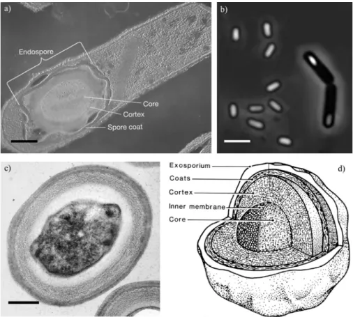
Bacterial spore (i.e., endospore), discovered in 1876 by Cohn1, Koch2,3 and Tyndall4
independently, is one of the hardiest dormant life forms on earth5 formed during the
resting stage in the life cycle of spore-forming genera such as Bacillus, Clostridium,
Sporosarcina and others (Figure 1.1). They are formed within vegetative cells during
sporulation, which is frequently triggered in response to adverse environmental
changes. Spores are renowned for their resistance against deleterious agents and
stresses, such as freezing, boiling, pressure, desiccation and attack by a wide variety
of toxic molecules6,7. Spore DNA is protected from the environment by a surrounding
spore coat comprised of calcium ions, dipicolinic acid and protein layers8-10. DPA
(dipicolinic acid, 2,6-pyridinedicarboxylic acid) is a unique chemical found only in
bacterial spores and can be released from the core into the surrounding by inducing
germination (e.g., with L-alanine11,12) or physical lysing (e.g., autoclaving,
microwaving13). With no detectable metabolism, bacterial spores can remain viable
for many years14. Some controversial reports claim that spores entombed in a bee
trapped in amber 25-40 million years ago have been revived14, and an even more
spectacular claim details the recovery of spores from 250-million-year-old halite
crystals15. The extreme resistance of bacterial spores has even been exploited to check
the performance of autoclaves16,17. Once favorable conditions return, spores undergo
germination and outgrowth to become vegetative cells that cause contaminations,
food spoilage, diseases, etc. Owing to their high resistance to physical extremes,
spores are ubiquitous and can be recovered from almost all extreme environments on
Earth.
In this thesis, I will focus on the study of Bacillus, which is a genus of rod-
shaped and Gram-positive bacteria under the Firmicutes division, with C + G values
ranging from 32% - 62%. A majority of the Bacillus species are strict aerobes or
facultative anaerobes producing endospores in the presence of oxygen. Several
species, e.g., B. infernus and B. arseniciselenatis, are found to be strictly
anaerobic18,19. A recent taxonomic reclassification has broken Bacillus into
Geobacillus, Paenibacillus, Lactobacillus, etc. Most of the species are mesophilic
with some psychrophiles and thermophiles. Bacillus species can be divided into three
groups based on morphology of the sporangia.
Group I encompasses ellipsoidal or cylindrical endospores situated centrally to
terminally in the progenitor mother cell with non-swollen sporangia. It contains most
of the human pathogenic species including B. anthracis, the causative agent of
anthrax, and B. cereus, causing food poisoning with symptoms similar to
staphylococcal food poisoning. A lot of benign soilborne endospores such as B.
subtilis and B. pumilis are also found in this group. Group II is composed of
ellipsoidal endospores situated centrally to terminally in a swollen sporangium. This
group is mainly characterized by insect pathogens such as B. larvae and B.
lentimorbus causing American foulbrood in honeybees and milky disease in Japanese
beetle, respectively. Group III consists of spherical endospores situated sub-terminally
to terminally in sporangia. Some of the psychrophilic Bacillus species are found in
this group, notably B. psychrophiles and B. longisporus.
Spores are interesting subjects to study from both a scientific and application
perspective. They are the ideal model for dormancy, resistance and survivability in the
nature. They account for a large majority of the microbial flora on Earth and take up
unique roles in homeland security, sterilization validation, and astrobiology.
Detection of pathogenic endospores has been an increasingly important issue
in the safety and security of each country. Most of the Bacillus species are harmless
soil-borne microorganisms. The most virulent species is Bacillus anthracis. Upon
contact with animals, it can cause either cutaneous anthrax, inhalational anthrax or
gastro-intestinal anthrax. Inhalational anthrax draws the most attention because of
convenient airborne spore delivery and high lethal rate. The lethal dose at which 50%
of an untreated exposed population would die (LD50) from inhalational anthrax is
believed to be between 2,000 and 55,000 spores, with a nominal value of 8,000 to
10,000 spores. Accidental release of anthrax from a military compound in Chkalovsky,
Russia in 1979 has led to the death of at least 70 people20. B. anthracis is undoubtedly
the notorious bacteria causing national upheavals during the anthrax attack. In 2001 B.
anthracis endospores have been weaponized in US postal offices to cause 22
inhalational anthrax cases, of which 5 people died. Spores entering lungs are carried
by macrophages to the mediastinal lymph where they germinate, outgrow and produce
lethal toxins. The incubation time (the time from the bioweapon exposure to the onset
of the first symptom and disease) is 2 to 3 days for anthrax. Real-time detection of
bioweapon release grants time and resources for emergency management to mitigate
possible consequences, such as by appropriate medical care, limiting the spread of
infection and timely decontamination.
Bacillus species are of great interests in sterilization control. B. cereus and B.
sphericus survive and pasteurization and are the cause of milk coagulates. B.
thuringiensis are natural insecticides. Residual endospores on spacecraft hardware
surfaces after disinfection post potential jeopardy to the data collected from other
planets. Understanding properties of endospores is important in preventing food
spoilage and aids in NASA’s objective in planetary protection.
Endospores are highly resilient dormant structures that can return to vegetative
cells when favorable conditions are restored after exposure to radiation or temperature.
Study of the viable number in endospores entombed in polar ices, suspended in frozen
lakes and embedded in permafrost provides clues for determining long-term viability.
1.2 Life cycle of a spore-forming bacteria
Endospores can lay dormant for long periods and remain viable, even while exposed
to harsh environmental conditions. When more favorable conditions are signaled by
the presence of water, nutrients, and germinants, endospores may break dormancy and
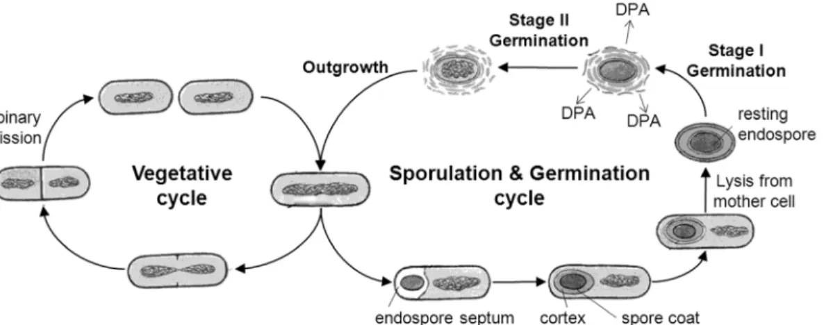
germinate to become metabolically active and multiply. Figure 1.2 outlines the life
cycle of an endospore-forming bacterium. As an endospore proceeds through
germination toward cell division, there are various stages, including spore activation,
stage I germination (during which DPA is released and water rehydrates the spore),
stage II germination (during which cortex hydrolysis occurs and metabolism begins),
and finally outgrowth (during which cell division occurs)21. Each of these stages in
the life cycle of endospore-forming organisms can be observed with the experimental
techniques described below. Certainly outgrowth, manifested as visible colonies on
growth media, is the most direct measurement of endospore viability. However,
colony formation requires approximately 20 cycles of replication over several days of
incubation before the colony becomes visible, and is therefore not amenable for rapid
endospore viability analysis. In contrast, observation of water influx with phase
contrast microscopy, DPA release with Tb-DPA luminescence assays, and ATP
production with the luciferin-luciferase bioluminescence assays provide rapid
measures of endospore viability, since these events occur much earlier in the
germination to outgrowth pathway.
The life cycle of endospore-forming bacteria comprises 3 different processes:
vegetative growth, sporulation and germination. Vegetative growth occurs when
nutrients are available and is characterized by cells growing logarithmically by
symmetric fission. When nutrients become limiting, these bacteria might initiate the
sporulation process. However, sporulation is not the only developmental option for
the vegetative cell that can also follow diverse routes, expressing genes needed for
general adaptation to adverse conditions, or genes needed to scavenge alternative
nutrients and to increase competitiveness against other species that are using the same
scarce energy resources. In such conditions various proteases and degradative
enzymes (amylases, xylanases, cellulases, etc.), alternate chemotaxis pathways,
different antibiotics and transport functions are expressed and/or activated. Some cells
become competent for uptake of exogenous DNA, which is introduced into the
recipient’s chromosome and may confer selective advantages to future generations
living under adverse conditions.
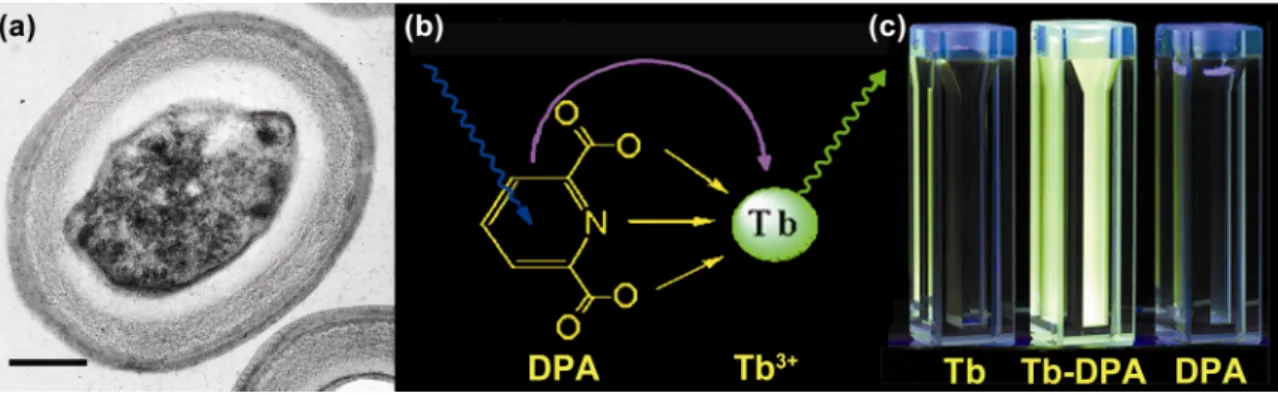
1.3 DPA-triggered terbium photoluminescence assay
DPA-triggered terbium photoluminescence assay forms the basis of spore detection in
my research. Tb3+ is a lanthanide ion, which is characterized by sharp and narrow
emission bands, as well as a millisecond-long excited state lifetime (Figure 1.3). The
native luminescence of Tb3+ is very weak attributed to its low oscillatory strength (≈
10-6) of the excitation bands22. The binding of a chelate, such as DPA, to Tb3+, when
excited with UV light, can transfer energy to the emissive state of Tb3+, leading to
great emission intensity enhancement (> 20,000 times)23. The original Tb3+ excitation
spectrum (λex = 270 nm) will be changed to a characteristic dual-peak spectrum (λex =
273, 279 nm) upon complexation with DPA.
The valence 4f electrons of Tb3+ are rarely involved in bonding because they
are shielded by the outer core 5s and 5p electrons. When Tb3+ absorbs a photon or is
otherwise supplied with a sufficient quantum of energy, it reaches an electronically
excited state. The low ligand-field splitting of the Tb3+ f electrons results in poor
nonradiative electronic transitions generally. The only significant nonradiative
deactivation of the emissive state of Tb3+ is through the O-H vibrations of coordinated
water molecules. This also explains why the lesser number of water molecules
coordinated the higher the lifetime Tb3+ or terbium chelate can attain. Energy lose via
radiative transition, with the emission of a photon as the electron transfer back into its
lower energy orbital, is known as fluorescence. These f-f transitions are, nevertheless,
Laporte forbidden because there is no change in parity. This results in Tb3+ absorption
and emission spectra being consisting of sharp and narrow bands, low molar
absorption coefficients in the near-UV and visible ranges (ε typically between 0.01
and 3 M-1 cm-1), as well as a millisecond-long luminescence lifetime. Therefore, Tb3+
exhibits a very weak emission upon direct near-UV excitation.
Absorption of photons, optimally in the UV range, promotes DPA from
ground state to a vibrationally excited singlet state. This excited singlet can lose its
excess energy by both radiative and nonradiative decay. Upon complexation with
DPA, the photophysical properties of Tb3+ are slightly perturbed owing to the well-
shielded 4f electrons. Sensitized luminescence can be observed due to energy transfer
from the lowest-lying DPA triplet excited state to the emissive 5D4 state of Tb3+. DPA
acts as a light harvesting antenna to receive UV excitation and then transfers the
energy to Tb3+. Because the energy gap between the Tb3+ ground state and emissive
states is very large (20,500 cm-1), the luminescence enhancement is very significant
according to the energy gap law.
DPA is thermodynamically and kinetically stable, thereby avoiding high
energy oscillator in the first coordination sphere. It also has a matched electronic
transition levels with Tb3+, in which an efficient intersystem crossing is possible due
to ΔE(1π π* - 3π π*) ≈ 5000 cm-1, demonstrating a large conjugated π electron system.
In additional to this, the energy difference, ΔE(3π π* - 5DJ) ≈ 2500 - 3500 cm-1, is well
above the 5D4 state of Tb3+ so that back transfer onto DPA triplet state is not possible.
Another favorable feature of Tb3+ is its retention in atomic properties upon
ligand binding due to its well-shielded 4f electrons. Only minimal changes are
observed in the emission spectra of Tb3+. Therefore, sensitized luminescence turn-on
can readily be observed by an up shift in the fluorescence spectra. Besides, excitation
spectrum of the sensitized terbium luminescence will mimic the excitation spectrum
of DPA energy donor moieties. This provides a basis for the distinctive identification
of the ligand responsible for the sensitization. In the case of DPA, a characteristic split
in the excitation spectrum is observed.
Background and autofluorescence signals are usually rapidly decaying
processes with lifetimes on the timescale of 1 – 100 nanoseconds24. Terbium
dipicolinate has a lifetime in the range of milliseconds25,26. Therefore, when excited
by UV, the emitted Tb-DPA intensity can be observed without interference from
autofluorescence and scattering light, resulting in a substantial increase in detection
sensitivity. Delayed luminescence intensity can thus be measured with a pulsed UV
excitation, xenon flashlamp or laser, with minimal background interference.
1.4 Outline of thesis
Brooks et al. defined indicators as “measures, variables, or indices that represent or mimic either the structure or function of ecological processes and systems across a disturbance gradient27.” In this thesis work, I evaluate the effectiveness of bacterial endospore as a biological indicator to characterize, track and predict changes in indoor air quality, spacecraft
bioburden level, and extreme ecosystems on Earth. Specifically, this goal is achieved via:
(1) development of new spectroscopy and microscopy methods to detect aerobic
endospores; (2) application of culture-based and culture-independent methods to
study endospores under various habitats. Chapter 2 is a literature review of
endospore detection methods, with a general introduction to culture-based and
culture-independent techniques. I have emphasized on detection tools that are relevant
to subsequent chapters. The rest of this thesis can be divided into 3 modules, namely,
air sampling, sterilization validation and astrobiology.
The first module is devoted to the detection of airborne endospores. A good
biological indicator has to be able to indicate impending changes and provide
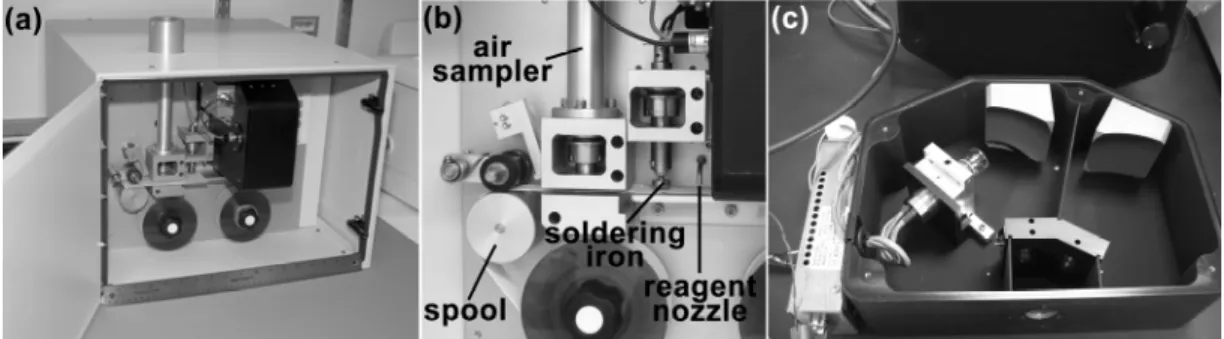
continuous assessment over a system. Chapter 3 presents the implementation of an
autonomous air sampler, known as the Anthrax Smoke Detector (ASD), to quantify
airborne endospores. The ASD is intended to serve as a cost-effective front-end monitor
for anthrax surveillance systems. The principle of operation is based on measuring
airborne endospore concentrations, where a sharp concentration increase signals an
anthrax attack. The combination of time-gated spectroscopy and the unique
photoproperties of terbium dipicolinate results in a sensitive and low detection limit.
The ASD-technology can also be extended to the development of a miniature air
sampling system to monitor indoor airborne endospore level in a closed system such
as spacecrafts and life-support space stations as an indicator of the general hygiene
level. Microbiological monitoring in confined spacecraft and space station environments
during long-duration manned missions is instrumental to guaranteeing the health, safety
and productivity of astronauts and crew members. Chapter 4 tests the hypothesis that
airborne endospore bioburden is an effective indicator of total biomass in a closed
system. A correlation characterized by a two-compartmental model has been
formulated from a laboratory controlled enclosure to everyday life examples such as
offices and laboratories. Future work will follow to miniaturize the current prototype
for a terminal integration with the life-support system.
The second module applies to the validation of sterilization. We focus on
developing rapid and sensitive methods to detect endospores as surrogates for the
measurement of total biomass on a surface in the context of planetary protection. A
microscopy version of the Tb-DPA luminescence assay, known as the microscopic
endospore viability assay (acronym: µEVA) has been introduced in Chapter 5, fully
validated with cultivation and phase contrast microscopy on enumerating pure
endospore suspension and monitoring the thermal and UV inactivation of endospores.
We have demonstrated a single endospore resolution with a linear measurement at
low inocula. The application of µEVA on assessing endospores on surfaces is
illustrated in Chapter 6, with an emphasis on the bioburden evaluation according to
planetary protection regulations. Side-by-side comparison tests have been conducted
using µEVA and the culture-based NASA standard assays in monitoring the surface
hygiene level based on endospore concentration on coupons made of spacecraft
materials and the Class 100 clean room in the Spacecraft Assembly Facility at JPL.
This method is expected to be deployed as a new bioburden assessment standard in
NASA for ensuring the cleanliness of spacecraft and associated facilities.
Chapters 7 through 9 cover the endospores analysis on environmental samples
in extreme environments. Essentially all the methods delineated in previous chapters
are developed again and tailored made for specific environmental samples. The goal
is to use endospores as the indicator microbe in the study of different ecological
systems to shed light on astrobiology questions. Chapter 7 describes a wide variety
of experiments involving method development, instrument building and model
formulation to prepare for the study of endospores under different extreme
environments on Earth as models for the eventual exobiological search of life in other
planets. So far, most of the assays are developed based on model laboratory
endospore strains, such as B. atrophaeus and B. subtilis. Instruments are developed
and modified to adapt to field analysis in desert and ice environments. New protocols
and calibration curves are obtained with various endospore viability assays in working
with environmental samples. Robustness of the assay has been improved by extending
the analysis from mesophilic to thermophilic and psychrophilic endospores. Novel
sporulation protocols have been developed to prepare pure and clean psychrophilic
endospores for preliminary experiments towards detection of endospores in the ice
biospheres. Microgermination and macrogermination models are formulated to give
us insights into more complex environmental endospores based on simple and
controlled model strains.
Chapter 8 describes the application of a combination of culture-based and
culture-independent assays on endospores in extreme cold biospheres on Earth,
including GISP2 (Green Ice Sheet Project 2) ice cores, Antarctic underground frozen
brine Lake Vida, and polar permafrost. The decontamination, sample handling and
analyses for each of these samples are depicted in detail in this chapter. Apart from
the Tb-DPA luminescence assays, many of other methods such as metabolic dyes,
bioluminescence and flow cytometry are explored to determine the viability of
resident microbes. 4 different viability ratios have been obtained independently based
on different measurables in terms of culturability, germinability and total microscopy
counts to characterize the microbial abundance and viability in each of these cold
habitats. In particular, a transect of 12 depths extending from 295 to 110,000 years old
have been analyzed to shed light on the longevity of the most resistant life form, i.e.
endospores, in the best storage repository on Earth.
Chapter 9 examines another terrestrial model to Mars – a hyper-arid
environment totally devoid of water. The Atacama Desert in Chile serves as the model
system because it is the driest desert on Earth. Previous studies have reported a near-
sterile state in the soils. My research group has been on a sampling expedition to the
Atacama Desert for 10 days in September 2007 to carry out a series of in situ and
sample return analyses with the goal to study the microbial diversity, abundance and
the response of the effect of water on the native microbes. New culture-independent
methods, such as phospholipid fatty acid and total organic carbon analyses were
performed on the desert soils along with Tb-DPA based assays. Significant
differences in culturability and germinability were obtained along a depth profile in a
soil pit corresponding to ages approximately equal to 25,000 and 35,000 years ago
based on radioactive carbon dating. A spike in microbial response was also observed
when sterile water was added into otherwise-sterile desert soils. Endospore isolates
obtained from the Atacama Desert were compared with laboratory strains and other
environmental isolates as an effort to investigate the long-debated ecological role of
endospores in a particular environment. In particular, we assess the feasibility of using
the abundance and viability of endospore as proxies to study the ecosystem in
Atacama.
Chapter 10 concludes the thesis by by proposing two future projects built on
the developed assays and tools. One of the projects aims to build a miniaturized
µEVA instrument for portable field deployment. As science evolves into a high-
throughput technology for the study of bacteria, new demands are continually being
placed upon analysis time and turnaround efficiency in quantitative methods. Chief
among these are increased detection sensitivity, portability, broad dynamic range and
compatibility with other methods and portability into spacecraft mission. In this future
work section, some of the current µEVA limitations, such as serial processing,
bulkiness and manual operation, are interfaced with automated microfluidic chip,
robotic high-throughput processing and a miniaturized design. The goal of the other
project is to incorporate several synergistic and complementary microbial detection
assays onto a microfluidic chip. It is expected that both deliverables will be integrated
into the life-detection system for future space explorations in due course.
1.5 References
1. Cohn, F. Untersuchungen uber Bacterien. IV. Beitrage zur Biologie der Bacillen. Beitr. Biol.
Pflanz. 2, 249-276 (1876).
2. Koch, R. Untersuchungen uber Bakterien V. Die Atiologie der Milzbrandkrankheit, begrundet auf die Entwicklungsgeschichte des Bacillus anthracis. Beitr. Biol. Pflanz. 2, 277-310 (1876).
3. Koch, R. Untersuchungen uber Bakterien VI. Verfahren zur Untersuchung, zum Conservieren und Photographieren. Beitr. Biol. Pflanz. 2, 399-434 (1877).
4. Tyndall, J. Further researches on the department and vital persitence of putrefactive and infective organisms froma a physical point of view. Phil. Trans. Royal Soc. 167, 149-206 (1877).
5. Roberts, T.A., Hitchins, A. D. Resistance of spores. The Bacterial Spore ed. Gould, G. W. and Hurst, A, 611-670 (1969).
6. Aronson, A.I. and Fitz-James, P. Structure and morphogenesis of the bacterial spore coat.
Bacteriological Reviews 40, 360-402 (1976).
7. Driks, A. and Setlow, P., Prokaryotic Development. (American Society of Microbiology, Washington, D.C., 2000).
8. Church, B.D. and Halvorson, H. Dependence of the heat resistance of bacterial endospores on their dipicolinic acid content. Nature 183, 124-125 (1959).
9. Byrne, A.F., Burton, T.H., and Koch, R.B. Relation of dipicolinic acid content of anaerobic bacterial endospores to their heat resistance. Journal of Bacteriology 80, 139-140 (1960).
10. Berg, P.E. and Grecz, N. Relationship of dipicolinic acid content in spores of Bacillus cereus to ultraviolet and gamma radiation resistance. Journal of Bacteriology 103, 517-519 (1970).
11. Foster, S.J. and Johnstone, K. Pulling the trigger: The mechanism of bacterial spore germination.
Molecular Microbiology 4, 137-141 (1990).
12. Johnstone, K. The trigger mechanism of spore germination. Journal of Applied Bacteriology 76, 17S-24S (1994).
13. Vaid, A. and Bishop, A.H. The destruction by microwave radiation of bacterial endospores and amplification of the released DNA. Journal of Applied Microbiology 85, 115-122 (1998).
14. Nicholson, W.L., Munakata, N., Horneck, G., Melosh, H.J., and Setlow, P. Resistance of Bacillus endospores to extreme terrestrial and extraterrestrial environments. Microbiology and Molecular Biology Reviews 64, 548-572 (2000).
15. Cano, R.J. and Borucki, M.K. Revival and identification of bacterial spores in 25-million-year-old to 40-million-year-old Dominican amber. Science 268, 1060-1064 (1995).
16. Vreeland, R.H., Rosenzweig, W.D., and Powers, D.W. Isolation of a 250 million-year-old halotolerant bacterium from a primary salt crystal. Nature 407, 897-900 (2000).
17. Dart, R.K., Microbiology for the Analytical Chemist. (The Royal Society of Chemistry, Cambridge, UK, 1996).
18. Switzer Blum, J., Burns Bindi, A., Buzzelli, J., Stolz, J.F., and Oremland, R.S. Bacillus arsenicoselenatis, sp. nov., and Bacillus selenitireducens, sp. nov.: two haloalkaliphiles from Mono Lake, California that respire oxyanions of selenium and arsenic. Archives of Microbiology 171, 19-30 (1998).
19. Boone, D.R. et al. Bacillus infernus sp. nov., an Fe (III)-and Mn (IV)-reducing anaerobe from the deep terrestrial subsurface. International Journal of Systematic and Evolutionary Microbiology 45, 441-448 (1995).
20. Meselson, M. et al, (1994), Vol. 266, pp. 1202-1208.
21. Setlow, P. Spore germination. Current Opinion in Microbiology 6, 550-556 (2003).
22. Richardson, F.S. Terbium(III) and europium(III) ions as luminescent probes and stains for biomolecular systems. Chemical Reviews 82, 541-552 (1982).
23. Hindle, A.A., Hall, E. A. H. Dipicolinic acid (DPA) assay revisited and appraised for spore detection. Analyst 124, 1599-1604 (1999).
24. Lakowicz, J.R., Principles of Fluorescence Spectroscopy. (Plenum, New York, 1983).
25. Jones, G. and Vullev, V.I. Medium effects on the photophysical properties of terbium(III) complexes with pyridine-2,6-dicarboxylate (supporting information). Journal of Physical Chemistry A 106, 8213-8222 (2002).
26. Jones, G. and Vullev, V.I. Medium effects on the stability of terbium(III) complexes with pyridine-2,6-dicarboxylate. Journal of Physical Chemistry A 106, 8213-8222 (2002).
27. Brooks, R.P., O'Connell, T.J., Wardrop, D.H., and Jackson, L.E. Towards a regional index of biological integrity: the example of forested riparian ecosystems. Environmental Monitoring and Assessment 51, 131-143 (1998).
Figure 1.1 | (a) Transmission electron micrograph of a bacterial spore embedded
within a Clostridium perfringens vegetative cell. Scale bar = 0.3 μm. (b) Bacterial
spores appear phase-bright observed under a phase contrast microscope and are
readily distinguished from the phase-dark rod-shaped vegetative mother cell. Scale
bar = 2 μm. (c) Cross section of a TEM of an endospore of Geobacillus
stearothermophilus. Scale bar = 0.2 µm. (d) Schematic representation of the internal structure of a typical bacterial spore, reproduced from Foster and Johnstone (1990).
Figure 1.2 | Life cycle of an endospore-forming bacterium.