ISSN: 1978-6417 Vol. 5 No. 1
Detection of Igm To Leptospira Agent With
Elisa and Leptodistick Method
M. Sabir1), Aristo2), Hasanuddin1), Sarifuddin2), dan M. Hatta3)
1)Department of Medical Microbiology, School of Medicine, Tadulako University.
2)Department of Medical Anatomy,School of Medicine, Tadulako University
3) Department of Medical Microbiology, School of Medicine, Hasanuddin University
E.mail: [email protected]//[email protected]
ABSTRAK
Sampel berupa serum diambil dari penderita demam yag dicurigai karena : leptospirosis,demam tifoid, malaria dan hepatitis. Pemeriksaan serum terhadap antibodi IgM spesifik untuk leptospira menggunakan pemeriksaan dipstik sederhana (leptodipstik). Sampel diperoleh dari bagian Mikrobiologi, pada rumah sakit universitas hasanuddin di makassar Indonesia, dan data hasil pemeriksaan sederhana ini dibandingkan dengan hasil prosedur diagnostik standar Rumah Sakit yang diterapkan oleh dokter di Rumah Sakit yang sama. Sebagai bagian dari penelitian ini, sebagian besar serum ,juga diperiksa dengan ELISA yang spesifik dan sensitif terhadap IgM leptospira pada Royal Tropical Institute di Amsterdam untuk dibandingkan dengan hasil pemeriksaan leptodipstik. Aplikasi leptodipstik memperlihatkan keberadaan antibodi IgM yang spesifik terhadap leptospira pada 21 dari 34 penderita yang telah didiagnosis leptospirosis dan 7 dari 368 penderita yang didiagosis selain leptospirosis. Hasil pemeriksaan dipstik memiliki korelasi yang baik dengan hasil pemeriksaan ELISA.
Kata kunci: IgM, ELISA,Leptodipstik, Leptospira.
INTRODUCTION
Leptospirosis is an acute febrile
illness caused by infection with
spirochaetal microorganisms of the genus leptospira of which more than 200 pathogenic strains are currently
known (3). Natural hosts of pathogenic
strains,which may cause infection in man, include wild animals, livestock and pets. Urine of infected animals is the main source of transmission and humans may become infected through wounded skin, mucous membranes and the conjunctivae. Exact incidence rates are not known for most regions of the world. The incidence of leptospirosis in
wet tropical countries can be as 5-20/100.000 per year.
Leptospirosis may manifest as
relatively mild flue-like symptoms or as a severe disease called Weil’s syndrome and symptoms may include renal failure,liver
impairment,meningitis and (lung)
hemorrhages. The fatality rate of severe cases is high. The clinical diagnosis of acute leptospirosis may easily be missed or the disease may be confused with other
major infectious disease including
influenza,malaria,hepatitis,bacterial
meningitis and viral hemorrhagic fever. Therefore, laboratory tests are important to
Several more or less complicated methods are available to make the laboratory diagnosis of leptospirosis. These include Microscopic Aglutination Test (MAT) and ELISA. Culture provide ultimate proof of leptospirosis but has a low efficacy. Recently two simple and
robust serological methods were
developed and evaluated. This is the
leptodipstik (4), which provide a high
sensitifity and specificity. The leptouria-test is the direct examination for leptospires in a patients urine-samples under a microscope, and is often used in some tropical hospitals as a diagnostic lab-test. In the literature this test is controversial since threads of fibrin and protein in a urine-sample can mimic leptospires which can give rise to
false-positive results (2).
Several undetermined fevers can mimic the onset or presence of leptospirosis (such as typhoid fever, hepatitis, dengue fever and malaria) and are important possible diseases which can be confused with this disease. During a recent outbreak of leptospirosis in Nicaragua the disease
was first mistaken for dengue fever (5).
collected from Department of
Microbiology at Hasanuddin University Hospital in Makassar, Indonesia. The total number of patients were 403. Of each patients was collected. In the
leptospirosis group (n=179) were
patients with clinical suspicion of
leptospirosis at admission.
Leptospirosis was suspected when patients; Suffered an acute febrile illness, myalgia (especially in the
calves) conjunctival suffusion,
icterohemorrhage, and hepatic disturbance. Showed renal involvement had a history of
exposure to an infected animal or
environment contaminated with animal urine. The other group (n=224) were patients which at admission had the suspicion of various other infectious disease including typhoi fever, malaria, hepatitis ad dengue fever or those with fever of unknown origin.
The direct examination for leptospires in a patient’s urine-sample is referred to as the lepto-uria test. The test was performed at the Hasanuddin University hospital (according to standard procedures). The leptouria-test was performed on all patietns with suspicion of leptospirosis and on some patients with the suspicion of other than leptospirosis.
A leptospira-specific IgM ELISA test
was performedon 237 out of 403 serum samples using routine procedure. The test is
highly specific and sensitive (7,8), And is often
used in the routine laboratory diagnosis of leptospirosis. The Igm ELISA was performed at the Royal Tropical Institute in Amsterdam, and the results were then used to compare with leptodipstik test result.
ELISA
The ELISA for detection of leptospira-specific IgM antibodies (IgM ELISA) was performed with antigen prepared from strain
Wijnberg as described (6,7). Sera with a titer
of 1:80 or higher were considered positive.
Leptodipstick Test Procedures
infections. The assay is performed by making a 1:50 dilution of serum (4 microliters) in the detection reagent (200 microliters) and incubating a wetted dipstick in this solution. Staining of the antigen band reveal the presence of specific IgM antibodies in the serum sample. The strength of the staining is important in the interpretation of the test result. A colored reference strip is used to compare the staining intensity which ranges from 0 (no reaction) to 4+ (best reaction). When evaluating the dipstick assay, a staining intensity of 2 + or higher are considered as a positive result.
Internal control
Each dipstick has an internal control band coated with anti-human IgM antibody. Coated antibody binds IgM molecules from the serum which are then stained by the detection reagent. The staining intensity of this iternal control band is rated 2+ for most of the sera tested, and is there to facilitate the interpretation of the assay results and also to make sure that a human serum is being tested each time.
Test and Storage Conditions
Dipstick incubation is performed
in an air-conditioned room (23-27 0 C)
for 3 hours. During the air-drying period of the dipsticks ‘cloudly’ dipsticks are seen in the beginning of the study. This is suspect caused by air dryness, because the dipsticks were being air dried in the air-conditioned room. Thouroughly tapping all excess water droplets from the dipsticks before leaving them to be air-dried is the key solution.
All dipstick strips, dipstick vials
are kept in the refrigerator (4 0 C ) for 1
hour before being used. All sera are
kept in the freezer (-10 0 C) for 1 hour
before being used.
Statistical evaluation
The variation between the different tests chapter result; test comparisons was determined by calculating kappa values with the standard error of kappa. Values vary between 0 to 1, where kappa values below 0.40 represent a slight agreement;0.40 and 0.80 represent a fair to good agreement; > 0.80 represent almost perfect agreement beyond chance.
RESULT
Of the 179 patients with clinical suspicion of leptospirosis 27 (15.1%) had a diagnosis of leptospirosis (table.1). The diagnosis was based on the clinical symptoms in addition to microscopical observation of leptospires in a urine-sample (leptouria-test). About 84.9% of the patients with clinical suspicion of leptospirosis had a diagnosis other than leptospirosis. The predominant diagnosis was typhoid fever. Of the 224 patients with various other diseases 7 (3.1%) had a diagnosis of leptospirosis.
The clinical diagnosis was compared to the results of the leptospira-specific IgM ELISA test. Of the 16 patients with a final diagnosis of leptospirosis 12 (75%) had a positive IgM ELISA test result (table 2).
Of the 34 patients with a diagnosis of leptospirosis 21 (61.8%) had a positive reaction the leptodipstick test (table 3). Only 7 patients with a diagnosis other than leptospirosis had a positive lepto dipstick result.
Table 1. Clinical diagnosis using leptouria test
Patient groups Final Diagnosis Total
Leptotospirosis Other
Leptospirosis suspect 27 152 179
Other Suspect 7 217 224
Total 34 369 403
Table 2. Clinical diagnosis in comparison to the leptospira-specific IgM ELISA Test
Final Diagnosis IgM ELISA Total
Positive Negative
Leptospirosis suspect 12 4 16
Other Suspect 2 219 221
Total 14 223 237
Table 3. Clinical diagnosis in comparison to the leptodipstick test
Leptodipstick Final Diagnosis Total
Leptotospirosis Other
Positive 21 7 28
Negative 13 361 374
Total 34 368 402
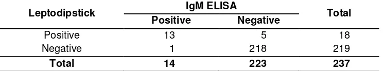
Table 4. Comparison between the leptodipstick test results and the ELISA IgM test results (in the detection of leptospira-specific IgM antibodies)
Leptodipstick IgM ELISA Total
Positive Negative
Positive 13 5 18
Negative 1 218 219
Total 14 223 237
DISCUSSION
The diagnosis of leptospirosis was based on the clinical symsptoms and the result of the leptouria test. Of the 403 patients in this study 34 (8.4%) have been diagnosed with leptospirosis (table 1). In a leptospira specific IgM ELISA test, leptospirosis should be considered if a sero-conversion or a
4-fold titer is observed in paired sera. However when only single sera is available, a single raised titer equal to or more than 1:80 may be considered consistent with leptospirosis.
Diagnostic results have been
used in the routine laboratory diagnosis leptospirosis. Of the 14 patients with a positive IGM ELISA 12 (85.7%) have a diagnosis of leptospirosis. For fear that doctors are biased by possible false positive leptouria test result it is important to compare the number of leptospirosis as a diagnosis with the number of negative IgM ELISA result. Of the 16 patients with a diagnosis of leptospirosis 4 (25%) have a negative IgM ELISA test result. This might indicate a slight bias for even more false-positive leptouria results.
When the results of the
eptodipstick are put into consideration 21 (61.8%) of the 34 patients with a diagnosis of leptospirosis were found positive and 7 of the 28 (25%) patients with a positive dipstick result have a diagnosis other than leptospirosis (table 3).
Comparison the of leptodipstick results with IgM ELISA test results (table 4),shows a very good correlation (observed agreement of 0.97; kappa
coeficient=0.80, standard error of
kappa=0.60) for the 237 patients tested by both methods. From both literature
(4), as well as results stated above, one
can argue that the leptodipstick is equally sensitive and a specific as the leptospira-specific IgM ELISA test to be used as a laboratory test for the sero-diagnosis of leptospirosis.
CONCLUSION
The leptodipstick test as a laboratory test for the sero-diagnostic of leptospirosis is equally sensitive and specific as leptospira specific IgM ELISA test.
REFERENCES
Faine S. A brief overview of the disease,leptospirosis. Leptospira and
Leptospirosis, First Edition,`CRC
Press. 1994, Chapter 14.
Farr RW. Leptospirosis. Clin Infect Dis 1995;21:1-8.
Gussenhoven GC, Van der Hoorn MAWG, Goris MGA, Terpstra WJ,Hartskerl RA, Mol BW, et.al. Leptodipstick, a
dipstick assay for detection of
leptospira-specific immunoglobulin M
antibodies in human sera. J.
Clin.Microbiol 1997;35:92-7.
Hatta.M, Smiths HL,Gussenhoven GC, Gooskens J. Introduction of a rapid dipstick assay for the detection of leptospira-specific immuno-globulin M antibodies in the laboratory diagnosis of leptospirosis in a hospital in
makassar, Indonesia. Southeast
Asian. J. Trop.Med.Public Health. 2000 Sept;31(3) :515-20.
Terpstra WJ, Ligthart GS,Schoone GJ. Serodiagnosis of human leptospirosis by Enzime-Linked
Immunosorbent-Assay (ELISA) .Zentralblatt
Bacteriol,Microbiol Hyg (Abteilung 1 Originale A)1980;247:400-5.
Terpstra WJ, Ligthart GS,and Schoone GJ. ELISA for the detection of specific IgM
and IgG in human
leptospirosis.J.Gen. Microbiol.
1985;131:377-85.
Zaki SR, Shieh WJ. Leptospirosis
associated with outbreak of acute
febrile illness and pulmonary
haemorrhage.Nicaragua, 1995. The Lancet Feb 1996;347;535-6
Zichowski WJ,Waitkins SA, Palmer MF. The use of ELISA in the diagnosis of
Human Leptospirosis.Isr.J.Vet.Med.