Thank you to all current and former members of the Mass Spectrometry Research Center, as colleagues and friends, you were a wonderful (and funny!) group of people. The authors realized the value of this observation and postulated the future application of wavelength-absorbing matrix molecules for the analysis of small organic molecules by laser desorption mass. The selection of a matrix compound can depend on the chemical properties of the analyte, sample conditions and the wavelength of the laser.
Much of the early work in IMS was done using secondary ion mass spectrometry (SIMS). The laser pulse causes both molecules to desorb from the sample surface and subsequent protonation of the analyte by the matrix occurs in the gas phase. The ability of the MALDI process to desorb and ionize diverse analyte species requires an equally flexible mass analyzer.
More detailed descriptions of the MALDI-TOF instrumentation and delayed extraction conditions can be found in several reviews.[46-48]. The fragment ions (i.e. product ions) are then detected and can be used to link back to the structure of the precursor ion. In the case of the QqTOF mass spectrometer, an additional radio frequency (RF)-only quadrupole (Q0) is added to provide collisional cooling and focusing of the ions from the MALDI source.
Early determinations of tissue distributions and PK/PD profiles of a new therapeutic compound play a major role in the drug discovery and development process and more sophisticated technologies are needed to overcome the apparent limitations of the currently available imaging modalities.
Develop a reliable, reproducible method for the direct tissue analysis of endogenous and exogenous compounds from whole-animal tissue
Employ the methods developed in Objective 1 to image the individual distributions of exogenous drug and subsequent metabolites in whole-
Employ methods developed in Objective 1 to evaluate the
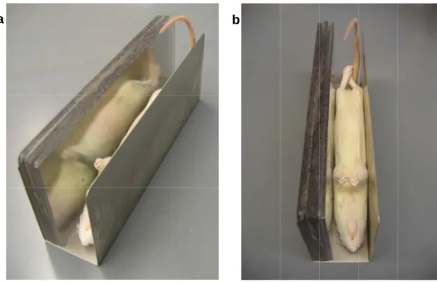
After successful administration of the pharmaceutical composition, the animal should be sacrificed for analysis by IMS. In the case of whole-body tissue sections, cardiac puncture is the preferred method of blood removal, as the integrity of the animal carcass will be preserved. Care must be taken during the freezing process to maintain a symmetrical shape of the carcass.
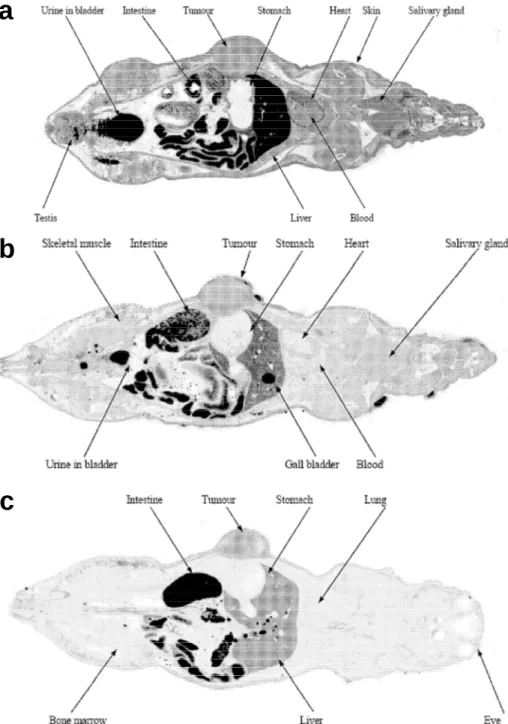
Depending on the parameters of the experiment, the carcass can be placed on the metal sample stage to collect coronal (back to belly), axial (nose to tail), or sagittal (left flank to right flank) whole-body sections (Fig. While still in the freezer, a wax platform is placed on the sample stage to raise the carcass to provide a margin from the metal stage to the blade of the cryomacrocut However, before whole-body tissue sectioning can be performed, the frame must be removed and the sample block must be equilibrated at -20 ºC for 1 hour to avoid cracking in the ice.
The tissue is pulled off the rice paper and onto the plate by placing a warm hand on the opposite side of the plate. An additional factor to consider when deciding on a matrix application approach is the spatial resolution of the imaging MALDI MS analysis. The resolution of a spray-coated tissue is limited by the laser diameter of the MALDI source.
Therefore, it is important to consider these interferences when optimizing the parameters of the MALDI MS/MS instrument, especially when choosing the precursor ion mass filtering window. Analysis of the spots by MALDI MS/MS showed signal variations for each of the compounds; but is normalized to the IS signal corrected for. Extraction efficiency (2) takes into account the affinity of the compound for the tissue type, as well as the efficient extraction of that compound from the tissue by the matrix solvent.
A similar approach can be taken to determine the extraction efficiency of any pharmaceutical compound of interest and application of the extraction normalization factors will provide quantitative information that can be derived from the MALDI MS/MS imaging results. During the tissue mounting step, four fiducials were placed in the corner of each MALDI plate of the sample. An optical image (1200 dpi jpeg) of the intact whole body section was obtained and then the plates were separated.
Using custom software, each MALDI MS/MS jpeg is loaded into the program along with motor coordinates from the instrument. In addition, the jpeg pixel coordinates for each of the fiducials from the optical image of the intact whole-body section are loaded.
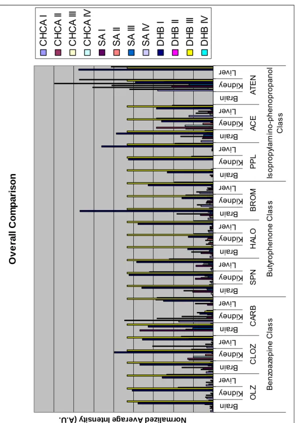
Isopropylamino- phenopropanol Class Butyrophenone Class Benzoazepine Class N
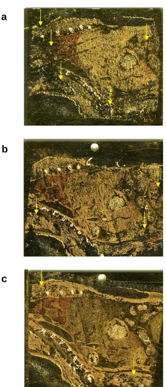
Stained matrix tissue sections were stored in a vacuum desiccator for ~12 h until MALDI MS/MS analysis. Comparison of WBA and MALDI MS/MS detection of olanzapine 2 hours postdose in a whole rat sagittal tissue section. a) Whole body autoradiography of [14C]OLZ showing the distribution of all labeled compounds [85], b) MALDI. MALDI MS/MS images obtained 24 hours post-dose indicate that most of the OLZ in the rat has been eliminated and are confirmed by the WBA images [85].
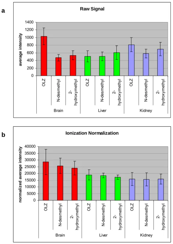
Signals amplified for visualization. a) whole body autoradiography of [14C] OLZ shows distribution of all labeled compounds [85], b) MALDI MS/MS detects individual OLZ and metabolite distribution. Plot of the average OLZ MALDI MS/MS signal from skeletal muscle across four sample plates demonstrates the reproducibility of the imaging methodology. The ratios of the HPLC-MS/MS values were compared with the ratios of the normalized OLZ MALDI MS/MS signal from the tissue brain, liver and kidney regions (Table 7).
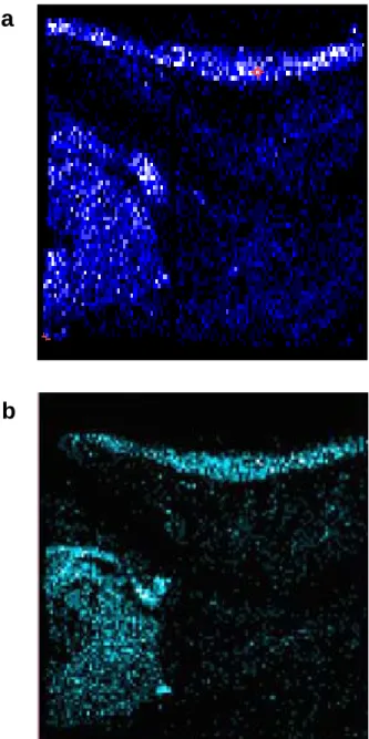
In the 2-hour MALDI MS/MS images, the drug is clearly localized in the brain and spinal cord of the target organs. On the basis of the MALDI MS/MS images, the 2-hydroxymethyl metabolite, produced by the CYP2D1-5 enzymes, was detected as the more abundant metabolite compared to N-desmethylolanzapine. The comparison of the imaging MALDI MS/MS OLZ signal in the target organ, brain and other major organs (liver and kidney) with HPLC.
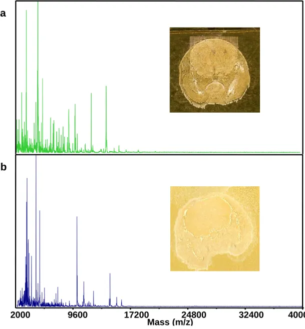
However, an analysis by MALDI MS/MS gains the advantage of preserving the spatial distribution of the analyte within a tissue sample. All matrix-coated tissue sections were allowed to dry under ambient conditions for ∼1 h before MALDI MS/MS analysis. After completion of MALDI MS/MS image acquisition, each plate was individually processed using oMALDI server 5.0 software.
To study the quantitative signal of the OLZ image, the regions of interest were extracted from the MALDI MS/MS images using oMALDI server 5.0 software. This physiological response can be seen in the temporal decrease of the calmodulin signal after chronic lithium administration by MALDI MS imaging. In addition, pharmacokinetic studies by MALDI MS/MS can be combined with PD studies using MALDI MS.
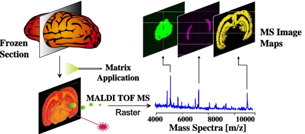
Much of the IMS methodology can be attributed to the established WBA protocols, which have been extended and optimized for the MALDI MS and MS/MS technologies. MALDI MS data were inserted and co-registered with the. corresponding image section in the block plane volume.