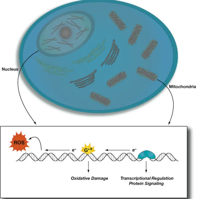
DNA-mediated charge transfer reactions are modulated by the structure and dynamics of the double helix. DNA-mediated oxidation of the [4Fe4S] cluster via a guanine radical intermediate leads to the formation of a [4Fe4S]3+ cluster as observed by electron paramagnetic resonance (EPR) and transient absorption spectroscopy. Thus, the auxiliary function of EndoIII could involve the DNA-mediated redox activity of the [4Fe4S] cluster in EndoIII.
Biological Contexts for DNA-mediated Charge Transport
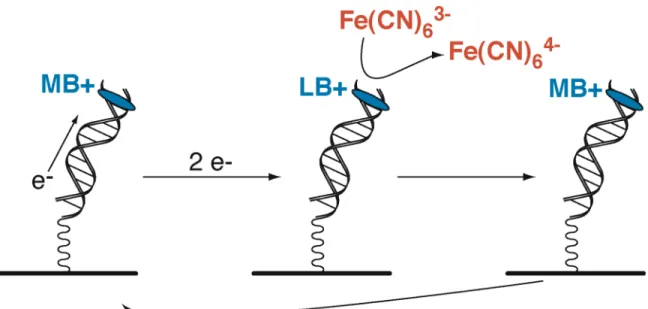
Electrochemical Detection of Lesions in DNA
DNA-bound Redox Activity of DNA-bound Repair Proteins
Electrochemical Investigation of Archaeal [4Fe4S] DNA Repair Proteins
Inactivation of genes in Escherichia coli
Biological Contexts for DNA Charge Transport Chemistry
Several studies on DNA CT in the presence of specific DNA-binding proteins have been performed. DNA repair proteins are another important class of DNA-binding proteins that can modulate or participate in DNA CT events. Within the cell, DNA CT can therefore play an important role in the DNA damage process by directing damage to specific sites.
Electrochemical Detection of Lesions in DNA
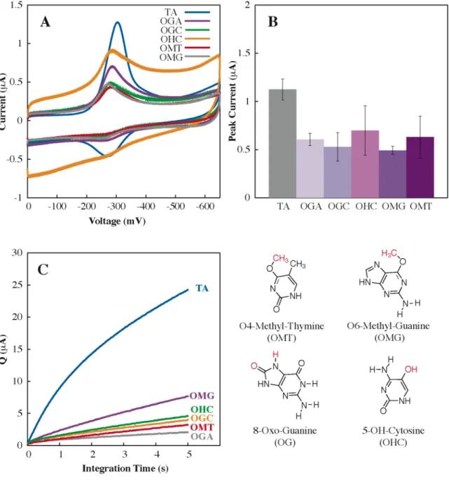
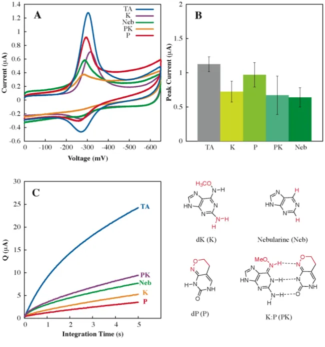
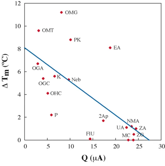
Perhaps most importantly, our understanding of the sensitivity and range of DNA CT chemistry in lesion detection provides a basis for the. Altering the Watson-Crick hydrogen bond interface produces a large loss in CT efficiency (OMT, OMG, Neb, P, and K), as it does. The efficiency of DNA-mediated CT does not appear to be dependent on the. thermodynamic stability of the helix.
DNA-bound Redox Activity of DNA Repair Glycosylases Containing [4Fe4S] Clusters
All three proteins were examined by EPR spectroscopy in the presence and absence of DNA using Co(phen)33+ as an oxidant. EPR measurements were performed at 10K to observe any changes in the oxidation state of the [4Fe4S] cluster. In the presence of DNA and [Co(phen)3]3+ (150 µM) this signal is also evident, but the intensity is much greater (~4-fold by integration).
EndoIII also does not exhibit an EPR signal without oxidant in the presence or absence of DNA. We also examined the oxidation of repair proteins by ferricyanide in the presence and absence of DNA. A similar enhancement in cluster oxidation was observed in the presence of DNA (data not shown).
However, this signal has a g-value of 2.02, indicating that the cluster is likely in the [3Fe4S]1+ configuration. Although some oxidation by Co(phen)33+ is found in the absence of DNA, significant improvements in oxidation are evident in the presence of DNA. Based on comparative g values, upon DNA binding in the presence of an oxidant, MutY, EndoIII, and AfUDG mainly promote the formation of the [3Fe4S]1+.
In the absence of electrode DNA modification, no redox activity can be detected and continues. DNA-modified electrodes containing an abasic site, the redox signals are. dramatically softened; these observations indicate that DNA base pair stacking mediates electron transfer to the protein, and the potentials determined are for the protein bound to DNA.
![Figure 3.1. Schematic illustration of the electrochemical measurement of DNA- DNA-binding proteins containing [4Fe4S] clusters at a DNA-modified Au electrode surface](https://thumb-ap.123doks.com/thumbv2/123dok/10407106.0/88.918.187.741.165.573/schematic-illustration-electrochemical-measurement-proteins-containing-clusters-electrode.webp)
Electrochemical Investigation of Archaeal DNA Repair Proteins Containing [4Fe4S] Clusters
As with archaeal UDG, the iron-sulfur cluster in XPD has not been evaluated in the DNA-bound form of the enzyme. The cysteines that bind the iron-sulfur cluster in MutY and EndoIII are separated by a CX6CX2CX7C pattern unique to these enzymes. Thus, the pathway for electron transfer to the iron-sulfur cluster in these proteins is likely DNA-mediated.
The role of the iron-sulfur cluster in these helicases is unknown, although it appears that the presence of an intact cluster is required for functional enzyme activity (21, 22). The iron-sulfur cluster in these proteins helps fulfill this need for greater DNA repair. It is perhaps a little less clear how XPD can use a redox-active iron-sulfur cluster in its function.
Perhaps the iron-sulfur cluster in XPD can be used to exploit DNA-mediated charge transport to localize XPC-associated lesions. Importantly, the iron sulfide group can be present in a wide range of XPD-related helicases found in a variety of organisms ( 21 ). Iron-sulfur clustering in this protein appears to be common to a large family of helicases present in many.
Perhaps the iron-sulfur cluster in archaeal UDG and DNA repair helicases has a similar function.

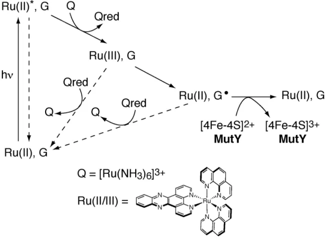
Protein-DNA Charge Transport: Redox Activation of a DNA Repair Protein by Guanine Radical
Schematic illustration of the flash-quench technique used to generate Ru(III) in situ and subsequently to oxidize DNA-bound MutY. 2dppz]3+ has been seen to promote the formation of the guanine radical but not the adenine radical cation. Additionally, we investigated the flash/quench response of the Ru-tethered duplex lacking the 30-mer strand.
Flash/quench experiments followed by EPR spectroscopy reveal spectra with g values characteristic of the oxidized clusters. We attributed this ability in the oxidation of the DNA-bound proteins to the shift in the oxidation potential. Interestingly, these data provide the first direct demonstration of [4Fe4S]3+ formation in MutY.
We find some evidence for the formation of the [4Fe4S]3+ cluster in the oxidation of DNA-bound uracil-DNA glycosylase from A. The EPR data show that the flash/quench reaction drives the oxidation of the [4Fe4S ]2+ cluster in DNA-bound MutY. DNA charge transport thus provides a route to redistribute the repair proteins to regions of the genome that contain DNA lesions.
Flash/quench experiments monitored by EPR spectroscopy reveal spectra with g and 2.02, characteristic of oxidized clusters.
Direct Electrochemistry of Endonuclease III in the Presence and Absence of DNA
After incubation with protein and cooling of the electrodes, electrochemical experiments are performed using the inverted droplet cell electrode configuration (40). Cyclic voltammetry of an electrode modified with DNA with an abasic site is shown in red (top left), with the abasic position corresponding to the complement of the italicized base. Consecutive positive scans lead to new broad, irregular signals at ~ -80 mV and ~ -710 mV versus NHE; in addition, the yellow color of the protein solution is lost.
These results are fully consistent with oxidative cleavage of the cluster in EndoIII without DNA. The potential difference between 3+/2+ and 2+/1+ pairs is somewhat smaller than expected (11) and may be an underestimate since we are at the edge of the potential window. DNA binding clearly stabilizes the oxidized 3+ form of the cluster, whereas without DNA, it is [4Fe4S]2+ that is more stable.
Instead, the ~200 mV shift in potential should correspond to a decrease in DNA binding affinity of more than three orders of magnitude between the 2+ and 3+ forms of the cluster. We have now identified the electrochemistry of EndoIII both with and without DNA on HOPG electrodes. DNA binding clearly promotes a shift in redox potential, activating the protein toward oxidation; subsequent reduction of the cluster to the 2+.
Furthermore, these data highlight the importance of the outer-sphere environment in regulating [4Fe4S] protein potentials (9, 12), as well as the utility of DNA-modified electrodes in probing the redox characteristics of protein binding with DNA. 1995) DNA Repair and Mutagenesis ASM Press.
Investigating the Role of the Rnf Operon in DNA Repair
It has also been proposed that the rnf operon might be involved in the maturation of protein-bound iron-sulfur clusters in nitrogen-fixing organisms. The relationship between the rnf gene products and EndoIII (or other [4Fe4S] cluster DNA repair enzymes) is unknown, although it has been theorized that genes cotranscribed often perform similar or related functions within the cell (32). EndoIII-associated mutations, a surprising result considering that knockout of the rnf operon should also eliminate EndoIII expression.
Therefore, further studies will be required to fully understand the relationship between the rnf operon and DNA repair. Thus, this result suggests that knockdown of the rnf operon has a mutation-suppressive effect on the cell, despite the loss of the DNA repair protein EndoIII. It is interesting to consider the result reported here in the context of the only other proposed role for rnf proteins in E.
Knockout of rnf genes has been demonstrated to slow the inactivation of soxS transcription ( 25 ), possibly resulting in a more constitutive state of the oxidative stress response. It is clear from these initial experiments that the relationship between the rnf and nth genes is complex and will not be fully elucidated by the simple experiments performed here. Finally, in-frame deletion of each of the rnf genes may reveal some of the specific roles of the individual genes.
To examine the relationship between rnf and EndoIII proteins, we inactivated the entire rnf operon in an EndoIII activity reporter strain of E.

Redox Signaling between DNA Repair Proteins for Efficient Lesion Detection: DNA Charge Transport within the Cell
In the case of MutY, an E.coli BER repair protein with a human homologue, there are 30 proteins in the E.coli cell (2) that interrogate 4.6 million bases (5); ratio of binding affinities for the target lesion, 8-. These estimates for T significantly underestimate the problem, since the interrogation time in the sliding model cannot be instantaneous, and sliding along the filament requires moving proteins, even water, which takes time. DNA-mediated charge transfer (CT) offers an alternative strategy to localize BER proteins near lesions.
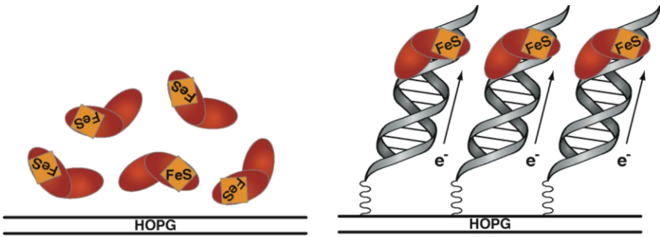
Oxidative damage to DNA has been demonstrated with oxidants covalently attached and spatially separated from oxidized sites in the DNA duplex at distances of > 200 Å with negligible loss in efficiency ( 16 – 18 ). When not bound to DNA, these proteins are found in the [4Fe4S]2+ state and are not easily oxidized or. However, for MutY and EndoIII, we demonstrated using DNA-modified electrodes that DNA binding shifts the 3+/2+ cluster potential more negative by > 200 mV DNA binding stabilizes the protein in the +3 form ( 30 ).
As illustrated in Figure 8.1 (b-d), this DNA-mediated redox signaling model involves binding to DNA by one protein in the 2+ state (donor), which will promote electron transfer from the donor protein to a distal protein (acceptor). bound to the helix and in the 3+ state. A model for DNA-mediated CT in DNA repair where DNA repair proteins, for example EndoIII (green) and MutY (orange), containing [4Fe4S]2+ clusters, bind DNA, activating them upon oxidation to the [4Fe4S ]3+ state. If instead a lesion site is present between the proteins (g), the DNA-mediated CT step is inhibited and the oxidized protein remains bound to DNA.
In this search mechanism, the sum of the DNA-mediated electron transfer steps between proteins constitutes a complete search of the genome with the end result being a redistribution of a low abundance of DNA repair proteins in the vicinity of lesions.
![Figure 8.1. A model for DNA-mediated CT in DNA repair where DNA repair proteins, for example EndoIII (green) and MutY (orange), containing [4Fe4S] 2+ clusters bind DNA, activating them towards oxidation to the [4Fe4S] 3+ state](https://thumb-ap.123doks.com/thumbv2/123dok/10407106.0/199.918.244.695.396.1003/figure-mediated-proteins-endoiii-containing-clusters-activating-oxidation.webp)


![Figure 1.6. A model for DNA CT in DNA repair. DNA-mediated redox activity in a class of DNA repair proteins that contain a [4Fe4S] cluster could allow these enzymes to use DNA CT as a damage detection strategy](https://thumb-ap.123doks.com/thumbv2/123dok/10407106.0/40.918.163.809.407.670/figure-mediated-activity-proteins-contain-cluster-detection-strategy.webp)