Thanks to the current Cliffel lab group: Kaixuan “Kysha” Xu, Grace Buckey, Nicholas Hortance, William Lowery, Olivia Owens, Pragun Tuladhar, Samantha Calvez, Matt Galazzo, and LTC John Williams II. Finally, I would like to thank my family, Andre, Nancy, grandparents, for their interest, even though you were not sure what was going on.
INTRODUCTION
The rate of mediated electron transfer must be fast if it occurs with other chemical species or at the electrode surface. While a good mediator is said not to interact with the biological compound in a way that changes its redox potential, mediators that react with other parts of the system can also cause problems.
ELECTROCHEMICAL METHODS
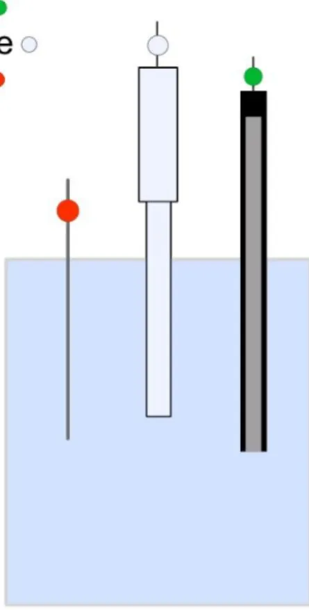
- Standard Electrochemical Cell
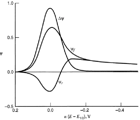
- Cyclic Voltammetry
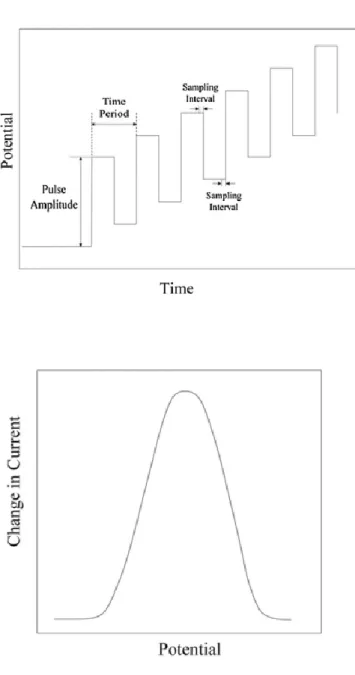
- Square Wave Voltammetry
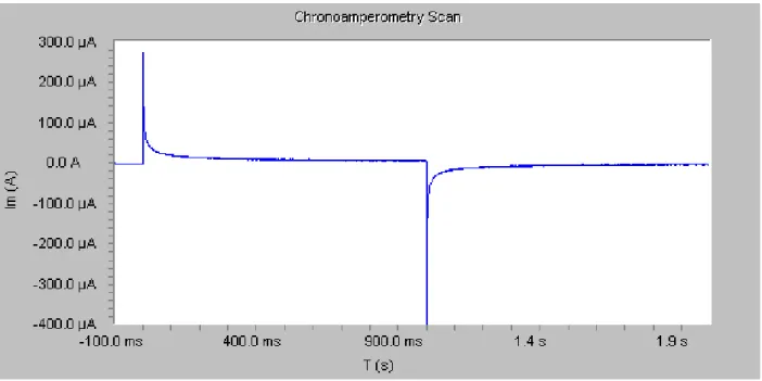
- Chronoamperometry or Amperometric i-t Curves
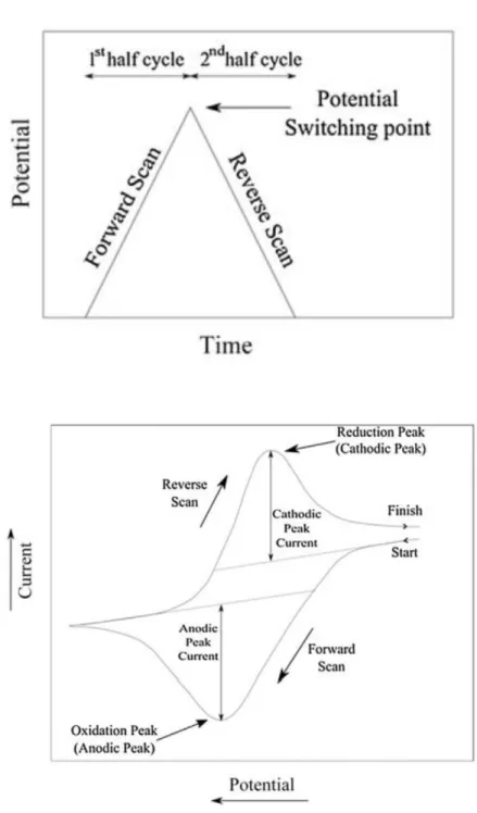
The power of CV as a characterization method comes from its waveform and applied potential vs. This allows the concentration of the analyte to be calculated if the analyte is at a typical diffusion rate and the number of electrons in the redox process is known.
OSMIUM REDOX POLYMER MEDIATOR FOR WIRED ENZYMATIC BIOSENSORS 15
- Introduction
- Materials and Methods
- Results and Discussion
- Conclusions & Future Outlooks
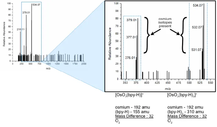
This compound is a redox polymer widely used in the field of biosensors.25-29 This literature includes the syntheses of the Os(bpy)2Cl-PVIAA polymer using an Os(bpy)2Cl2 precursor to facilitate loading of the redox couple onto the PVIAA backbones. . Several inches of the membrane were cut from the roll and the end clamped with a magnetic dialysis clip. Thus, we believe that the identity of the organometallic that was consistently generated during the oxygen-contaminated synthesis of Os(bpy)2Cl2 is OsO2(bpy)2Cl2.
Peaks shown in the ~1.1–1.3 ppm range represent hydrogens at primary, secondary, and tertiary carbons in the polymer chain backbone. As shown, the 0-6 ppm shifts in both spectra show similar characteristics of the PVI backbone. The spectrum of f-Os-PVI shows a further shift of the basic imidazole peak due to oxygen, which was found in the product.
For example, the integrals for the imidazole protons and the osmium affected protons were selected during the treatment of the 1HNMR spectrum of Os-PVI. Since the concentrations of the Os-PVI and f-Os-PVI samples were the same, we can directly compare the two. For the fluorescence of the organometallic polymers when excited in the UV range (360 nm), f-Os-PVI could emit light in the visible spectrum.
STATIC AND MICROFLUIDIC INDIRECT COMPETITION ENZYMATIC
Optimal p-aminophenol Concentration Determination for Multi-platform Early Sepsis
- Introduction
- Materials and Methods
- Results & Discussion
- Conclusions & Future Outlooks
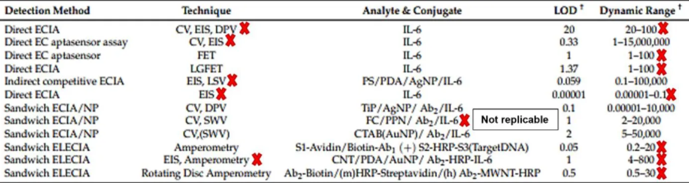
Papers in IL-6 are interested in publishing the sensor work they have, as opposed to whether it provides a path toward an appropriate sensor for a clinically relevant IL-6 sensor. Alkaline phosphatase (AP) is an enzyme that cleaves a phosphate group from the compound it acts on.50-51 It works optimally at pH 9; however, it can function at pH 7.4, the pH value most commonly found in the body.52 AP is often used as a label for biological research. However, the lack of inclusion in the sensor review for IL-6 shows that it is not a commonly used sensor for the IL-6 community (although it can be recognized that no review can be exhaustive).58-59 Several additional uses for AP/PAPP -combination is used explicitly for PAP or PAPP.
The typical pH in the human body is pH 7, where the blood is specifically within the range of Outside this range, the blood moves towards being considered acidic or alkaline, with metabolic acidosis officially defined as pH 7.00 and below. 67 Metabolic acidosis due to chronic use of paracetamol is where physicians are primarily concerned, therefore investigation of this compound outside biologically relevant pH is not necessarily relevant to that community.68-69. The difference comes from the location of the antibodies, which are immobilized on magnetic beads. IL-6 in the sample will compete for antibody binding sites with IL-6 bound to AP.
However, the IL-6 in the sample will not convert PAPP to PAP, creating the electrochemical signal. These solutions were always made fresh every day and are used consistently throughout the experiments unless otherwise stated. More concentrations were tested in the CV experiments than in the other experiments; however, their samples were prepared in the same manner, starting with a 1 mM stock solution in the KCl/PBS buffer and then diluting to the concentration of 1000 µM and from there performing serial dilutions to achieve the desired concentrations for analysis .
The difference in slope values between the equations generated from the linear regression indicates the sensitivity of the two techniques. Due to the thickness of the film, the reduced electron transfer due to the impedance will cause the current to decrease as shown in the CV of higher concentrations.
STATIC SANDWICH ELECTROCHEMICAL IMMUNOASSAY (sECIA) FOR
Determination of layer weakness in ferrocene particle sandwich immunoassay
- Introduction
- Materials and Methods
- Results and Discussion
- Conclusions & Future Outlooks
The porosity of the particles and their distance from the electrode surface led to inconsistency during use - adhesion of the base antibody film to the electrode surface eroded during repeated rinsing. Attachment of the antibody to the electrode occurred via a layer-by-layer deposition reported by Rusling et al.75 using 10 µL of solution. The square wave voltammetry performed on the samples showed the effectiveness of the two preparations.
A reduction in spacing between non-sonicated data compared to sonicated data is expected. EDC/NHS supplementation is consistent with this hypothesis and the data reflect this. However, the addition of the antibody should increase the upstream resistance due to its bulky, insulating nature.
This could potentially be that the number of antibodies trying to bind to the surface of the electrode are not successful and instead tear off previously placed parts of the film. The adaptation of the research of the previous authors was not suitable for the new application. The mechanism of action of this sensor is due to the diffusion of ferrocene from inside the solid electrolyte to outside.

CONCLUSION & FUTURE DIRECTIONS
Summary
Outlook
Further investigation of PAP in the designed sensor device and the determined sensor concentration should be investigated. It is also possible that using a pulse pre-step to clean the electrode between runs could also solve the problems. As evidenced by the projects described in this thesis, more care must be taken during the research and publication of research activities.
In Chapter 3, the difficulties in synthesizing the Os polymer could have been mitigated if earlier researchers had taken due care to fully characterize their materials and share those results through additional information. In the entire Os polymer target literature, only one article shared a 1H NMR, an essential and necessary characterization method for any organic or organometric synthesis. This oversight may call into question whether the earlier papers made the target or whether they were using something else entirely.
In addition, Chapter 4 details how specific sensor platforms or techniques would be useful for applications far beyond their experimental parameters. Much of the literature on PAP is under acidic/basic conditions or in non-aqueous environments, but claims to be useful for biological sensing purposes.
J.; Rajagopalan, R.; Heller, A., Glucose electrodes based on [Os(bpy)2Cl]+/2+ cross-linked poly(1-vinylimidazole) films. J.; Rajagopalan, R.; Heller, A., "Wired" enzyme electrode for the amperometric determination of glucose or lactate in the presence of interfering substances. O.; Kavanagh, P.; Leech, D., Oxidation of glucose by enzyme-mediated osmium redox polymer electrodes operating at low potential and in oxygen, for application in enzymatic fuel cells.
O.; Güven, O., Synthesis and characterization of poly(N-vinylimidazole-co-acrylonitrile) and determination of monomer reactivity ratios. Tertiš, M.; Ciui, B.; Suciu, M.; Săndulescu, R.; Cristea, C., A label-free electrochemical aptasensor based on gold nanoparticles and polypyrrole for the detection of interleukin 6. Tu, Z.; Zhang, J.; Yang, G.; He, G., Characterization of an alkaline phosphatase-labeled UidA (Gus) probe and its use in testing transgenic tritordeum.
Yin, H.; Ma, Q.; Zhou, Y.; Ai, S.; Zhu, L., Electrochemical behavior and voltammetric determination of 4-aminophenol based on graphene-chitosan composite film modified glassy carbon electrode. Wang, J.; Jin, B.; Cheng, L., Investigation of redox mechanism of p-aminophenol in non-aqueous media by FT-IR spectroelectrochemistry. Nematollahi, D.; Shayani-Jam, H.; Alimoradi, M.; Niroomand, S., Electrochemical oxidation of acetaminophen in aqueous solutions: Kinetic evaluation of hydrolysis,.
METHYLENE BLUE THIN-FILM HEMOGLOBIN SENSOR FOR THE
- Introduction
- Materials and Methods
- Results and Discussion
- Conclusions & Future Outlooks
Compounds such as riboflavin, poly(Nile blue), and methylene blue are traditional electrochemical mediators used to facilitate electron transfer from hemoglobin to the electrode surface by acting as an electron shuttle. A methylene blue film was formed on the electrode surface to provide a platform for catalytically mediated electron transfer. This method is often used to aid in the electrochemical analysis of proteins whose metal centers are not easily accessible to the electrode surface.78 To add the methylene blue film to the working electrode, it was kept under stirring for 60 seconds.
The freshly isolated iron hemoglobin (Hb2+) adheres to the behavior expected for the enzyme, catalytically enhancing the methylene blue reaction and showing a current response on methylene blue. The newly isolated iron hemoglobin (Hb3+) appears to have suppressed the catalytic activity of methylene blue, as indicated by the current response within methylene blue. , catalytically inhibiting methylene blue. In Figure 8.1A, hemoglobin behaves as expected with CV approximately following the methylene blue films during the oxidative half of the scan.
Ag/AgCl instead of the E1/2, the returning reductive half of the scan results in the hemoglobin deviating from the methylene blue CVs rather than forming peaks at the reductive peak potential, instead the hemoglobin becomes flat lines. This indicates that the freshly isolated Hb2+ helps in the catalysis of the methylene blue redox, increasing the current. The suppression in Figure 8.1B, C, and D ultimately suggests that the hemoglobin displaces the methylene blue from the surface and adsorbs it in place.