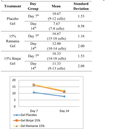
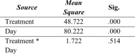
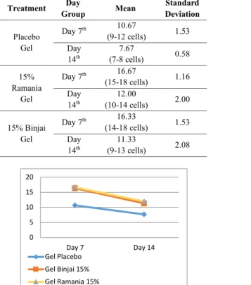
The effect of Binjai Leaves Extract Gel and Ramania Leaves Extract Gel on the number of fibroblast cells. Binjai leaves gel extract and ramania leaves gel extract have an effect on the number of fibroblasts on days 7 and 14. The average value of the number of fibroblast cells in the healing of incision wounds on the back wound of the rat on days 7 and day 14 is illustrated in Table 1 and Figure 1.
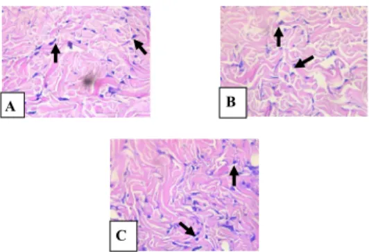
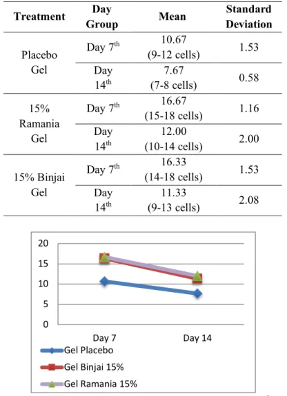
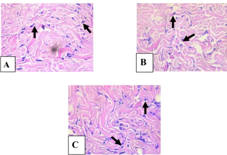
Average of fibroblast cells in incisional wound healing of rats on day 7 and day 14. Histopathology of fibroblast cells in incisional back wound healing of rat on day 7: (A) Placebo gel group, (B) 15% Binjai Leaves Gel Extract group, (C) 15 % Ramania Leaves Gel Extract group. There was a significant difference based on treatments and based on days in the number of fibroblast cells.
The result of this study proves that binjai (Mangifera caesia) leaf extract gel and ramania (Bouea Macrophylla Griffith) leaf extract gel were able to increase the number of fibroblasts on day 7 and decrease the number of fibroblasts on day 14. Based on Figure 1, it is shown that that the average number of fibroblast cells on day 7 and day 14 showed significant difference between placebo gel and treatment groups such as binjai leaf extract gel at 15% concentration and ramania leaf gel extract at 15% concentration. The placebo gel group has the number of fibroblast cells lower than binjai leaf extract gel at 15% concentration and ramania leaf extract gel at 15% concentration.
Based on statistical results and discussion shows that there is effect of binjai leaf extract gel at concentration of 15% and ramania leaf extract gel at concentration of 15% to the number of fibroblast cells in rat incision back which will increase on day 7 and decrease on day 14.
Introduction
Materials & method
Results
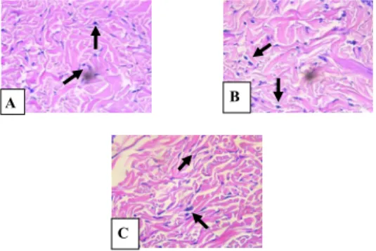
Histopathology of fibroblast cells in incisional spinal wound healing of rat on day 14: (A) Placebo Gel Group, (B) 15% Binjai Leaves Gel Extract Group, (C) 15% Ramania Leaves Gel Extract Group Figure 3 shows the histopathology of fibroblast cells number in incisional spinal wound of rat in placebo gel group with a total of 7-8 cells, binjai leaf extract gel at the concentration of 15% group with a total of 9-13 cells, and finally ramania leaf extract gel at the concentration of 15% group with a total of 10-14 cells. Two-way anova test was performed and shows that there is significant difference based on treatments (p<0.05) and based on days (p<0.05). Meanwhile, the significance value in the interaction between treatment and day was greater than 0.05, which means that there was no significant difference between treatment and day in the number of fibroblast cells.
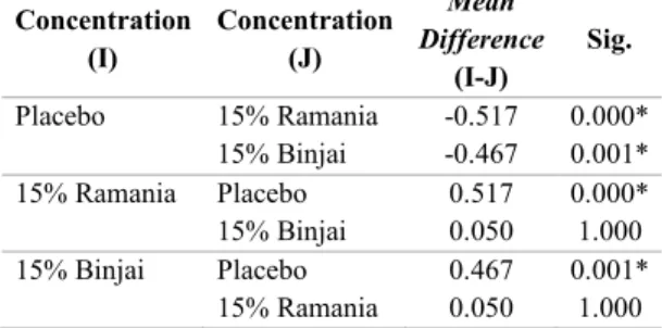
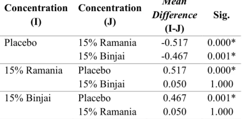
Since there was a significant difference, data analysis was followed by Post-Hoc Bonferroni to statistically identify a significant difference. Based on the table, there was no significant difference between binjai leaf extract gel at 15% group concentration and ramania leaf extract gel at 15% group concentration (p=1,000). Based on the mean difference between placebo gel group and ramania leaf extract gel at 15% concentration group (0.467) and between placebo gel and ramania leaf extract group at 15% concentration group (0.517), it is revealed that leaf extract gel ramania at 15% concentration has a better effect compared to binjai leaf extract gel at 15% concentration.
The result based on day shows that there was significant difference between day 7 and day 14. The result of this study proves that binjai leaves extract gel and ramania leaves extract gel were able to increase the number of fibroblasts on day 7 and decrease the number of fibroblasts on day 14. Based on research by Kurnia (2015), who used Camellia sinensis, flavonoid will support lymphocyte proliferation and interleukin-1 (IL-1) which will stimulate T-cell proliferation and differentiation to T-helper1 (Th1).
Flavonoid will support the increase in insulin-like growth factor-1 (IGF-1) as a mediator of fibroblast proliferation. This study result based on figure 1 shows that there is a difference in the average of fibroblast cell number on day 7. Ramania leaves extract gel at the concentration of 15% group and binjai leaves extract gel at the concentration of 15% group shows better result in comparison with placebo gel group.
According to a study by Ismiardianita et al., (2019), which used Clausena extract containing flavonoid in extraction wounds of rats, it is found that the number of fibroblasts decreased on day 14 compared to day 7. After the peak on day 7, the fibroblast decrease because the wound healing process proceeds well and the fibroblast will be more progressive in synthesizing the collagen in the maturation phase. Another cause of the decrease in the number of fibroblast cells is due to the phenotype change from fibroblast to myofibroblast.
Conclusion
In addition to growth factors, flavonoid can produce cytokines, such as IL-1, IL-4, IL-8, to support fibroblast chemotaxis and keratinocytes.18,20. Seven days after wound formation, the number of fibroblasts will reach their peak and replace the macrophage in the inflammatory phase.21 Macrophage-releasing growth factors, such as platelet-derived growth factor (PDGF), fibroblast growth factor (FGF), endothelial growth factor (EGF), transforming growth factor-α ( TGF-α) and TGF-β, which stimulate the proliferation of fibroblasts and generate the formation of extracellular matrix (ECM).22 Fibroblast will occupy the fibrin matrix with the help of matrix metalloproteinase (MMP) and replace it with glycosaminoglycan (GAG). The extracellular matrix will be replaced by type III collagen which is also produced by fibroblasts.1 Type III collagen is usually found for tissue repair and reaches its peak on the 5th to 7th day after wound formation.22.
Myofibroblast helps the wound contract and reconnect.23 Myofibroblasts are connected by cell-to-cell and cell-to-matrix and express α-Smooth Muscle Action (α-SMA) through repetitive contraction to stimulate collagen fibers in the produce an area of injury. 1,22,25 At this stage, collagen type III will be replaced by collagen type I, which has a band shape and greater tensile strength and density in new tissue.24 Only about 80% of the tensile strength will be restored due to the ability of collagen fibers om can only regain 80% of his normal strength before the injury.1.
Peran ekstrak etanol topikal daun mengkudu (Morinda citrifolia L.) dalam penyembuhan luka berdasarkan CD34 dan ekspresi imunoekspresi kolagen pada tikus Wistar. Potensi ekstrak teh hijau (Camellia sinensis) dalam meningkatkan jumlah sel soket fibroblas pasca pencabutan gigi pada tikus wistar. Jumlah fibroblas pada tikus luka bakar derajat dua dengan pemberian gel ekstrak etanol biji kakao dan perak sulfadiazin.
Efektivitas pemberian gel Binahong (Anredera cordifolia) 5% terhadap jumlah sel fibroblas pada gigi kelinci percobaan (Cavia cobaya) pasca pencabutan. Pengaruh gel dengan ekstrak daun Binjai (Mangifera Caesia) dan gel dengan ekstrak daun Ramania (Bouea.
The Effect Of Binjai Leaves Extract Gel (Mangifera Caesia) And Ramania Leaves Extract Gel (Bouea
Cells In Incisional Wound Of Male Rats (Rattus Norvegicus)
Discussion
The cut wound in the mouse will be followed by the wound healing process which consists of 3 phases, which is inflammation, proliferation and remodeling [2] Fibroblast cell can be found in the connective tissue and play an important role to form the extracellular matrix (ECM) to the wound. closure [15]. Flavonoids function as secondary antioxidants to balance the amount of oxidant and antioxidant through the hydrogen donor to activate Nrf2 in order to increase the activation of endogenous antioxidant, such as superoxidase dismutase (SOD), catalase (CAT) and glutathione peroxidase (GPX. Flavonoid will protect the body so that the wound healing process can continue and promote the proliferation of fibroblasts and collagen synthesis [7], [8].
Based on the research of [19], which used Camellia sinensis, flavonoid will support lymphocyte proliferation and interleukin-1 (IL-1) which will stimulate T cell proliferation and differentiation into T-helper1 (Th1). Growth factors that macrophages release, such as platelet-derived growth factor (PDGF), fibroblast growth factor (FGF), endothelial growth factor (EGF), transforming growth factor-α (TGF-α), and TGF-β , which stimulate fibroblast proliferation and generate extracellular matrix (ECM) formation [22]. According to a research by [24], which used the flavonoid-containing Clausena extract in the rat extraction wound, it was reported that the number of fibroblasts decreased on the 14th day compared to the 7th day.
Myofibroblasts are connected by cell-to-cell and cell-to-matrix and express α-Smooth Muscle Action (α-SMA) through repetitive contractions to produce collagen fibers in the area of injury. At this stage, type III collagen will be replaced by type I collagen, which has a ribbon shape and has stronger tensile strength and density in new tissue [24]. Only about 80% tensile strength will be recovered due to the ability of collagen fibers to only recover 80% of their normal pre-injury strength [1].
Berdasarkan hasil statistik, gel ekstrak daun Ramania (Bouea microphylla Griffith.) pada konsentrasi 15% mempunyai hasil yang lebih baik dibandingkan gel ekstrak daun binjai (Mangifera caesia) pada konsentrasi 15% dan gel plasebo. Pengaruh pemberian Ibuprofen pra operasi terhadap distribusi sel inflamasi kronis pada proses penyembuhan luka pasca pencabutan gigi. Penggunaan krim ekstrak batang dan daun Seruhan (Peperomia pellucida L.H.B.K) dalam proses penyembuhan luka bakar pada tikus putih (Rattus norvegicus).
Effect of gum gel Anti-density banana plant stem on local collagen fibers Wound healing process after tooth extraction Marmota. Daun Kelor Extract Gel Formula (Moringa oleifera Lam.) Sebagai Anti Inflammasi Topical Pada Tikus (Rattus novergicus). Effectiveness of Clausena Excavate Methanol Extract on fibroblast number and collagen fiber density after tooth extraction.