MICROGLIA IN THE CEREBRAL AND CEREBELLAR CORTICES IN
INDIVIDUALS WITH AUTISM
Thesis by Nicole A. Tetreault
In Partial Fulfillment of the Requirements For the Degree of
Doctor of Philosophy
California Institute of Technology Pasadena, CA
2013
(Defended May 23, 2013)
ii
© 2013 Nicole A. Tetreault All rights reserved.
iii ACKNOWLEDGMENTS
This work was supported by grants from the Simons Foundation (SFARI
#137661), the James S. McDonnell Foundation, and by NIH grant MH089406. The brain tissue and related anonymous phenotypic information was obtained from the NICHD Brain and Tissue Bank for Developmental Disorders. Special thanks to Dr. Ronald Zielke, Robert Johnson and Melissa Davis for providing the brain tissue and anonymous clinical records; our study would not have been possible without their dedicated service.
Thank you to all of the tissue donors and donor family members, this work is made possible by your donations. Thanks to the additional granting agencies for their support:
College Women's Club of Pasadena, the Kanal Foundation, and the Caltech SURF program.
I have an immense amount of gratitude for all of the individuals who encouraged, guided, and supported me along the way to complete the work described in thesis. Thank you to my mentor and friend John Allman, for his daily discussions, which were filled with insight and enthusiasm. This thesis could not have been done without John's continued support, patience and vision. I was an extremely fortunate graduate student to have a mentor that allowed me to work independently as well as being available to answer questions, big and small, even if he was on top of a mountain. Thank you to Barbara Wold, for her guidance, expertise in the field of molecular biology and continued encouragement. Barbara, thank you for always encouraging me to ask the big questions
iv guidance, insightful discussions, and scientific rigor. Thank you to Ralph Adolphs for valuable input in experimental design, continued encouragement and enthusiasm. Thank you to all my friends and members of the Allman lab: Atiya Hakeem, Soyoung Park, and Sue Jiang; and previous lab members: Karli Watson and Virginie Goubert. Atiya, we have worked many years together and I have always valued our scientific discussions, shared interests in traveling and our passion for animal conservation. Soyoung, you have always been a pleasure in the lab and I am grateful for our time together; you brighten the lab with your smile. Sue-Jiang, thank you for working so diligently on the counts and tracings; working with you has been outstanding. Thank you to Libby Allman for her expertise in autism and for taking the time to evaluate the medical records of our subjects.
Thank you to all the Wold lab members for their time, guidance and training. Thank you to Brian Williams for his patience, advice in experimental design and training in
molecular techniques. Brian, you are a generous and kind teacher. Thank you to the Caltech graduate office, including Natalie Gilmore, Felicia Hunt and Joseph E. Shepherd.
Thank you to Liz Ayala, Barbara Besse and Patricia Mindorff for guiding me along the way. Thank you to my good friend, Tara Gomez, for always encouraging me and inspiring me to be a strong female scientist.
Thank you to my incredible family, extended family and friends, for continued encouragement, love and support. I have tremendous gratitude for both of my parents, Dave and Loretta Tetreault, for continually inspiring me, being my first teachers, and encouraging me to ask questions. Dad, thank you for always telling me to do what I love and that the rest will follow. You were and are right. Mom, thank you for all of your
v
courage, passion and strength; you taught me to always believe and see the best in people. I know living with Parkinson’s disease is a tremendous challenge, and for you, I will continue to ask the most valuable scientific questions. Many thanks to my brothers and sister: David, Scott, Phil and Donna, for teaching me how to work productively in a group, to persevere, and to listen with an open mind, which is crucial in science. You are all remarkable siblings and I would not be who I am without you. Thank you to my in- laws, John and Kathy DeClercq, Shana and Deborah, Marissa and Jake, for always being interested in my studies of autism, for our discussions on the latest findings in
neuroscience and for their constant encouragement.
My greatest gratitude is to Billy DeClercq, my best friend and husband, for his continued patience, encouragement, and love. Billy, you have been with me every step of the way, beginning with our first marathon together, to studying together in the libraries at UC Davis and UCLA, our seven eventful years at Caltech and beyond. Thanks to Spencer DeClercq, my son, my light, and my little scientist, for the constant reminder that life is an experiment. Asking the question is just the beginning.
vi ABSTRACT
In this thesis, we explore the density of the microglia in the cerebral and cerebellar cortices of individuals with autism to investigate the hypothesis that neuroinflammation is involved in autism. We describe in our findings an increase in microglial density in two disparate cortical regions, frontal insular cortex and visual cortex, in individuals with autism (Tetreault et al., 2012). Our results imply that there is a global increase in the microglial density and neuroinflammation in the cerebral cortex of individuals with autism.
We expanded our cerebellar study to additional neurodevelopmental disorders that exhibit similar behaviors to autism spectrum disorder and have known cerebellar pathology. We subsequently found a more than threefold increase in the microglial density specific to the molecular layer of the cerebellum, which is the region of the Purkinje and parallel fiber synapses, in individuals with autism and Rett syndrome.
Moreover, we report that not only is there an increase in microglia density in the
molecular layer, the microglial cell bodies are significantly larger in perimeter and area in individuals with autism spectrum disorder and Rett syndrome compared to controls that implies that the microglia are activated. Additionally, an individual with Angelman syndrome and the sibling of an individual with autism have microglial densities similar to the individuals with autism and Rett syndrome. By contrast, an individual with Joubert syndrome, which is a developmental hypoplasia of the cerebellar vermis, had a normal density of microglia, indicating the specific pathology in the cerebellum does not
vii necessarily result in increased microglial densities. We found a significant decrease in Purkinje cells specific to the cerebellar vermis in individuals with autism.
These findings indicate the importance for investigation of the Purkinje synapses in autism and that the relationship between the microglia and the synapses is of great utility in understanding the pathology in autism. Together, these data provide further evidence for the neuroinflammation hypothesis in autism and a basis for future
investigation of neuroinflammation in autism. In particular, investigating the function of microglia in modifying synaptic connectivity in the cerebellum may provide key insights into developing therapeutics in autism spectrum disorder.
viii
TABLE OF CONTENTS
Acknowledgements... iii
Abstract... vi
Chapter 1: Introduction Autism Spectrum Disorder...1
Genetic Studies of Autism...3
Neuroinflammation in Autism...7
Microglia ...8
Maternal Infection in Autism...11
Gastrointestinal Abnormalities in Autism...13
Rett Syndrome, Angelman Syndrome and Fragile X Syndrome...14
Genetics and Inflammation in Mental Illness: Schizophrenia, Sickness Behavior, and Depression...17
Conclusion...19
References...22
Chapter 2: Microglia in the Cerebral Cortex in Autism Copyright Acknowledgement ...30
Abstract...31
Introduction...31
Methods ...32
Tissue Samples ...32
Sectioning and Immunocytochemistry ...32
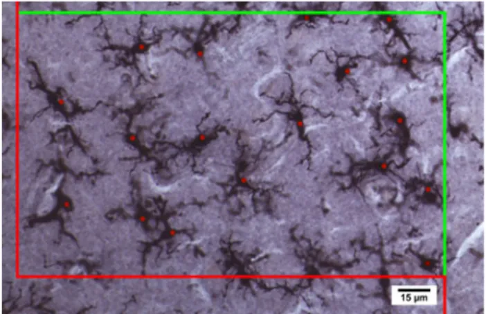
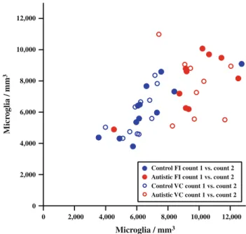
Quantification of Microglial Densities...33
Statistical Analysis...36
Results...36
Discussion ...40
Are Microglia Predators or Protectors? ...42
Microglial Defects as Causes of Disease...42
Visual Abnormalities in Autism...43
Summary ...44
Acknowledgements...44
References...44
ix
Chapter 3: Microglial and Purkinje Cell Densities in the Cerebellar Cortex in Autism, Rett, Angelman, and Joubert Syndromes
Abstract...47
Introduction...48
Methods ...51
Tissue Samples...51
Sectioning and Staining...52
Quantification of Microglial Densities...52
Microglia Shape Analysis...53
Purkinje Cell Counts...54
Purkinje Cell Shape Analysis...54
Granule Cell Counts...55
Statistical Analysis...55
Results...56
Discussion ...61
Microglia and Neuroinflammation...61
Purkinje Cells...66
The Cerebellum in Autism...70
Conclusion ...74
Chapter 4: Summary and Conclusions...76
Is elevated of microglia density in autism a biomarker for the disorder? ...83
Would an anti-inflammatory treatment aid in reducing the symptoms in autism? ...84
References ...86
Tables ...98
Table 1 ...99
Table 2 ...101
Table 3 ...109
Table 4 ...110
Table 5 ...111
x
Figures ...115
Figure 1 ...116
Figure 2 ...117
Figure 3 ...118
Figure 4 ...119
Figure 5 ...120
Figure 6 ...121
Figure 7 ...122
Figure 8 ...123
Figure 9 ...124
Appendix ...125
Photomicrographs: Microglia in all the Cases ...126
Photomicrographs: Purkinje Cells in all the Cases...140
Raw Data for Cerebellar Cell Counts ...149
Raw Data for Microglial Shape Analysis ...150
Raw Data for Purkinje Cell Shape Analysis...151
CHAPTER 1: INTRODUCTION
Autism Spectrum Disorder
Autism spectrum disorder is heterogenous clinically and etiologically where the diagnosis is entirely based on the behavioral phenotype (Miles et al., 2011). It is a neurodevelopmental disorder described by two core features: a lack in social communication and restricted repetitive behaviors, often diagnosed by age three.
(American Psychiatric Association, DSM 5, 2013). The diagnosis of the disorder is at present entirely clinical, based on differing aspects of behavior and its developmental time course. This includes a number of specifiers (extensions to a diagnosis that further clarify its course, severity, or special features). Diagnosis in the clinic can include additional disorders such as attention deficit hyperactivity disorder, Rett syndrome, seizures, other disorders and neurological symptoms that are comorbid with autism (American Psychiatric Association, DSM 5, 2013). Since autism spectrum disorder is a heterogeneous disorder, it may involve additional symptoms including seizures, sensory abnormalities such as hypersensitivity, motor deficits, and gastrointestinal alterations (Danielsson et al., 2005; Leekam et al., 2007; Vilensky et al., 1981; D’Eufemia et al., 1996; Horvath et al., 1999; De Magistris et al., 2010). Seizures occur in 38% of individuals with autism, and the frequency of seizures has great impact on the individuals’ lives (Danielsson et al., 2005). Sensory abnormalities have been often described in autism spectrum disorder. These include pain insensitivity and
hypersensitivity to tactile, auditory, and visual stimuli, which can be debilitating
(Bemporad et al., 1979; Grandin & Scariano, 1986; Cesaroni & Garber, 1991). Over 90%
1
sensory disabilities are extensive, multimodal and continuous across age and ability in individuals with autism (Leekam et al., 2007). In addition, individuals with autism experience a number of motor deficits, which may include gait disturbances, various postural instabilities, greater clumsiness, and altered motor coordination (Vilensky et al., 1981; Bauman & Kemper 2005; Molloy et al., 2003). A number of individuals with autism experience gastrointestinal disturbances and 43% of individuals with autism in one sample had altered intestinal permeability compared to neurotypicals, who had none (D’Efufemia et al., 1996). Of these gastrointestinal disturbances, the most severe
symptoms include constipation and diarrhea, and constipation is associated with severity of the language and social impairment in individuals with autism (Gorrindo et al., 2012;
Chandler et al., 2013). Emerging evidence of increased autoantibodies may indicate an inflammatory state of the intestines and may alter the mucosal barrier and integrity, which may lead to gastrointestinal issues in individuals with autism (Coury et al., 2012).
As of 2012, about 1 in 50 children in the United States have been diagnosed with autism or autism spectrum disorder (up from 11 per 1,000 in 2008) (Blumberg et al., 2013; Rutter et al., 2005). This increase in prevalence has raised tremendous concern among parents, clinicians and the scientific community, leading to the suggestion that environmental factors may contribute to the epidemic of autism (Miles et al., 2011).
Currently, the cause of autism remains unknown. Considerable advances in the genetic causes of autism can be attributed to identifying known genetic mutations and disorders that predispose to the development of autism (Miles et al., 2011). Approximately 20-25%
of autism spectrum disorder can be identified by a genetic cause, leaving 75-85% as presently unknown causes (Miles et al., 2011). Moreover there are a number of
2
environmental exposures during fetal development that increase the risk of autism that include maternal infection and immune activation, elevated metabolic conditions such as diabetes and obesity and elevated levels of C reactive protein in the mother’s serum (Brown et al., 2004; Patterson et al., 2011; Krakowiak et al, 2012; Brown et al., 2013). It is possible that an interaction of genes and environmental exposures contribute to the increased incidence in autism. Insights from three areas of study are invaluable for understanding the underlying mechanisms of autism: first, examination of autopsy tissue of individuals with autism for pathological and genetic analysis; second, patient studies investigating behavior and genetics in living individuals with autism; and third,
investigations using mouse models of autism for both genetic causes of autism and environmental contributions to developing autism. Integrating these research areas provides promise for better diagnostics, behavioral treatments and therapeutics for individuals with autism.
Genetic Studies of Autism
There is now strong evidence that genetic alterations can contribute strongly to the autism phenotype. First, there are data on overall heritability. If one of a pair of monozygotic twins has autism, there is an 88% likelihood that the other will have a form of autism, whereas if the twins are dizygotic, the chances are 31% that the other twin will have autism (Rosenberg et al., 2009). A more recent study by Hallmayer and colleagues (2011) describes a 77% concordance for monozygotic male twins with autism spectrum disorder and a 31% concordance among male dizygotic pairs, as well as a 50%
3
pairs, and attributes the variance in liability to shared environmental factors and a
moderate genetic heritability (Hallmayer et al., 2011). Second, single gene disorders, referred to as “syndromic autism,” present autism-like features; these include fragile X, Rett syndrome, tuberous sclerosis complex, and Timothy syndrome (Miles et al., 2011).
The genetic causes of autism account for 20-25% of autism cases based on data from genome-wide association studies of autism, single gene mutations in autism, and copy number variations (CNVs), i.e., chromosomal deletions or duplications (Miles et al., 2011). The most common chromosomal abnormalities in autism are found on the maternally-derived 15q duplications of the Prader Willi/Angelman region, which accounts for 1-3% of cases with autism (Wang et al., 2013). The most commonly implicated gene in this region is UBE3A. Clinically-relevant CNVs were found in eight out of 29 individuals with autism (27.5%) using a 1Mb genome-wide array (Jacquemont et al., 2006). Sebat and colleagues (2007) described de novo copy number variations in 10% of children in which they are the sole family member with autism, 2% from autism in multiple family members compared to 1% of neurotypicals, implicating de novo germline mutation as a substantial risk factor for autism (Sebat et al., 2007). Genes encoding neuronal cell-adhesion molecules, including NRXN1, NLGN1 ASTN2, CNTN4, were enriched with CNVs in autism, and genes involved in the ubiquitin pathways, including UBE3A, PARK2, RFWD2 and FBXO40, were affected by CNVs not observed in controls using a whole genome CNV study of 550,000 single nucleotide polymorphism markers in 859 individuals with autism and 1,409 neurotypicals (Glessner et al., 2009). In some monozygotic twins who are discordant for autism, the UBE3A gene is methylated in the autistic twin, which suppresses the expression of the gene (Wang et
4
al., 2013). To date, the SFARI website (https://gene.sfari.org/autdb/) reports 252
annotations for CNVs and 582 genes thought to be associated with autism (Basu et al., 2007) which is 2% of the entire genome. Thus, multi-genetic alterations, as well as genetic and environmental interactions during critical periods of development, may contribute to autism.
Additionally, investigators are studying the genetics in autism by analyzing the transcriptome, the set of all mRNA molecules. The number of transcripts and transcript abundance for each gene is measured in various tissues and cells to determine genes and possible networks of genes that may be dysregulated in autism. Microarray analysis of cerebellar tissue, prefrontal cortex and caudate-putamen reveal an increase in transcripts related to AMPA-type glutamate receptors in individuals with autism compared to neurotypicals (Purcell et al., 2001). Lymphoblastoid cell lines from five male twins discordant for autism based on language impairment revealed an increase in transcripts for the pro-inflammatory cytokines and a decrease in transcripts for those that are involved in brain development, neuronal differentiation and axon guidance in the individuals with autism (Hu et al., 2006).
Lymphoblastoid cell lines genomic profiling in individuals with autism compared to controls reveals a small number of genes expressed predominately in natural killer cell mediated cytotoxicity (Gregg et al., 2008). These findings indicate that a number of genes involved in natural killer cell proliferation are elevated in individuals with autism that can contribute to the autism phenotype. Gene expression profile analysis of superior temporal gyrus, auditory cortex, shows an increased transcript level of many immune system-
5
individuals with autism compared to controls (Garbett et al., 2008). Additionally, there is a decrease in genes involved in neuronal development and neurite outgrowth in autistic cases compared to neurotypicals (Garbett et al., 2008). The expression patterns appear to be associated with late recovery of an autoimmune disorder, rather than an innate
immune response (Garbett et al., 2008). Importantly, this study was limited to a small number of individuals with autism (N=6); thus, replication with a larger sample size would be useful would be useful for testing the auto-immune hypothesis.
In a more recent study using microarray analysis of gene expression in individuals with autism compared to controls, Voineagu et al. (2011) describe a module of elevated immune and microglial genes and deduce that the increased gene expression of the immune and microglia genes is a non-genetic etiology since these genes have not been found in genome-wide association studies, which points to the genes being causally related to autism. This implies that the elevated immune and microglial genes are possibly a result of internal or external environmental influences on inflammation in autism (Voineagu et al., 2011). These data describe alterations in the expression of genes involved in synapse formation and inflammation in the developing brain of individuals with autism. Since only 20-25% of the incidence of autism is accounted for by genetics, and the etiology is largely unknown, environmental factors likely warrant further exploration. In particular, it is not clear how inflammation alters synaptic development and how disrupted synaptic development of neural network formations can influence inflammatory genes. One explanation advanced by Paul Ashwood and colleagues (2006) is that "immune dysregulation could result in the generation of localized or systemic inflammation and/or the release of immunomodulatory molecules that could influence,
6
alter, or modify neurodevelopment and/or neuronal function, especially at critical times of development." Specifically, there could be inflammatory events at critical time periods during fetal and postnatal development that could alter proper network formation and maintenance dysregulating the normal brain development. Since then, substantial work has been done that addresses this general set of possibilities as discussed below.
Neuroinflammation in Autism
There have been numerous reports implicating neuroinflammation or an increase in production of microglial cells in the central nervous system in autism spectrum
disorder (Vargas et al., 2005; Pardo et al., 2005; Zimmerman et al., 2005; Voineagu et al., 2011; Chez & Guido-Estrada, 2010; Wei et al., 2011; Morgan et al., 2010; Tetreault et al., 2012; Suzuki et al., 2013). Neuroinflammation is best described as activated
microglia and astrocytes and increased production of cytokines such as interleukin-6 (IL- 6) and tumor necrosis factor-alpha (TNF-alpha), which are all measures of an
inflammatory state (Monnet-Tschudi et al., 2011).
Vargas and colleagues’ (2005) innovative study of human autopsy tissue reported an increase in cytokines in the brains of individuals with autism compared to
neurotypicals. In addition, individuals with autism have a significant increase in
cytokines in the cerebral spinal fluid and in the frontal cortex compared to neurotypicals (Zimmerman et al., 2005; Vargas et al., 2005; Li et al., 2009). Wei and colleagues (2011) found an increase in IL-6 in the cerebellum of individuals with autism and hypothesized that the microglia are altering the cerebellar granule cell excitatory synapses (Wei et al., 7
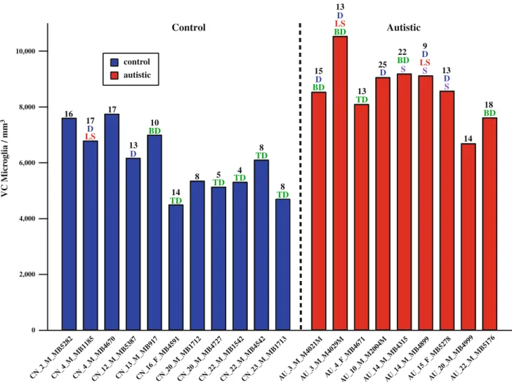
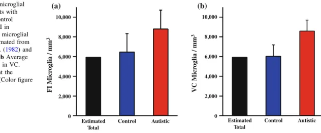
stain for microglia of human leukocyte antigen receptor (HLA-DR) in the cerebellum of individuals with autism compared to controls (Vargas et al., 2005). However, this method did not measure the microglial cell densities, sizes and shapes. Thus, quantification of microglia and shape analysis are essential to studying the state of microglia activation in the brains of individuals with autism. Morgan and colleagues (2010) reported an increase in microglia in dorsal lateral prefrontal cortex in individuals with autism and observed microglial processes retracting and thickening suggesting the microglia were in an activated state. Similarly, we reported an increase in the microglial density in fronto- insular cortex and visual cortex in individuals with autism. Since these are two disparate cortical areas, this indicates a global increase in the microglial density in individuals with autism (Tetreault et al., 2012). These data support the hypothesis that neuroinflammation, specifically, increased density of microglia, is involved in autism spectrum disorder. Our results for molecular and granule cell layers of cerebellar cortex for individuals with autism and related disorders compared to neurotyical subjects are reported in Chapter 3 of this thesis.
Microglia
Microglia reside throughout the brain and have long been thought to mainly comprise the brain’s immune system, which is distinctly separated from the body’s immune system by the blood-brain barrier. In the quiescent state, microglia actively survey their environment and make contact with synapses (Wake et al., 2009). Microglia phagocytose invading microorganisms and debris to defend against infection, clear damaged tissue, and dispose of metabolic waste (Graeber & Streit 1990, 2010).
8
Moreover, microglia are essential for normal brain development and are critical for synaptic plasticity (Paolicelli et al., 2011; Schafer et al., 2012).
Microglia arise from cells in the yolk sac in embryogenesis and later in life from cells in the bone marrow. Microglia originate in the yolk sac during embryogenesis and can be detected in the brain rudiment from embryonic day eight in mice where microglia continually rise steadily during the first two postnatal weeks, when the majority of microglia are born (Alliot et al., 1999). More recently, using a tamoxifen-inducible Cre recombinase under the control of Runx-1, a hematopoietic transcription factor, confirmed that microglia originate from embryonic Runx-1-expressing yolk sac hematopoietic progenitors between 7.25 and 7.5 days post-conception (Ginhoux et al., 2010).
Moreover, the authors reported that in the healthy mouse brain that peripheral myeloid cells do not contribute to new microglia to achieve maintenance numbers in the adult brain (Ginhoux et al., 2010). Additionally, Kierdorf described the yolk sac progenitors that give rise to microglia and identified them as early 8 days post conception (Kierdorf et al., 2013). Mouse microglia originated from primitive c-kit+ yolk sac precursors developing into CD45+ c-kitlo CX3CR1− cells prior to maturation and migrate into the embryonic maturing brain as CD45+ c-kit- CX3CR1+ cells and proliferate into the microglia (Kierdorf et al., 2013). These studies provide evidence that the majority of adult microglia in the health brain arise during early development.
To defend against infection, microglia phagocytose invaders, damaged tissue and metabolic waste (Graeber & Streit 1990, 2010). It is conceivable that maternal
inflammation elicits an activation of microglia, which can be a risk factor for autism 9
microglia recruit monocytes into the brain through the release of TNF-alpha (D’Mello et al., 2009). When microglia are activated by an infection, traumatic brain injury, stroke, or neurodegenerative disease, they enter the phagocytic phase, engulfing and removing debris in the brain in contrast to the quiescent stage where microglia are primarily engaged in surveying their environment (Lin & Bergles, 2004; Wake et al., 2009).
Microglia appear to have functions separate from immune activity. Increasing evidence points to a key role in neurodevelopment and neural plasticity through the mechanism of synaptic pruning (Paolicelli et al., 2011; Schafer et al., 2012).
Visualization of fluorescently labeled microglia in the mouse brain using two-photon imaging shows that microglia actively survey their environments by retracting and expanding their processes in response to neuronal stimuli (Nimmerjahn et al., 2005;
Davalous et al., 2005 and Wake et al., 2009). A mouse model of neurodegeneration resulting from brain ischemia shows microglia increase contact with synapses that have reduced activity (Wake et al., 2009).
During the peak critical period in postnatal development of the visual system, upon visual stimulation microglia contact dendritic spines, synaptic terminals, and clefts in layer II and IV of the visual cortex (Tremblay et al., 2010). In the developing visual system, microglia are necessary for phagocytic synapse elimination in the mouse retino- geniculate pathway for normal development, which is dependent on the microglia phagocytic signaling pathway of the complement receptor CR/C3, a property of the innate immune system (Schaffer et al., 2012). Together these data exhibit that microglial phagocytosis is crucial for normal brain development and circuit formation. As a result of these studies, a number of questions arise: Is the neuroinflammation an indicator of
10
dysregulated synaptic connectivity? Or is the neuroinflammation in response to systemic infection? Or could it be both? Since autism is a heterogenous disorder, could the neuroinflammation represent a dysregualtion in the synaptic connectivity and a systemic infection? Or could it be due to a history of systemic infections?
Maternal Infection in Autism
There is evidence that maternal viral infection in the first trimester and maternal bacterial infection in the second trimester have significant association with autism (Atladóttir et al., 2010). Individuals with autism have elevated levels of a number of inflammatory cytokines in amniotic fluid compared to neurotypicals (Abdallah et al., 2011). There is also a significant association between elevated levels of C-reactive protein (indicative of the inflammatory response) in maternal serum and the likelihood that her child was autistic (Brown et al., 2013). In addition, a subset of people with autism have a continuous pro-inflammatory pathology in the brain and cerebral spinal fluid, and it is hypothesized that maternal infection or a systemic infection may lead to inflammation and autism spectrum disorder (Chez & Guido-Estrada, 2010). Prenatal infection and immune dysfunction are biologically plausible potential causes of autism (Patterson et al., 2011). Using a mouse model of maternal immune activation (MIA), a single injection of interleukin-6 (IL-6) during mouse pregnancy results in offspring with behavioral deficits similar to those in autism, including deficiencies in prepulse inhibition (PPI) and latent inhibition (LI), but these deficits do not occur when IL-6 is eliminated from the MIA model using genetic methods or blocking antibodies (Smith et al., 2007).
11
and exploratory and social deficits as well as the alterations in gene expression in the brains MIA offspring (Smith et al., 2007). Similarly, MIA in IL-6 knock-out mice do not exhibit behavioral deficits as seen in the MIA wild-type offspring (Smith et al., 2007).
Together these data indicate that the cytokine IL-6 crucial for moderating behavioral and transcriptional changes in the MIA mouse model.
In a mouse model of MIA, polyinosine:cytosine (poly(I:C)) injection elicits a viral infection response, where the offspring demonstrate behaviors similar to autism,
including deficits in social communication and interaction, reduction in ultrasonic vocalizations (USVs), and increased stereotyped/repetitive behaviors such as excessive marble burying and self-grooming (Malkova et al., 2012; Hsiao et al., 2011).
Additionally, the MIA offspring exhibit a reduction in the linear density of Purkinje cells specific to lobule VII of the cerebellum, which is a hallmark of autism neuropathology, indicating that the alteration of the Purkinje cells is induced by maternal activation of the immune system occurring during embryonic development (Shi et al., 2009). The MIA offspring after a poly(I:C) injection exhibit changes in a number of cytokines in the brains and sera in a region and age-specific manner (Garay et al., 2012). The MIA- induced cytokines do not breach the blood brain barrier, permit immune cell infiltration or lead to an increase in microglia (Garay et al., 2012). Although these results argue that there is an increased level of cytokines in brain regions similar to those reported in autism, there is no elevation in the microglia in this mouse model.
In another model of maternal inflammation, administering a lipopolysaccharaide (LPS) injection and a concurrent single treatment of an IL-1 receptor antagonist results in
12
normal brain development in the mouse, providing promise for treatments that are
protective for normal fetal brain development (Girard et al., 2010).
Gastrointestinal Abnormalities in Autism
Gastrointestinal disturbances occur in individuals with autism including inflammatory bowel syndrome, reflux esophagitis, chronic gastritis, chronic
duodenitisdisaccharide malabsorptionmay (Horvath et al., 1999). Again, the question arises as to whether it is causative or correlative, relative to the behavioral phenotype that defines autism. Based on parent questionnaires and evaluations by pediatric
gastroenterologists, diagnosis of autism was concordant with a clinical diagnosis of any (95%) gastrointestinal disorder, most commonly (85%) constipation, which was
associated with younger age of onset and increased social impairment (Gorrindo et al., 2012). Using parent reports, individuals with autism have increased history of vomiting and diarrhea and nearly half of the individuals with autism described having an individual lifetime gastrointestinal symptom (Chandler et al., 2013). Altered intestinal permeability was reported in nine of the 21 (43%) children with autism compared to none of the 40 neurotypicals, indicating an increased passage of peptides through the gut mucosa in individuals with autism (D'Eufemia et al., 1996). It has been hypothesized that
individuals with autism have a “leaky gut,” or high intestinal permeability, that is caused by a disruption in the tight junctions of the gut that have their origin during infancy (D'Eufemia et al., 1996; De Magistris et al., 2010). The gut, like the blood-brain barrier, has tight junctions, which when disrupted could allow for absorption of substances that
13
individuals with autism (36.7%) and their first degree relatives (21.2%) show increased intestinal permeability (De Magistris et al., 2010). In addition, individuals with autism that have gastrointestinal problems exhibit elevated levels of autoantibodies that bind to the intestinal mucosa on the basement membranes of epithelial cells (Torrente et al., 2004). Autoantibodies may indicate an inflammatory state of the intestines and could in turn alter the mucosal barrier and integrity leading to gastrointestinal problems in individuals with autism (Coury et al., 2012). This could be another line of evidence for systemic infection and chronic inflammation in individuals with autism, where the gut and brain together are susceptible to systemic inflammation. Specifically, Buie and colleagues (2010) describe a set of beneficial diagnostics for individuals with autism spectrum disorders, including guidelines for routine pediatric testing of abdominal pain, chronic constipation, and gastroesophageal reflux disease. Coury and colleagues (2012) describe the importance of ascertaining whether individuals with autism differ from controls in several gastrointestinal features such as the microbiome, metabolites, inflammation, neurotransmitters, immune response and mucosal integrity, which may lead to possible biomarkers for individuals that may be a risk for developing autism.
Rett Syndrome, Angelman Syndrome and Fragile X Syndrome
Syndromic autism is described as a single gene disorder that presents autism-like features. Ubiquitin-protein ligase E3A (UBE3A), methyl CpG binding protein 2
(MECP2), and fragile X (FMR1) are genes associated with syndromes in which individuals present the core behaviors of autism (Jellinger et al., 1988; Bonati et al., 2007; Farzin et al., 2006).
14
Rett syndrome is caused by a mutation of the methyl CpG binding protein 2 (MECP2) and is almost exclusively found in females. Since MECP2 is found on the X chromosome, females have a normal copy of the gene; by contrast, the condition in males is generally fatal early in life. It accounts for 1% of children diagnosed with autism (Lintas & Persico, 2009). Individuals with Rett syndrome present a lack of
communication, increased stereotyped behaviors, severe mental retardation, and
gastrointestinal disorders. It has been reported that individuals with Rett syndrome have neuropathology involving increased numbers of astrocytes in the cortex, a decrease in dendritic spine density in the cerebral cortex, and a decrease in the number of cerebellar Purkinje cells (Armstrong et al., 2005). MECP2 knockout mice exhibit features of Rett syndrome, including a decreased life span, reduced body weight, reduced brain weight, and behaviors of Rett syndrome which can be assayed in mice using behavioral tests to exhibit the extent of the disease (Chen et al., 2011; Guy et al., 2001).
Angelman syndrome is caused by a deletion on chromosome 15 Q, 2Q 13, which most often is the ubiquitin E3 ligase (UBE3A) gene (Wang et al., 2013). UBE3A is an imprinted gene in which only the maternal copy is expressed in the brain (Smith et al, 2011). Angelman syndrome is a neurodevelopmental disorder characterized by ataxia, severe learning impairments, and epilepsy. Individuals with Angelman syndrome commonly meet the diagnositic criteria for autism based on the autism diagnostic observation scale (ADOS) (Bonati et al., 2007). The UBE3A maternal knockout mouse model involves deficits in reversal learning, impairments in motor function, hypoactivity, and reduced rearing and marble burying (Huang et al., 2013). By increasing the UBE3A
15
exhibits core autism behaviors including defective social interaction, impaired
communication and increased stereotypic behaviors as well as altered gluatamatergic synaptic transmission, resulting in reduced excitatory transmission (Smith et al., 2011).
Thus, increasing or decreasing the expression of UBE3A has important behavioral consequences relevant to autism (McNamara & Isles, 2013). These mouse models have promise for studying the behavioral features in autism, allowing histological comparative analysis, and genetic manipulations, thus providing insights for the genetic and
environmental factors contributing to autism.
Fragile X syndrome is caused by mutations in the FMR1 gene and accounts for 1- 3% of the cases of autism. The mutation is an expansion in of the CGG trinucleotide on the FMR1gene to a full mutation size of 200 or more repeats, children with autism have been reported to have 55-200 CGG repeats (Reddy et al., 2005). Fragile X- associated tremor/ataxia syndrome (FXTAS) is progressive degenerative movement disorder with the features of tremors, cerebellar gait ataxia, parkinsonism and cognitive decline with an onset at age 50 (Hall & O’Keefe, 2012). Often, these individuals have family members with fragile X and it occurs in both males and females and often the female carriers have 45-54 CGG (Hall & O’Keefe, 2012). Individuals with fragile X syndrome exhibit a marked Purkinje cell loss, axonal swelling and glial cell loss in the cerebellum (Tassone et al., 2004). The female CGG knock-in mice, which are fragile X carriers exhibit deficits for learning a skilled forelimb reaching task compared to wild-type littermates, and that these deficits worsen with increasing CGG repeat lengths (Diep et al., 2012). The FMR1 knockout mice exhibit features similar to fragile X and specifically represent behaviors of
16
autism such as impaired social interaction and repetitive behavior (Bernardet et al., 2006).
Genetics and Inflammation in Mental Illness: Schizophrenia, Sickness Behavior, and Depression
A recent study of genome-wide single-nucleotide polymorphism (SNP) found shared SNPs for five major neuropsychiatric disorders including schizophrenia, bipolar disorder, autism, major depression and attention deficit hyperactivity disorder, based on genetic data from 60,000 subjects worldwide (Cross-Disorder Group of the Psychiatric Genomics Consortium et al., 2013). The authors report that SNPs within two L-type voltage-gated calcium channel subunits, CACNA1C and CACNB2, had genome-wide significance, a model selected analysis supported these loci in several disorders, and pathway analysis supported a role for calcium channel signaling genes for all five disorders (Cross-Disorder Group of the Psychiatric Genomics Consortium et al., 2013).
This finding provides promise for a greater understanding of the disease process in mental illness and particularly for investigation for therapeutics in the diseases. It provides a common genetic link across the disorders, highlighting the complexity of differentiating the pathologies across the different mental illnesses. Moreover, each of these diseases has a neuroinflammatory component similar to autism, so understanding the genetics and neuroinflammation in these neurodevelopmental and psychiatric disorders is essential for developing treatments.
A genome-wide microarray study of left superior temporal cortex in
17
altered genes related to the immune system may compromise synaptic functioning in the region (Schmitt et al., 2011). Moreover, individuals with schizophrenia exhibit executive dysfunction, and it is proposed that schizophrenia has a strong neurodevelopmental component (Brown et al., 2007). An epidemiological study of maternal infection in schizophrenia revealed that mid-gestational influenza is associated with schizophrenia (Brown et al., 2000; Brown et al., 2004). A study of autopsy tissue found increased activation of microglia labeled by HLA-DR in a subset of cases with schizophrenia (Bayer et al., 1999). Microglia analysis in postmortem tissue in the frontal and temporal lobes of individuals with schizophrenia shows evidence for both microglial activation and degeneration (Wierzba-Bobrowicz et al., 2004). In addition, Wierzba-Bobrowicz and colleagues (2005) found evidence of degeneration in microglia cells from schizophrenic individuals, including reduction of the cytoplasm, mitochondrial damage, fragmenting, and thinner and shortened microglial processes (Wierzba-Bobrowicz et al., 2005). A number of postmortem studies show greater evidence of microglia activation and infiltration by other immune cells in the brain in individuals with schizophrenia (Radewicz et al., 2000; Steiner et al., 2006; Drexhage et al., 2011). In schizophrenia, inflammation and possible maternal immune activation plays a key role in the neuroinflammation in the brain.
Another result of a systemic infection is “sickness behavior” which drives an increase in signals to the brain causing changes in metabolism, social withdrawal, appetite suppression and a general ill feeling. The main pro-inflammatory cytokines involved are interleukin-1 beta (IL1B) and tumor necrosis factor alpha (TNF-alpha) (Exton et al., 1997; Hart et al., 1998; Perry et. al., 2010). Sickness behavior is another
18
example of how systemic infection and inflammation can alter both the inflammatory response and the brain and behavior. There are a number of parallels between sickness behavior and major depression, such as the somatic features of fatigue, loss of appetite, sleep disturbances, and depressed mood (Dantzer et al., 2009). Elderly individuals with depression exhibit elevated levels of inflammatory cytokines, including IL-6, in the blood (Trzonkowski et al., 2004). In a postmortem study of microglia in depression there is evidence for microglial activation in patients with depression who completed suicide, compared to patients who died via other methods and healthy controls (Steiner et al., 2008).
It is important to note that each of these mental illnesses is associated with neuroinflammation and immune dysregulation, which is a common pathology in autism spectrum disorder. Understanding the connection of the neuroinflammation in mental illness could advance the therapies for these diseases.
Conclusion
In this thesis, we explore the density of the microglia in the cerebral and cerebellar cortices of individuals with autism to investigate the hypothesis that
neuroinflammation is involved in autism. We chose fronto-insular cortex (FI) because multiple lines of evidence have previously implicated FI in autism (Allman et al., 2005, Di Martino et al., 2009, Santos et al., 2011). Vargas et al. (2005) reported that
inflammation was widespread throughout the brains of individuals with autism and found an increase in inflammatory cytokines in the cerebellum and cerebral cortex in cases with 19
In our cerebellar study we included additional neurodevelopmental disorders that exhibit similar behaviors to autism spectrum disorder and have known cerebellar
pathology. We found increased microglial density in two disparate cortical regions, frontal insular cortex and visual cortex, in individuals with autism (Tetreault et al., 2012).
Our results imply that there is a global increase in the microglial density and
neuroinflammation in the cerebral cortex of individuals with autism. We subsequently found a more than threefold increase in the microglial density specific to the molecular layer of the cerebellum, which is the region of the Purkinje and parallel fiber synapses, in individuals with autism and Rett syndrome. Moreover, we report that not only is there an increase in the microglia density in the molecular layer, but also, the microglial cell bodies are significantly larger in perimeter and area in individuals with autism spectrum disorder and Rett syndrome compared to controls, which implies that the microglia are in the activated state.
In addition, we report that an individual with Angelman syndrome and the sibling of an individual with autism have microglial densities similar to the individuals with autism and Rett syndrome. By contrast, an individual with Joubert syndrome, which is a developmental hypoplasia of the cerebellar vermis, had a normal density of microglia, indicating the specific pathology in the cerebellum does not necessarily result in
increased microglial densities. Finally, we found a significant decrease in Purkinje cells specific to the cerebellar vermis in individuals with autism thus providing direct
quantitative evidence for abnormal cerebellar circuitry in this conditions.
These findings indicate the importance of investigation of the Purkinje synapses in autism and that the relationship between the microglia and the synapses is of great
20
utility in understanding the pathology in autism. Together, these data provide further evidence for the neuroinflammation hypothesis in autism and a basis for future
investigation of neuroinflammation in autism. In particular, investigating the function of microglia in modifying synaptic connectivity in the cerebellum may provide key insights into developing therapeutics in autism spectrum disorder.
21
REFERENCES
Abdallah M.W., Larsen N., Grove J., Nørgaard-Pedersen B., Thorsen P., Mortensen E.L., et al. (2011) Amniotic fluid inflammatory cytokines: Potential markers of
immunologic dysfunction in autism spectrum disorders. World J Biol Psychiatry, 2011 Dec 19. [Epub ahead of print.]
Alliot F, Godin I, Pessac B. (1999) Microglia derive from progenitors, originating from the yolk sac, and which proliferate in the brain. Brain Res Dev Brain Res., 117, 145-52.
Allman, J., Watson, K., Tetreault, N., & Hakeem, A. (2005) Intuition and autism: a possible role for von Economo neurons. Trends in Cognitive Science, 9, 367-373.
American Psychiatric Association (2000) Diagnostic and statistical manual of mental disorders: DSM-IV. Washington, D. C.
Armstrong CL, Vogel MW, & Hawkes R. (2005) Development of Hsp25 expression compartments is not constrained by Purkinje cell defects in the Lurcher mouse mutant. J Comp Neurol., 491, 69-78.
Ashwood, P., Wills, S., & Van der Water, J. (2006) The immune response in autism: A new frontier for autism research. Journal of Leukocyte Biology, 80, 1-15.
Atladóttir, H. O., Thorsen, P., Østergaard, L., Schendel, D. E., Lemcke, S., Abdallah, M., et al. (2010) Maternal infection requiring hospitalization during pregnancy and autism spectrum disorders. Journal of Autism and Developmental Disorders, 40, 1423-1430.
Basu S.N., Kollu R., & Banerjee-Basu S. (2007) AutDB: a gene reference resource for autism research. Nucleic Acids Res. 37(Database issue):D832-6.
Bauman, M. L., & Kemper, T. L. (2005) Neuroanatomic observations of the brain in autism: a review and future directions. Journal of Developmental Neuroscience, 23, 183-187.
Bayer T. A., Buslei R., Havas L., & Falkai P. (1999) Evidence for activation of microglia in patients with psychiatric illnesses. Neurosci Lett, 271, 126-8.
Bemporad J. R. (1979) Adult recollections of a formerly autistic child. J Autism Dev Disord., 9, 179-197.
Blumberg, S. J. et al. (2013) Changes in prevalence of parent-reported autism spectrum disorder in school-aged U.S. children: 2007 to 2011–2012. National Health Statistics Reports, 65.
Bonati, M. T., Russo, S., Finelli, P., Valsecchi, M. R., Cogliati, F., Cavalleri, F., et al.
(2007) Evaluation of autism traits in Angelman syndrome: a resource to unfold autism genes. Neurogenetics, 8, 169-178.
22
Brown A.S., Schaefer C.A., Wyatt R.J., Goetz R., Begg M.D., Gorman J.M., et al. (2000)
Maternal exposure to respiratory infections and adult schizophrenia spectrum disorders: a prospective birth cohort study. Schizophr Bull. 26, 287-95.
Brown A.S, Begg M.D., Gravenstein S., Schaefer CA.., Wyatt R.J., Bresnahan M., et al.
(2004) Arch Gen Psychiatry. 61, 774-80.
Brown A..S. & Derkits E.J. (2007) Prenatal infection and schizophrenia: a review of epidemiologic and translational studies. Am J Psychiatry. 167, 261-80.
Brown, A. S., & Patterson, P. H. (2011) Maternal infection and schizophrenia:
implications for prevention. Schizophr Bull, 37, 284-290.
Brown A. S., Sourander A., Hinkka-Yli-Salomäki S., McKeague I. W., Sundvall J. &
Surcel H. M. (2013) Elevated maternal C-reactive protein and autism in a national birth cohort. Mol Psychiatry, Jan 22. [Epub ahead of print.]
Buie T., Fuchs G.J., Furuta G.T., Kooros K., Levy J., Lewis J.D., et al. (2010) Recommendations for evaluation and treatment of common gastrointestinal problems in children with ASDs. Pediatrics, 125 Suppl 1, S19-29.
Cesaroni L. & Garber M. (1991) Exploring the experience of autism through firsthand accounts. J Autism Dev Disord., 21, 303-313.
Chandler S., Carcani-Rathwell I., Charman T., Pickles A., Loucas T., Meldrum D., et al.
(2013) Parent-reported gastro-intestinal symptoms in children with autism spectrum disorders. J Autism Dev Disord, Feb 1. [Epub ahead of print.]
Chen, R. Z., Akbarian, S., Tudor, M., & Jaenisch, R. (2011) Deficiency of methyl- CpGbinding protein-2 in CNS neurons results in a Rett-like phenotype in mice.
Nature Genetics, 27, 327-331.
Chen, S. K., Tvrdik, P., Peden, E., Cho, S., Wu, S., Spangrude, G., et al. (2010) Hematopoietic origin of pathological grooming in Hoxb8 mice, Cell, 141, 775- 785.
Chez, M. G., & Guido-Estrada, N. (2010). Immune therapy in autism: historical experience and future directions with immunomodulatory therapy.
Neurotherapeutics, 7, 293-301.
Coury D.L., Ashwood P., Fasano A., Fuchs G., Geraghty M., Kaul A., et al. (2012) Gastrointestinal conditions in children with autism spectrum disorder: developing a research agenda. Pediatrics, 130 Suppl 2, S160-8.
Cross-Disorder Group of the Psychiatric Genomics Consortium, Smoller J. W., Craddock N., Kendler K., Lee P. H., Neale B. M., et al. (2013) Identification of risk loci with shared effects on five major psychiatric disorders: a genome-wide analysis.
Lancet. 20;381(9875), 1371-9. Epub 2013 Feb 28.]
D'Mello, C., Le, T., & Swain, M. G. (2009) Cerebral microglia recruit monocytes into the 23
D'Eufemia P., Celli M., Finocchiaro R., Pacifico L., Viozzi L., Zaccagnini M., Cardi E.,
Giardini O. (1996) Abnormal intestinal permeability in children with autism. Acta Paediatr. 8, 1076-9.
Danielsson S., Gillberg I.C., Billstedt E., Gillberg C. & Olsson I. (2005) Epilepsy in young adults with autism: a prospective population-based follow-up study of 120 individuals diagnosed in childhood. Epilepsia, 46, 918-23.
Dantzer R. (2009) Cytokine, sickness behavior, and depression. Immunol Allergy Clin North Am, 29, 247-64.
Davalos, D., Grutzendler, J., Yang, G., Kim, J. V., Zuo, Y., Jung, S., et al. (2005) ATP mediates rapid microglial response to local brain injury in vivo. Nat Neurosci, 8, 752-758.
De Magistris L, Familiari V, Pascotto A, Sapone A, Frolli A, Iardino P, et al. (2010) Alterations of the intestinal barrier in patients with autism spectrum disorders and in their first-degree relatives. J Pediatr Gastroenterol Nutr, 51, 418-24.
Di Martino, A., Ross, K., Uddin, L., Sklar, A., Castellanos, F., & Milham, M. (2009) Processes in autism spectrum disorders: an activation likelihood estimation metaanalysis. Biological Psychiatry, 65, 63-74.
Diep A.A., Hunsaker M.R., Kwock R., Kim K., Willemsen R. & Berman R.F. (2012) Female CGG knock-in mice modeling the fragile X premutation are impaired on a skilled forelimb reaching task. Neurobiol Learn Mem. 97, 229-34.
Drexhage R.C., Weigelt K., van Beveren N., Cohen D., Versnel M.A., Nolen W.A., et al.
(2011) Immune and neuroimmune alterations in mood disorders and schizophrenia. Int Rev Neurobiol. 101, 169-201.
Exton, M. S. (1997) Infection-induced anorexia: active host defense strategy. Appetite, 29, 369-83.
Farzin F., Perry H., Hessl D., Loesch D., Cohen J., Bacalman S., et al. (2006) Autism spectrum disorders and attention-deficit/hyperactivity disorder in boys with the fragile X premutation. J Dev Behav Pediatr. 27(2 Suppl), S137-44
Garbett K., Ebert P.J., Mitchell A., Lintas C., Manzi B., Mirnics K., Persico A.M. (2008) Immune transcriptome alterations in the temporal cortex of subjects with autism.
Neurobiol Dis. 30, 303-11.
Garay P.A., Hsiao E.Y., Patterson P.H., McAllister A.K. (2012) Maternal immune activation causes age- and region-specific changes in brain cytokines in offspring throughout development. Brain Behav Immun. 31, 54-68.
Girard, S., Tremblay, L., Lepage, M., & Sébire, G. (2010) IL-1 receptor antagonist protects against placental and neurodevelopmental defects induced by maternal inflammation. Journal of Immunology, 184, 3997-4005.
24
Ginhoux F., Greter M., Leboeuf M., Nandi S., See P., Gokhan S., et al. (2010) Fate
mapping analysis reveals that adult microglia derive from primitive macrophages.
Science, 5, 841-5.
Glessner, J. T., Wang, K., Cai, G., Korvatska, O., Kim, C. E., Wood, S. et al. (2009) Autism genome-wide copy number variation reveals ubiquitin and neuronal genes. Nature, 459, 569-573.
Gorrindo P., Williams K.C., Lee E.B., Walker L.S., McGrew S.G., Levitt P. (2012) Gastrointestinal dysfunction in autism: parental report, clinical evaluation, and associated factors. Autism Res, 5, 101-8.
Grandin T. & Scariano M. (1986) Emergence: Labelled autistic.
Graeber, M. B., & Streit, W. J. (2010) Microglia: biology and pathology. Acta Neuropathologica, 119, 89-105.
Graeber, M. B., & Streit, W.J. (1990) Microglia: immune network in the CNS. Brain Pathology, 1, 2-5.
Gregg J. P., Lit L., Baron C. A., Hertz-Picciotto I., Walker W., Davis R. A., et al. (2008) Gene expression changes in children with autism. Genomics, 91, 22-9.
Guy, J., Hendrich, B., Holmes, M., Martin, J. E., & Bird, A. (2001) A mouse Mecp2-null mutation causes neurological symptoms that mimic Rett syndrome. Nature Genetics, 27, 322-326.
Hall D.A. & O'Keefe J.A. (2012) Fragile x-associated tremor ataxia syndrome: the expanding clinical picture, pathophysiology, epidemiology, and update on treatment. Tremor Other Hyperkinet Mov, Epub 2012 May 11.
Hallmayer J., Cleveland S., Torres A., Phillips J., Cohen B., Torigoe T., et al. (2011) Genetic heritability and shared environmental factors among twin pairs with autism. Arch Gen Psychiatry, 68, 1095-102.
Hart, B. L. (1998). Biological basis of the behavior of sick animals. Neuroscience and Biobehavioral Reviews, 12, 123-37.
Horvath K., Papadimitriou J.C., Rabsztyn A., Drachenberg C., & Tildon J.T. (1999) Gastrointestinal abnormalities in children with autistic disorder. J Pediatr. 135, 559-63.
Hsiao E. Y., McBride S. W., Chow J., Mazmanian S. K., & Patterson P.H. (2012)
Modeling an autism risk factor in mice leads to permanent immune dysregulation.
Proc Natl Acad Sci USA. 109, 12776-81.
Hu V.W., Frank B.C., Heine S., Lee N.H., & Quackenbush J. (2006) Gene expression profiling of lymphoblastoid cell lines from monozygotic twins discordant in
25
Huang H.S., Burns A.J., Nonneman R.J., Baker L.K., Riddick N.V., Nikolova V.D., et al.
(2013) Behavioral deficits in an Angelman syndrome model: effects of genetic background and age. Behav Brain Res. 243, 79-90.
Jacquemont M.L., Sanlaville D., Redon R., Raoul O., Cormier-Daire V., Lyonnet S., et al. (2006) Array-based comparative genomic hybridisation identifies high frequency of cryptic chromosomal rearrangements in patients with syndromic autism spectrum disorders. J Med Genet., 43, 843-9. [Epub 2006 Jul 13.].
Jellinger, K., Armstrong, D., Zoghbi, H. Y., & Percy, A. K. (1988) Neuropathology of Rett syndrome. Acta Neuropathologica, 76, 142-158.
Kierdorf K., Erny D., Goldmann T., Sander V., Schulz C., Perdiguero E. G., et al. (2013).
Microglia emerge from erythromyeloid precursors via Pu.1- and Irf8-dependent pathways. Nat. Neurosci. 16, 273–280.
Leekam S. R., Nieto C., Libby S. J., Wing L., Gould J. (2007) Describing the sensory abnormalities of children and adults with autism. J Autism Dev Disord, 37, 894- 910.
Li, X., Chauhan, A., Sheikh, A. M., Patil, S., Chauhan, V., Li, X. M., et al. (2009).
Elevated immune response in the brain of autistic patients. Journal of Neuroimmunology, 207, 111-116.
Lin, S. C., & Bergles, D. E. (2004) Synaptic signaling between neurons and glia. Glia, 47, 290-298.
Lintas C. & Persico A.M. (2009 ) Autistic phenotypes and genetic testing: state-of-the-art for the clinical geneticist. J Med Genet. 46:1-8.
Malkova N. V., Yu C. Z., Hsiao E. Y., Moore M. J., & Patterson P. H. (2012) Maternal immune activation yields offspring displaying mouse versions of the three core symptoms of autism. Brain Behav Immun., 26, 607-16. Epub 2012 Jan 30.
Miles J.H. (2011) Autism spectrum disorders--a genetics review. Genet Med, 13, 278-94.
Molloy, C. A., Dietrich, K. N., & Bhattacharya, A. (2003) Postural stability in children with autism spectrum disorder. Journal of Autism and Developmental Disorders, 33, 643-652.
Monnet-Tschudi, F., Defaux, A., Braissant, O., Cagnon, L., & Zurich, M. G. (2011) Methods to assess neuroinflammation. Current Protocols in Toxicology, Chapter 12:Unit12.19.
Morgan, J. T., Chana, G., Pardo, C. A., Achim, C., Semendeferi, K., Buckwalter, J., et al.
(2010) Microglial activation and increased microglial density observed in the dorsolateral prefrontal cortex in autism. Biological Psychiatry, 68, 368-376.
Nimmerjahn, A., Kirchhoff, F., & Helmchen, F. (2005) Resting microglial cells are highly dynamic surveillants of brain parenchyma in vivo. Science, 308, 1314-18.
26
Paolicelli R.C., Bolasco G., Pagani F., Maggi L., Scianni M., Panzanelli P., et al. (2011)
Synaptic pruning by microglia is necessary for normal brain development.
Science, 333, 1456-8. Epub 2011 Jul 21.
Pardo, C. A., Vargas, D. L., & Zimmerman, A. W. (2005) Immunity, neuroglia and neuroinflammation in autism. International Review of Psychiatry, 17, 485-495.
Patterson P.H. (2009) Immune involvement in schizophrenia and autism: etiology, pathology and animal models. Behav Brain Res, 204, 313-21. Epub 2008 Dec 24.
Patterson P. H. (2011) Maternal infection and immune involvement in autism. Trends Mol Med.,17, 389-94. Epub 2011 Apr 7.
Patterson P. H. (2012) Maternal infection and autism. Brain Behav Immun., 26, 393.
Epub 2011 Oct 7.
Perry, V. H. (2010) Contribution of systemic inflammation to chronic neurodegeneration.
Acta Neuropathologica, 120, 277-86.
Purcell A. E., Jeon O. H., Zimmerman A. W., Blue M. E., & Pevsner J. (2001) Postmortem brain abnormalities of the glutamate neurotransmitter system in autism. Neurology. 57, 1618-28.
Radewicz K., Garey L. J., Gentleman S. M., & Reynolds R. (2000) Increase in HLA-DR immunoreactive microglia in frontal and temporal cortex of chronic
schizophrenics. J Neuropathol Exp Neurol., 59, 137-50.
Rosenberg R.E., Law J.K., Yenokyan G., McGready J., Kaufmann W.E.,& Law
P.A.(2009) Characteristics and concordance of autism spectrum disorders among 277 twin pairs. Arch Pediatr Adolesc Med, 163, 907-14.
Rutter, M. (2005) Incidence of autism spectrum disorders: changes over time and their meaning. Acta Paediatrica, 94, 2-15.
Santos, M., Uppal, N., Butti, C., Wicinski, B., Schmeidler, J., Giannakopolous, P., et al.
(2011) Von Economo neurons in autism: a stereological study of frontoinsular cortex in children. Brain Research, 1380, 206-217.
Schafer, D. P., Lehrman, E. K., Kautzman, A. G., Koyama, R., Mardinly, A. R., &
Yamasaki R. (2012) Microglia sculpt postnatal neural circuits in an activity and complement-dependent manner. Neuron, 74, 691-705.
Schmid, C. D., Melchior, B., Masek, K., Puntambekar, S. S., Danielson P. E., Lo, D. D., et al. (2009) Differential gene expression LPS/IFN activated microglia and macrophages: In vitro versus in vivo. Journal of Neurochemistry, 109, 117-125.
Schmitt A., Leonardi-Essmann F., Durrenberger P.F., Parlapani E., Schneider-Axmann T., Spanagel R., et al. (2011) Regulation of immune-modulatory genes in left
27
Schulz C., Gomez Perdiguero E., Chorro L., Szabo-Rogers H., Cagnard N., Kierdorf K.,
et al. (2012). A lineage of myeloid cells independent of Myb and hematopoietic stem cells. Science 336, 86–90.
Sebat J., Lakshmi B., Malhotra D., Troge J., Lese-Martin C., Walsh T., et al. (2007) Strong association of de novo copy number mutations with autism. Science., 316, 445-9. Epub 2007 Mar 15.
Shi, L., Smith, S. E., Malkova, N., Tse, D., Su, Y., & Patterson, P. H. (2009) Activation of the maternal immune system alters cerebellar development in the offspring.
Brain, Behavior, and Immunity, 23, 116-123.
Smith, S. E., Li, J., Garbett, K., Mirnics, K., & Patterson, P. H., (2007). Maternal immune activation alters fetal brain development through interleukin-6. The Journal of Neuroscience, 27, 10695-702.
Smith S.E., Zhou Y.D., Zhang G., Jin Z, Stoppel D.C., & Anderson M.P. (2011)
Increased gene dosage of Ube3a results in autism traits and decreased glutamate synaptic transmission in mice. Sci Transl Med, 5,103ra97.
Steiner J., Mawrin C., Ziegeler A., Bielau H., Ullrich O., Bernstein H. G., et al. (2006) Distribution of HLA-DR-positive microglia in schizophrenia reflects impaired cerebral lateralization. Acta Neuropathol. 112, 305-16. Epub 2006 Jun 17.
Suzuki, K, Sugihara, G, Ouchi ,Y, Nakamura, K, Futatsubashi, M, Takebayashi, K et al.
(2013) Microglial activation in young adults with autism spectrum disorder.
JAMA Psychiatry, 70, 49-58.
Steiner J., Bielau H., Brisch R., Danos P., Ullrich O., Mawrin C., et al. (2008)
Immunological aspects in the neurobiology of suicide: elevated microglial density in schizophrenia and depression is associated with suicide. J Psychiatr Res., 42,151-7. Epub 2006 Dec 15.
Tetreault, N. A., Hakeem, A. Y., Jiang, S., Williams, B. A., Allman, E., Wold, B. J., &
Allman, J. M. (2012) Microglia in the cerebral cortex in autism. Journal of Autism and Developmental Disorders, 42, 2569-2584.
Tassone F., Hagerman R.J., Garcia-Arocena D., Khandjian E.W., Greco C.M., Hagerman P.J. (2004) Intranuclear inclusions in neural cells with premutation alleles in fragile X associated tremor/ataxia syndrome. J Med Genet. 41(4), e43.
Theoharides T. C. & Doyle R. (2008) Autism, gut-blood-brain barrier, and mast cells. J Clin Psychopharmacol., 28, 479-83.
Torrente F., Anthony A., Heuschkel R. B., Thomson M. A., Ashwood P., Murch S. H.
(2004) Focal-enhanced gastritis in regressive autism with features distinct from Crohn's and Helicobacter pylori gastritis. Am J Gastroenterol., 99, 598-605.
Tremblay, M. È., Lowery, R. L., & Majewska, A. K. (2010) Microglial interactions with synapses are modulated by visual experience. PLoS Biology, 8(11), e1000527.
28
Trzonkowski P., Myśliwska J., Godlewska B., Szmit E., Łukaszuk K., Wieckiewicz J., et
al. (2004) Immune consequences of the spontaneous pro-inflammatory status in depressed elderly patients. Brain Behav Immun., 18, 135-48.
Vargas, D. L., Nascimbene, C., Krishnan, C., Zimmermann, A. W., & Pardo, C. A.
(2005) Neuroglial activtion and neuroinflammation in the brains of patients with autism. Annals of Neurology, 57, 67-81.
Vilensky, J. A., Damasio, A. R., & Maurer, R. G. (1981) Gait disturbances in patients with autistic behavior: a preliminary study. Archives of Neurology, 38, 646-649.
Voineagu, I., Wang, X., Johnston, P., Lowe J. K., Tian, Y., Horvath, S., et al. (2011) Transcriptomic analysis of autistic brain reveals convergent molecular pathology.
Nature, 474, 380-84.
Wake, H., Moorhouse, A. J., Jinno, S., Kohsaka, S., & Nabekura, J. (2009) Resting microglia directly monitor the functional state of synapses in vivo and determine the fate of ischemic terminals. The Journal of Neuroscience, 29, 3974-3980.
Wang J. C., Vaccarello-Cruz M., Ross L., Owen R., Pratt V. M., Lightman K., et al.
(2013) Mosaic isochromosome 15q and maternal uniparental isodisomy for chromosome 15 in a patient with morbid obesity and variant pws-like phenotype.
Am J Med Genet A. 2013 May 17:0. [Epub ahead of print.]
Wei H., Zou H., Sheikh A.M., Malik M., Dobkin C., Brown W.T. et al. (2011) IL-6 is increased in the cerebellum of autistic brain and alters neural cell adhesion, migration and synaptic formation. J Neuroinflammation,. 8, 52.
Wierzba-Bobrowicz T., Lewandowska E., Kosno-Kruszewska E., Lechowicz W., Pasennik E., & Schmidt-Sidor B. (2004) Degeneration of microglial cells in frontal and temporal lobes of chronic schizophrenics. Folia Neuropathol., 42,157- 65.
Zimmerman, A., Jyonouchi, H., Comi, A., Connors, S., Milstien, S., Varsou, A., &
29
CHAPTER 2: MICROGLIA IN THE CEREBRAL CORTEX IN AUTISM
Originally published as Tetreault, N. A., Hakeem, A. Y., Jiang, S., Williams, B.
A., Allman, E., Wold, B. J., & Allman, J. M. (2012) Microglia in the cerebral cortex in autism. Journal of Autism and Developmental Disorders, 42, 2569-2584.
Springer and the original publisher, Journal of Autism and Developmental Disorders, volume 42, year 2012, page 2569-2584, “Microglia in the cerebral cortex in autism.” Tetreault, N. A., Hakeem, A. Y., Jiang, S., Williams, B. A., Allman, E., Wold, B. J., & Allman, J. M, and figures therein, are copyrighted materials of Springer
Science+Business Media, LLC 2012, and are included in this thesis with kind permission from Springer Science and Business Media.
30