Opal Phytoliths
in Southeast Asian Flora
Lisa Kealho fer and Dolores R. Piperno
SMITHSONIAN INSTITUTION PRESS Washington, D.C.
1998
A B S T R A C T
Kealhofer, Lisa, and Dolores R. Piperno. Opal Phytoliths in Southeast Asian Flora.
Smithsonian Contributions to Botany, number 88,39 pages, 49 figures, 5 tables. 1998.-One of the major uses of phytolith analysis is the reconstruction of regional environmental histories. As a relatively new subset of paleoecology, reference collections and studies of phytolith distributions and morphology are still relatively few. This article summarizes a study of phytolith form and distribution across a broad spectrum of 77 families of both monocotyledons and dicotyledons. A total of 800 samples from different plant parts of 377 species were analyzed, with diagnostic phytoliths occurring in nine monocotyledon and 26 dicotyledon families. These diagnostic types are described and illustrated herein. Poaceae phytoliths were not included in this review because they warrant more detailed and systematic description. The wide distribution of diagnostic phytoliths across all basic habitats described for Thailand, demonstrated herein, indicates that phytolith analysis has great potential for paleoecological reconstruction.
OFFICIAL PUBLICATION DATE is handstamped in a limited number of initial copies and is recorded in the Institution's annual report, Annals of the Smithsonian Institution. SERIES COVER DESIGN: Leaf clearing from the katsura tree Cercidiphyllum japonicum Siebold and Zuccarini.
Library of Congress Cataloging-in-Publication Data Kealhofer, Lisa.
p. cm. - (Smithsonian contributions to botany ; no. 88)
Opal phytoliths in Southeast Asian flora / Lisa Kealhofer and Dolores R. Piperno.
Includes bibliographical references.
1. Angiosperms-Cytotaxonomy-Thailand. 2. Angiosperms-Cytotaxonomy-Asia, South- eastern. 3. Phytoliths-Thailand. 4. Phytoliths-Asia, Southeastern. I . Piperno, Dolores R. 11. Title. 111. Series.
QK364.K435 1998
571.6'82'095Mc21 98-38793
CIP
@ The paper used in this publication meets the minimum requirements of the American National Standard for Permanence of Paper for Printed Library Materials 239.48-1984.
Page
Introduction
. . .
1Methods
. . .
1Acknowledgments
. . .
12 Monocotyledons
. . .
2Order ALISMATALES
. . .
2Order ARECALES
. . .
2Order CYPERALES
. . .
5Order ZINGIBERALES
. . .
6Order LILIALES
. . .
6Order ORCHIDALES
. . .
6Monocotyledons Summary
. . .
6Dicotyledons
. . .
9Subclass ASTERIDAE
. . .
9Subclass CARYOPHYLLIDAE
. . .
9Subclass DILLENIIDAE
. . .
9Subclass HAMAMELIDAE
. . .
13Subclass MAGNOLIIDAE
. . .
17Subclass ROSIDAE
. . .
17Dicotyledons Summary
. . .
20Conclusion
. . .
22Literature Cited
. . .
38Results of Phytolith Analysis
. . .
...
111Opal Phytoliths
in Southeast Asian Flora
Lisa Kea l h ofer and Dolores R. Piperno
Introduction
Phytolith analysis was first used in archaeology in the early 1970s and came of age in the 1980s in New World archaeology (Rovner, 1971 ; Piperno, 1988; Pearsall, 1989). Phytolith analysts working in other parts of the world have contributed important but often isolated studies of specific families or issues (e.g., Fujiwara et al., 1985; Rosen, 1989). Recently, we have begun to build a comparative phytolith collection for Southeast Asian flora. Much of the phytolith work to date focuses on the reconstruction of prehistoric agricultural systems; however, phytolith studies are becoming increasingly important for issues in geology, paleoecology, and paleontol- ogy (Piperno, 1988; Piperno and Ciochon, 1990; Piperno and Pearsall, 1994; Kamanina, 1997; Madella, 1997; Pinilla et al.,
1997).
The comparative collection reviewed herein was created to study environmental change and the development of land use in Southeast Asia (e.g., Kealhofer and Piperno, 1994; Kealhofer, 1996a, 1996b; Kealhofer and Penny, in press). Phytolith analysis, only recently applied in Old World tropical studies, is one of the primary means of palaeoecological and human ecological analysis (e.g., Mercader et al., in press; Runge and Runge, 1997). In this paper, we present the initial results of an investigation of phytolith production and morphology in Southeast Asian flora, focusing in particular on Thai flora. We also assess the utility of phytolith analysis for the reconstruc- tion of vegetation patterns and plant use in this critical region.
Lisa Kealhofer, Department of Anthropology, The College of William and Mary, Williamsburg, Virginia, United States. Dolores R. Piperno, Smithsonian Tropical Research institute, Box 2072, Balboa, Republic of Panama (mailing address: Smithsonian Tropical Research Institute,
Unit 0948, APO A A 34002-0948).
Reviewers: Arlene Miller-Rosen, Department of Archaelogy, Ben Gurion University, P 0. Box 653, Beer Sheva, 84 105 Israel; Deborah Pearsall, Department of Anthropology-American Archaeology, Uni- versity of Missouri, Columbia, 103 Swallow Hall, Columbia, Missouri 6521 1, United States.
METHODS.-Three hundred seventy-seven species from 77 families were analyzed for opal phytoliths. The species analyzed were chosen on the basis of three criteria: (1) known phytolith production in related genera or families from other regions of the world (Piperno, 1988); (2) known economic uses (Yen, 1982; Jacquat, 1990; Harlan, 1992); and (3) the species’
specificity of habitat, and therefore its potential as an environmental indicator (Craib, 1912, 1913; Ogawa et al., 196 1 ; Smitinand, 1968; Kiichler and Sawyer, 1967; Stott, 1986; Maloney, 1992).
The 377 species represent 17 monocotyledon families (1 1 orders, four subclasses) and 59 dicotyledon families (27 orders, five subclasses). Samples were taken from specimens at the United States National Herbarium (USNH) or were field- collected in northeastern Thailand (J.C. White, private collec- tion (JCW)). Herbarium samples were from Thai plants when possible, but when a Thai sample was unavailable, samples from other East and Southeast Asian locales were used. USNH vouchers are identified by herbarium sheet number.
The species were subsampled by leaf, inflorescence, fruit, and root when possible. Piperno (1988) showed that size and form differences often distinguish phytoliths from different plant parts, and these can have taxonomic and economic significance. Standard phytolith extraction techniques (wet ashing), as described by Piperno (1 988) and Kealhofer (1 996b), were followed. Samples were mounted on slides, and the slides were scanned three times using a light microscope, 50 mm per scan, at x400, to count and identify assemblages of phytolith forms. Diagnostic phytolith types were photographed at x ~ O O - X ~ O O . (Figures 1-48 herein were reduced 1 l%.)
The basic phytolith forms (i.e., silicified tissues) identified by Piperno (1985, 1988) also were found in this assemblage, including epidermis cells, hair-base cells, hair cells, stomata, mesophyll cells, sclerenchyma cells, and vascular tissue. The forms identified and discussed herein are based on these previous descriptions, with added distinctions as appropriate.
ACKNOWLEDGMENTS.-This research was made possible by a grant from the Andrew W. Mellon Foundation to the Smithsonian Tropical Research Institute, Paleoecology Sec
-
1
2 SMITHSONIAN CONTRIBUTIONS TO BOTANY
tion. The National Science Foundation also partially supported the collection and analysis of material for this study. We would particularly like to thank Vince Pigott for his generous assistance. Guiselle Mora and Roque Viera provided valuable technical support for this project. The Thai Fine Arts Department, the people of Huai Pong, Thailand, Joyce White, and the staff at both the United States National Herbarium and the Smithsonian Tropical Research Institute (STRI) also contributed significantly to this undertaking. Particular thanks are due to the Photographic Services Department at STRI.
Results of Phytolith Analysis
Eight hundred samples were analyzed from 377 species. Of these, 154 samples (47 monocotyledons, 107 dicotyledons) contained diagnostic phytolith forms, representing nine mono- cotyledon and 26 dicotyledon families. This distribution reinforces the utility of phytoliths as significant indicators of many types of vegetation, not only as indicators of grasses.
The diversity of phytolith types present in grass species warrants a separate presentation; therefore, the grass species included in this project are not discussed herein but will be discussed in a subsequent publication on Southeast Asian Poaceae. When the Gramineae (Poaceae) are excluded, 29 monocotyledon species with diagnostic phytoliths remain.
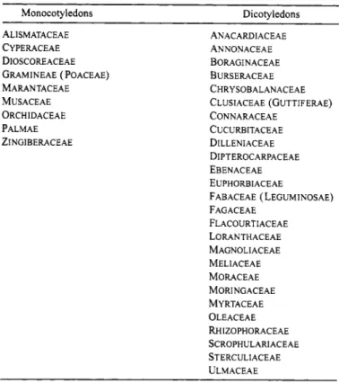
Only those species with diagnostic phytolith forms are discussed below; species studied that had no phytoliths or had nondiagnostic phytoliths are summarized in Table 5. Results are presented by taxon. Table 1 summarizes the families in which diagnostic phytoliths occur.
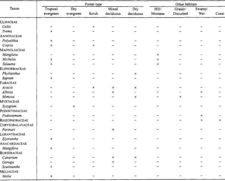
Economic and ecological information is discussed, where pertinent and available, based on general botanical reviews (Mabberly, 1987; Heywood, 1993) and specific discussions of Thai flora (Craib, 1912, 1913; Ogawa et al., 1961; h and Sawyer, 1967; Smitinand, 1968; Smitinand and Larsen, 1972, 1975; Maxwell, 1975; Ogino, 1976; Bunyavejchewin, 1983; Stott, 1984, 1986; Maloney, 1992). Table 2 presents the primary habitats of the genera and families with diagnostic phytoliths.
MONOCOTYLEDONS
Eleven of 19 orders in four of five subclasses of monocotyle- dons were studied: Alismatales, Arales, Arecales, Cyperales, Eriocaulales, Liliales, Najadales, Orchidales, Poales, Typhales, and Zingiberales. Nine families in these orders have diagnostic phytoliths (Table 3). As noted above, phytoliths in Poaceae will be described elsewhere. No diagnostic phytoliths were found in Arales (Araceae), Eriocaulales (Eriocaulaceae), Typhales (Typha), or Najas sp. of Najadales (Table 5). The diagnostic forms are described below.
Order ALISMATALES
The species of family Alismataceae are of particular interest for environmental reconstruction because they are common in
TABLE 1.-Families with diagnostic phytoliths.
~~
Monocotyledons Dicotyledons
ALISMATACEAE CYPERACEAE DIOSCOREACEAE
GRAMINEAE ( POACEAE)
MARANTACEAE MU s A c E A E ORCHIDACEAE PALMAE ZINGIBERACEAE
~~
ANACARDIACEAE ANNONACEAE BORAGINACEAE BURSERACEAE CHRYSOBALANACEAE
CLUSIACEAE (GUTTIFERAE)
CONNARACEAE CUCURBITACEAE DILLENIACEAE DIPTEROCARPACEAE EBENACEAE
EUPHORBIACEAE
FABACEAE ( LEGUMINOSAE)
FAGACEAE FLACOURTIACEAE LORANTHACEAE MAGNOLI ACEAE MELIACEAE MORACEAE MORINGACEAE MYRTACEAE OLEACEAE RHIZOPHORACEAE SCROPHULARI ACEAE
STERCULIACEAE ULMACEAE
freshwater-swamp habitats. The two species analyzed each contain two types of spheres. Both species have folded spheres;
additionally, Caldesia has a sphere 8 ym in diameter, with pentagonal facets, and Ranalisma has dimpled spheres (Figure l), ranging from 10 pm to 20 pm in diameter.
Order ARECALES
Family Palmae was studied in this order, with species chosen from seven of the eight Palmae subfamilies found in the Old World. Phytoliths are common to abundant in Palmae species.
Palm phytoliths commonly are distinctive spinulose to tabular spheres (Figure 2), but some subfamilies have conical to hat-shaped phytoliths (e.g., Caryota and Nypa; Table 3, Figure 3). Except for these more diagnostic genera, the spinulose spheres vary little in shape from genus to genus, although sphere diameter varies between leaves and inflorescences in a few cases (e.g., Borassus and Calamus). Subfamilies also seem to produce spheres in overlapping but different size ranges (Table 3). For example, Borassus spheres were the largest phytoliths encountered, with inflorescence spheres ranging from 20 pm to 27 pm in diameter. Phoenicoideae spheres were the smallest phytoliths seen, ranging from 4 pm to 6 ym in diameter. Without study of more Palmae species no definite subfamily characterizations can be made; however, these data suggest a further study could reveal more diagnostic Palmae characteristics.
FIGURES 1-4 (x356).-1, Ranalisrna rostratum (Alismataceae): dimpled sphere from leaf sample, USNH 1425829. 2, Areca catechu (Palmae): spinulose spheres from fruit sample, USNH 2455123. 3, Curyota mitis (Palmae): conical phytoliths from leaf and inflorescence samples, USNH 210777 I . 4, Cyperus corymbosus (Cyperaceae): regular, sharp peaks on leaf epidermis sample, JCW 285.
4 SMITHSONIAN CONTRIBUTIONS TO BOTANY TABLE 2.-Environment-specifc genera and families with diagnostic phytoliths in Southeast Asia. Presence at
the family level is indicated only when the family is associated in general with the given habitat.
Taxon
~~ ~
MONOCOTYLEDONS ALISMATACEAE PALMAE
Areca Calamus COCOS Corypha Livistona Phoenix Rhapis [POACEAE]
NYPa
CYPERACEAE
MARANTACEAE Phiynium MUSACEAE
ZINGIBERACEAE Costus Curcuma Zingiber
DIOSCOREACEAE ORCHIDACEAE
Aerides Coelogyne Dendrobium BORAGINACEAE
OLEACEAE Ligustrum SCROPHULARIACEAE
Centranthera MORINGACEAE
Moringa DILLENIACEAE
Dillenia EBENACEAE
Diospyros STERCULIACEAE CLUSIACEAE
Calophyllum DIPTEROCAWACEAE
Hopea Shorea CUCURBITACEAE
Momordica Trichosanthes FLACOURTIACEAE
Casearia Flacourtia Hydnocarpus FAGACEAE
Lithocarpus Quercus Artocarpus Broussonetia Ficus Streblus DICOTYLEDONS
MORACEAE
Forest type Other habitats
Tropical Dry Mixed Dry Hill/ Grassy/ Swamp/
evergreen evergreen Scrub deciduous deciduous Montane Disturbed Wet Coast
TABLE 2 . 4 o n t i n u e d . Taxon
ULMACEAE Celris Trema ANNONACEAE
Polyalihia Uvaria Manglieta Michelia Talauma Phyllanihus Sapium FABACEAE
Acacia Albizia Mimosa Syzygium Podostemum MAGNOLIACEAE
EUPHORBIACEAE
MYRTACEAE PODOSTEMACEAE
RHIZOPHORACEAE CHRYSOBALANACEAE
Parinari LORANTHACEAE
Elytranihe ANACARDIACEAE
Mangifera BURSERACEAE
Canarium Garuga Scutinanthe MELIACEAE
Melia
Forest type Other habitats
Hill/ Grassy/ Swampi
Tropical Dry Mixed Dry
evergreen evergreen Scrub deciduous deciduous Montane Disturbed Wet Coast
The typical Palmae habitat is the understory of tropical rain forests; however, individual species often have more specific habitats. For example, Nypa is found in mangrove habitats.
Phoenix species occur in dry seasonal forests, swamp habitats, and mixed forests. Calamus, a climbing palm, is characteristi- cally found in tropical rain forests but also is found in tall evergreen forests in northern Thailand. In the dry evergreen forest, Areca, Calamus, Corypha, Livistona, and Rhapis live near stream banks.
Order CYPERALES
Leaf and inflorescence samples of five species of Cyperaceae were studied (Table 3). Cyperaceae achenes (seeds) were not sampled; however, they often produce the most diagnostic phytolith forms. Except for Scirpus petelotii, phytoliths were common to abundant in both leaf and inflorescence samples.
Although various forms are present in these species (see Table
3), the diagnostic Cyperaceae phytolith is known as a “hat” or cone. Ollendorf (1992) discussed the typology of hat forms found in Cyperaceae but did not distinguish achene forms. The criteria described shape (top view), number of apices, sculptur- ing, presence of satellites, and cone occurrence either individu- ally or on a platelet (Ollendorf, 1992:102). The leaf and inflorescence forms of the five species studied are distinguish- able based on hat diameter (at base) or maximum width and on the shape of the hat (circular or square). Both Scirpus and Eleocharis species have square and round hat shapes, whereas Cyperus hats are round. Cyperus commonly has numerous satellites, either around the perimeter or distributed evenly across the cone. The epidermal tissue of Cyperus also has some diagnostic characteristics, with regular, sharp peaks on flat tissue (Figure 4).
Cyperaceae species are herbaceous perennials, mainly found in marshes and swamps and disturbed, damp to wet habitats (Table 2). Cyperus in northern Thailand also occurs on
6 SMITHSONIAN CONTRIBUTIONS TO BOTANY
damp, marshy ground in evergreen forests and is occasionally associated with rice fields.
Order ZINGIBERALES
Ten species from three families of Zingiberales, Maranta- ceae, Musaceae, and Zingiberaceae, were analyzed (Table 3).
In Marantaceae, distinctive folded and nodular spheres were found in leaf samples. Phytoliths are abundant in this family.
Spheres are commonly 10-12 pm in diameter, but one species also has spheres of 5 pm (Figure 5). Inflorescence samples, on the other hand, had both hollow and infilled conical shapes.
Diameters of these varied from 14 pm to 22 pm. Anticlinal- epidermis forms also are characteristic of the genus Phrynium.
The Marantaceae are most common in the New World, but two genera, Phrynium and Cucurligo, are common in Thai dry evergreen forests. Ph y n i u m is well represented in the herbaceous layer of evergreen forests. Phrynium pamiyorum also is a forb in higher-elevation tall evergreen forests in northern Thailand (Table 2).
Musaceae phytoliths are notable for their highly diagnostic trough shapes (Figure 9). These shapes are abundant in leaves of Musa sp., but no diagnostic forms were found in the inflorescence samples. One sample also had multifaceted- polyhedral shapes.
The Musaceae are known as “jungle weeds” and are characteristic of disturbed habitats in forests from India to Malesia. Musa is found in lowland dry evergreen forests. Musa acuminata also often grows in ravines in montane evergreen forests (Table 2).
In Zingiberaceae, the most common phytolith form is a folded, decorated sphere. Size ranges vary by species (Figures 6, 7), but without further analysis it is unclear if size is species or genus diagnostic. Sphere diameters are often quite uniform within species, varying by only 1-2 pm. The intensity of folding makes these spheres quite distinctive. Other nondiag- nostic phytolith forms, particularly tracheids, are common.
Large multifaceted polyhedrals occur in Zingiber sp. and Curcuma sp. Inflorescences of Zingiber sp. also have unusual, decorated ovoids (Figure 8).
Zingiberales species in general are found in lowland tropical habitats. Zingiberaceae are perennial aromatic plants with multiple economic uses, including spices, medicines, and dyes.
Curcuma is found from evergreen to deciduous forests in northern Thailand, whereas Zingiber primarily grows in damp evergreen forests. Costus is often a forb in tall evergreen forests.
Order LILIALES
Although phytoliths are rare in Dioscoreaceae, a family known for its economic species of yams (Dioscorea spp.), leaves in one species, Dioscorea membranacea, has small (5-6 pm in diameter), tabular spheres (Figure lo). These could be
confused with the small, ovoid, rough palm or orchid phytoliths because their diminutive size makes their decoration difficult to discern. Palm species, however, also have more diagnostic spinulose forms that allow them to be definitely identified. Two other Liliales families, Pontederiaceae and Taccaceae, either do not have phytoliths or have nondiagnostic phytoliths (Table 5).
Dioscoreaceae species are herbaceous climbers in tropical forests, although temperate species do occur. Many Dioscorea species are climbers in hillside seasonal forests. Dioscorea membranacea is found in lowland mixed forests in Thailand (Table 2).
Order ORCHIDALES
Orchidaceae species produce small, rugulose, spherical and conical phytoliths (Table 3). Three species studied herein produce spheres in unique size ranges (4-6 pm, 8-10 pm,
10-14 pm in diameter; Figure 11). There also are size differences between leaf and inflorescence spheres in Aerides.
One species, CoelogyneJleuryi, has distinctive truncated cones (8-10 pm in diameter at base; Figure 12). Other than a silicified epidermis, few additional phytolith forms were found in these species. Two species, in Eria and Vanilla, do not have phytoliths.
As with the Palmae, this family warrants more detailed investigation of phytolith variability among genera and species.
Although Orchidaceae is a cosmopolitan family, the distribu- tion of individual species is often ecologically constrained (see Table 2).
MONOCOTYLEDONS SUMMARY
The monocotyledon families studied herein can be identified using one or two of their diagnostic phytolith forms. Most commonly these forms are spherical or conical and show considerable potential to be diagnostic at least to genus. All monocotyledon families need detailed definition of phytolith variability within and between subfamilies, tribes, and species.
This is particularly important for environmental reconstruction because a phytolith identified to genus in a sediment sequence provides more detailed environmental information than does family attribution.
Although the diversity and abundance of silica present in a plant varies in relation to soil characteristics and plant senescence (Jones and Handreck, 1967; Iler, 1979; Mc- Naughton et al., 1985), this rarely affects the presence of diagnostic phytolith forms. Even without considering the Poaceae, phytoliths from diagnostic monocotyledon genera can be used to distinguish a diverse set of specific habitats, as shown in Table 2. Orchidaceae and Zingiberaceae phytoliths have already aided in the identification of lowland forests, and phytoliths from Cyperaceae and Palmae genera have been used to identify coastal and swamp habitats in central Thailand (Kealhofer and Piperno, 1994; Kealhofer, in press).
FIGURES 5-8.-5, Phrynium pawiflorum (Marantaceae) (x 178): folded, rugulose, conical phytoliths from inflorescence sample, USNH 241 1368. 6, Costus speciosus (Zingiberaceae) ( ~ 3 5 6 ) : folded, decorated sphere from leaf sample, USNH 2395216. 7, Elettaria cardamomum (Zingiberaceae) (x356): folded, decorated sphere from bract sample, USNH 206300. 8, Zingiber sp. (Zingiberaceae) (x356): decorated ovoids from inflorescence sample, USNH 2395209.
8 SMITHSONIAN CONTRIBUTIONS TO BOTANY
FIGURES 9-12 (x356).-9, Musa sp. (Musaceae): trough shapes from leaf sample, USNH 1512179. 10, Dioscorea membranacea (Dioscoreaceae): tabular spheres, 5-6 pm in diameter, from leaf sample, USNH
1701266. 11, Dendrobium crumenaturn (Orchidaceae): rugulose spheres from leaf sample, USNH 221 1836. 12,
Coelogvnefleuryi (Orchidaceae): rugulose, conical phytoliths from leaf sample, USNH 2532095.
DICOTYLEDONS
Five of the six subclasses of dicotyledons studied herein have diagnostic phytoliths: Asteridae, Dilleniidae, Hamameli- dae, Magnoliidae, and Rosidae (Table 4). Dicotyledon phy- tolith forms most commonly include hair cells or associated cells (e.g., hair-base cells and cystoliths); tissues related to respiration, such as stomata, tracheids, sclereids, and multifac
-
eted polyhedrals; and spheres, often from epidermal and subepidermal tissue. The sclereids, hair forms, multifaceted polyhedrals, and spheres have thus far proved to be the most diagnostic of the phytolith forms identified.
Subclass ASTERIDAE
Boraginaceae species (Lamiales) have short, armed hair cells and spheroids (Figure 13). The hair cells found in Cordia are of medium length, heavily armed, and side attached, lying flush with the epidermis. The spheres in Ehretia are very smooth and are associated with epidermal cells. Their size range is highly variable, from 6 pm to 22 pm in diameter, although most are 10-12 ym.
This family is most common in temperate regions; however, Southeast Asian genera are predominantly tropical trees.
Lamiaceae and Verbenaceae species (Lamiales) either did not produce phytoliths or contained a few rare, nondiagnostic forms (see Table 5 ) .
Ligustrum (Scrophulariales: Oleaceae) contained an unusual hemispherical clump of subepidermal cells that may be diagnostic (Figure 14).
This shrubby genus has a disjunct distribution in Europe and AsiaAndomalesia. In Thailand, Ligustrum is found in higher elevation evergreen forests.
The leaves of one genus of Scrophulariaceae, Centranthera, also have diagnostic armed-hair phytoliths. This species was collected in deciduous tropical forests. Another Scrophulariales family, Lentibulariaceae, does not produce phytoliths.
The species in other orders of Asteridae sampled, Gen- tianales (Apocynaceae and Asclepiadaceae), Rubiales (Ru- biaceae), and Solanales (Convolvulaceae and Solanaceae), did not yield diagnostic phytoliths (Table 5 ) .
Subclass CARYOPHYLLIDAE
In the two families sampled, Amaranthaceae and Basel- laceae, phytoliths were not found in leaves or in fruits (Table 5 ) .
Subclass DILLENIIDAE
In Dilleniidae, diagnostic phytoliths were identified in six orders: Capparidales, Dilleniales, Ebenales, Malvales, Theales, and Violales (Table 4). Thus far, families in Violales, specifically Cucurbitaceae and Flacourtiaceae, have yielded the most species with diagnostic phytolith forms. Several families
within the Malvales, Theales, and Violales do not produce diagnostic phytoliths.
Phytolith production in Capparidales is not consistent, CIeome (Capparidaceae) does not produce phytoliths, but Moringa (Moringaceae) inflorescences have very distinctive armed hair cells, similar to those of Streblus (Moraceae).
Among the Dilleniales, Dillenia has unusual hair cells and hair bases. Both D. ovata and D. robusta have hair cells with either square distal or square proximal ends (see Figure 16).
Dillenia robusta has an additional hair-cell type with a proximal end shaped like an arrow point (Figure 17). Both species have angular, fused hair-base cells that may be diagnostic as well (Figure 18).
Dillenia species are tropical trees and are most common from China to Australia. In Southeast Asia they are found in evergreen to mixed deciduous forests, and certain species are persistent in degraded dry dipterocarp (seasonal) forests (Blasco, 1983).
Both Diospyros (Ebenaceae) and Madhuca (Sapotaceae) in Ebenales produce phytoliths. Diospyros ebenum has large (19-20 pm in diameter) and small (7-9 pm in diameter) spheres both in leaf and seed samples. Madhuca phytoliths are predominantly found in vascular tissue, including sclereids and tracheids. Some epidermal tissue also is silicified.
Madhuca and Diospyros species are both used for timber and fruit. Diospyros is often found in mixed deciduous and semievergreen forests. Diospyros siamensis was identified in a disturbed, open seasonal forest in northern Thailand.
In Malvales few families produce phytoliths. A few species of Sterculiaceae have rare, diagnostic types. Eriolaena has a rugulose sphere 15 pm in diameter, Pterospermum and Sterculia species both produce sclereids, and Sterculia has other vascular- and hair-related forms. Melochia has a rare, unusual form of linked spheres (Figure 19).
The species sampled from other Malvales families, Bom- bacaceae, Malvaceae, and Tiliaceae, produce phytoliths only rarely, and none are diagnostic (Table 5 ) .
In Theales, the genus Calophyllum in Clusiaceae (Guttiferae) has abundant phytoliths. A spherical form, shaped somewhat like a seed, is the most distinctive shape (Figure 20). Another diagnostic, slightly spinulose, elongate epidermal form also is common and has an unusual spike on one of its longer sides (Figure 21). In six other genera of Clusiaceae, phytoliths are absent or are not diagnostic (Table 5 ) ; however, Mammea produces tiny (4 pm in diameter) spheres, and Ochrocarpus has tiny sclereids.
Species of Calophyllum, like those of Dillenia, are predomi- nantly tropical trees, the timber of which is useful for its durability and ease of working. The species analyzed herein are most common in hill evergreen forests.
The family Dipterocarpaceae (Theales), known for habitat- specific tropical trees, yielded few diagnostic phytoliths.
Leaves from two genera, Shorea and Hopea, however, did have rare, decorated, spherical phytolith forms that are at least
10 SMITHSONIAN CONTRIBUTIONS TO BOTANY
FIGURES 13-16.--13, Cordia grandis (Boraginaceae) (x 178): short, armed hair cells from leaf sample, USNH 1213307. 14, Ligustrum robustus (Oleaceae) (x356): subepidermal hemispherical clump From leaf sample, USNH 1214625. 15, Centranthem hispida (Scrophulariaceae) (x 178): armed hair from leaf sample, USNH 294 1665. 16, Dillenia ovata (Dilleniaceae) (x 178): square proximal hair cells from leaf sample, USNH 1700567.
FIGURES 17-20.-17, Dillenia robustu (Dilleniaceae) (x356): proximal end of arrow-point hair cell from leaf sample, USNH 1700660. 18, Dillenia ovafa (Dilleniaceae) (x 178): angular, fused hair-base cells from leaf sample, USNH 1700567. 19, Melochia umbellafa (Sterculiaceae) (x 178): stalk-linked spheres from leaf sample, USNH 3083385. 20, Culophyllum burmanii (Clusiaceae) ( ~ 3 5 6 ) : seed-like sphere from leaf sample, USNH 1668055.
12 SMITHSONIAN CONTRIBUTIONS TO BOTANY
FIGURES 2 1-24.-21, Calophyllum burmanii (Clusiaceae) ( x 178): spinulose phytolith (spike end) from leaf sample, USNH 1668055. 22, Hopea odorata (Dipterocarpaceae) (x356): decorated sphere from leaf sample, USNH I701 199. 23, Shorea obtusa (Dipterocarpaceae) (x356): decorated sphere from fruit sample, USNH 22 1 181 9. 24, Solena heterophylla (Cucurbitaceae) ( x 178): armed hair cells from leaf sample, USNH 2553066.
genus-specific (Figures 22, 23) and have been found in sediments (Kealhofer, 1996b). The Vatica and Dipterocarpus species investigated, however, did not have diagnostic phyto- lith forms.
Dipterocarp species, most abundant in Malesia, also are widespread in Thailand (dipterocarp species comprise 45% of Thai forest species). The culturally modified dry dipterocarp forest type is defined by a few dominant Dipterocarpaceae species (Pentacme siamensis, Shorea obtusa, Dipterocarpus tuberculatus, D. obtusifolius). Ogawa and coworkers (1 961) identified two dipterocarp forest subhabitats: Pentacme-Shorea (drier) and Dipterocarpus tuberculatus-Dipterocarpus obtu- sifolius (wetter). In general, the Pentacme-Shorea habitat is species poor, including only a few grass species (such as Arundinaria spp.), Cycas siamensis, Phoenix humilis, P:
acaulis, and relatively infrequent arboreal species. Composition varies regionally. Different Dipterocarpaceae (particularly Dipterocarpus spp.) are found in mixed and evergreen forests, but their relative abundance is much lower as species diversity increases (Ogawa et al., 1961:67). Hopea odorata is common in dry evergreen forests.
Elatinaceae species (Theales) do not produce diagnostic phytoliths.
By far the most abundant and distinctive phytoliths of the Dilleniidae are found in the Violales, particularly in Cucurbita- ceae and Flacourtiaceae (Table 4). The Cucurbitaceae are known for their segmented hair cells, some of which are armed.
Eight genera of Cucurbitaceae from two subfamilies were analyzed. Only Momordica did not produce armed or seg- mented hair cells in their leaves. None of the inflorescences produced diagnostic phytoliths. No fruits were analyzed, but distinctive, faceted spheres have been identified with certain parts of the fruit (Bozarth, 1986).
Citrullus, Cucumis, Gymnopetalum, Lufla, and Trichosan- thes all have distinctive segmented hair cells of various sizes, whereas Solena and Mukia have armed hair cells (Figures 24, 25). Other diagnostic features include curved hair tips (Gymnopetalum) and unique hair bases (Citrullus, Cucumis).
Momordica contains cystoliths, often seen linked at the top to form a tripodal arrangement (Figure 26). In only one genus, Solena, were two species examined, and these appeared to have species-specific forms. Further analyses within these genera may yield more phytolith forms diagnostic at the species level.
These Cucurbitaceae genera are all from the Asian tropics and are most commonly lianas, or climbing vines. They are often weeds in disturbed habitats. Members of this family were some of the earliest species domesticated.
Samples from members of Flacourtiaceae (Violales) only sporadically produced phytoliths. Nondiagnostic phytoliths are common in Flacourtiaceae leaves, and diagnostic forms are relatively rare. The fruits of several species contain spherical shapes, possibly associated with epidermal cells. These spheres are 10-14 pm in diameter, with a dimple (Casearia) or with flat tissue attached (Hydnocarpus). Scolopia flowers have a
rare, spheroid, kidney-bean-shaped phytolith. In leaves, epider- mal forms also are diagnostic in a few cases, particularly for Hydnocarpus (Figure 27). Zdesia has an unusual, multifaceted- polyhedral form (Figure 28).
The Flacourtiaceae are fairly cosmopolitan trees and shrubs with wide distribution in the tropics and subtropics. Casearia and Flacourtia are both found in dry mixed forests and occasionally in dry deciduous forests. Hydnocarpus is a common shrub in tropical evergreen to dry evergreen forests.
Only a few species, namely Scolopia and Casearia (usefil timber) and Flacourtia (edible fruit), have economic uses.
Samples from species in two other families in the Violales, Caricaceae and Passifloraceae, did not yield diagnostic phyto- liths (Table 5 ) .
Subclass HAMAMELIDAE
Three of the four Hamamelidae families analyzed revealed characteristic phytoliths: Fagaceae (Fagales) and Moraceae and Ulmaceae (Urticales) (Table 4). The one Juglandaceae species tested did not produce diagnostic phytoliths (Juglandales;
Table 5).
In Fagaceae (Fagales), the leaves of Lithocarpus have distinctive, faceted, spherical polyhedrals (Figure 29) and spheres. Quercus samples produced spheres from the nut-shell.
The Fagaceae are, in general, a temperate family (oaks), but in the tropics they appear in montane evergreen forests. Three Quercus species and one Lithocarpus species have been identified as particularly common on the higher slopes of montane forests in northern Thailand (Kuchler and Sawyer, 1967). Other Quercus species, such as Q. mespilifoliodes and Q. kerrii, however, are found in dry dipterocarp forest understory.
The order Urticales was more intensively investigated because it is more extensively represented in the tropics. Two families, Moraceae and Ulmaceae, demonstrated a wide variety of phytoliths. In both families, hair cells and cystoliths are the most diagnostic forms.
Among the Moraceae, three Artocarpus species were studied. Leaves of all species of Artocarpus revealed armed hair cells (Figure 30). One species of Ficus and one species of Streblus also have diagnostic armed hair cells, and these are the only two genera with diagnostic phytoliths in either fruit or inflorescence parts. Streblus has various armed and unarmed hair cells (Figures 3 1,32). Broussonetia and Malaisia hair cells also are distinctive but are not armed (Figure 33). Some species contain up to five different types of hair-cell phytoliths, with characteristic attachment locations, curvature, striations, and other features. Other genera, such as Taotrophis, have simpler but still diagnostic hair cells. Ficus annulata, Broussonetia kasinoki, Artocarpus elasticus, and Morus alba leaves all contain cystoliths as well (e.g., Figure 34). Ficus hispida epidermis has unusual hexagonal epidermal(?) cells, and F.
annulata fruit epidermal cells have a wrinkled surface (Figure 35).
14 SMITHSONIAN CONTRIBUTIONS TO BOTANY
FIGURES 25-28.-25, Mukia maderaspatana (Cucurbitaceae) (x 178): armed hair cells from leaf sample, USNH 2039897. 26, Momordica charantia (Cucurbitaceae) (x 178): top-linked cystoliths from leaf sample, USNH 2039869. 27, Hydnocarpus anthelmintheca (Flacourtiaceae) (x356): epidermal phytoliths from leaf sample, USNH 1427948.28, Idesiapolycarpa (Flacourtiaceae) ( ~ 3 5 6 ) : multifaceted polyhedral from leaf sample, USNH 2986601.
FIGURES 29-32.-29, Lithocurpus acuminatissima (Fagaceae) (x356): multifaceted polyhedral from leaf sample, USNH 1701025.30, Artocarpus elusticus (Moraceae) (x 178): armed hair cells from leafsample, USNH 29391 50.
31, Streblus asper (Moraceae) (x356): armed hair cells from leaf sample, USNH 2064818. 32, Streblus asper (Moraceae) (x356): armed hair cell from leaf sample, USNH 20648 18.
16 SMITHSONIAN CONTRIBUTIONS TO BOTANY
FIGURES 33-36.-33, Broussonetia kasinoki (Moraceae) ( x 178): unarmed hair cells from leaf sample, USNH 1597387. 34, Arrocarpus elasticus (Moraceae) (x 178): cystolith from leaf sample, USNH 2939 150. 35, Ficus annulara (Moraceae) ( x 178): wrinkled epidermal cells from leaf sample, USNH 2602693.36, Celtis cinnamomea (Ulmaceae) (x356): unusual cystoliths from leaf sample, USNH 1212982.
Silicified epidermal cells, hair bases, stomata, tracheids, mesophyll, and nondiagnostic hair cells are common in this fami 1 y
.
Members of the Moraceae, and particularly Ficus species, are widespread but are often associated with river or stream habitats in gallery forests. Moraceae species include a variety of economically important tropical plants. Both Artocarpus and Ficus species grow in evergreen to semievergreen forests as well as often being found in middle and higher elevations.
Ficus is a huge genus, with a broad variety of trees, shrubs, and lianas. Broussonetia is often in the shrub understory of evergreen forests, and Streblus has been identified in open disturbed seasonal forests in northern Thailand.
Many characteristics of the Moraceae also were found in the Ulmaceae. Hair-cell, hair-base, and cystolith forms are com- mon in both leaves and fruit (Table 4). Gironniera has armed hair cells, and the two species of Celtis have unusual cystoliths (Figure 36). A spiny, irregular phytolith form was found in Celtis. Two Trema species have distinctive, small, anticlinal epidermal cells (Figure 37). Trema orientalis also revealed an unusual, pitted, striated phytolith form (Figure 38). Gironniera leaf epidermis is distinctive. No phytoliths were found in Holoptelea leaves.
The tropical and subtropical tribe Celtideae of Ulmaceae is composed predominantly of trees, with some shrubs. Many of these genera produce decay-resistant timber. Celtis is common in scrub forests, whereas species of Trema are often found in evergreen forests.
Subclass MAGNOLIIDAE
Six of the eight orders of Magnoliidae were sampled, but only one, Magnoliales (3 1 species), produced diagnostic phytoliths (Table 4). No phytoliths were found in Aristolochia- les. Some samples from the families Illiciales, Laurales, Piperales, and Ranunculales did produce phytoliths, but they were rare to uncommon.
Among the Magnoliales, only three families are present in Thailand. Two of these families, Magnoliaceae and Annon- aceae, contain characteristic phytoliths. Species sampled from the third family, Myristicaceae, did not produce silica bodies.
In Annonaceae, the characteristic phytoliths are relatively large, multifaceted polyhedrals (Figure 39). Artabotlys has multifaceted spheres, and other multifaceted polyhedrals were found in Polyalthia species and in Sageraea elliptica. Sageraea phytolith forms are unique in having tiny spinulose protrusions as decoration. Sclereid forms are present in Annonaceae, and diagnostic phytoliths occur in leaves of Goniothalamus, Sageraea, and Uvaria (Figure 40). Uvaria fruit has large, irregular spheroids (1 8 pm in diameter). Vascular-tissue and stomata forms also are well represented in these species.
Annonaceae species include both trees and shrubs, several of which produce important edible fruit. All species studied herein are most common in tropical, lowland evergreen forests and
often occur in mixed deciduous forests. Polyalthia grows in tropical evergreen forests. Uvaria was identified in scrub forests, but it also is found in evergreen forests.
Magnoliaceae species also have multifaceted bodies, particu- larly Michelia. Distinctive decorated and faceted epidermal phytolith forms were found in Talauma and Manglieta (Figure 41). Phytolith production, however, seems to vary by species because no diagnostic phytoliths were found in Michelia Jloribunda.
The Magnoliaceae are most abundant in southeast Asia, and certain species are used for timber. Genera analyzed include trees of evergreen forests; Michelia is often found at the edge of lowland forests as well as in (mossy) montane evergreen forests, whereas Talauma is associated with streams or hills, and Manglieta is typical in the diverse, higher-slope forests of northern Thailand.
Subclass ROSIDAE
The last subclass studied, Rosidae, comprises 18 orders, 11 of which were sampled. Species in four of these orders do not produce diagnostic phytoliths: Apiales, Celastrales, Podos- temales (see below), and Rhamnales (Table 5). Phytolith production is not consistent in those orders that do produce phytoliths. For example, in Santalales, some families have phytoliths, and others do not.
In the Euphorbiaceae family (Euphorbiales), phytoliths were common in three of the four genera sampled, Sapium, Phyllanthus, and Manihot. In Manihot esculenta leaves, slightly irregular spheres, 6-10 pm in diameter, were common (Figure 42). Fruit samples of Phyllanthus also produced spheres, but these are nodular and 6-15 pm in diameter. The epidermal cells of Sapium also are diagnostic, with an irregular, rough surface decoration (Figure 43).
Euphorbiaceae is a large and cosmopolitan family, but its richest concentration of genera is in Indomalaysia. Two of the genera with diagnostic phytoliths are economically useful, particularly Manihot esculenta (cassava, a New World species), an important edible root crop, and Phyllanthus, used medici- nally and for dyes. Sapium is most common in tall broadleaf evergreen forests.
In the Fabales, many species of Leguminosae (Fabaceae) produce abundant phytoliths, but few forms are diagnostic (Tables 4, 5 ) . Three subfamilies of Fabaceae were analyzed:
Caesalpinoideae, Mimosoideae, and Papilionoideae. None of the species tested in Caesalpinoideae produced diagnostic phytoliths.
In the Mimosoideae, the stomata may need further investiga- tion because they seem to take some unusual shapes (Albizia, Pithecellobium; Figure 44). Acacia also may have diagnostic hair cells and hair-base cells. The epidermis in many species, particularly in subfamily Papilionoideae, also is distinctive.
Tracheid or hair-base-cell forms are common to all subfamilies in which silica is present (-50% of those sampled).
18 SMITHSONIAN CONTRIBUTIONS TO BOTANY
FIGURES 37-40.-37, Trema cannabina (Ulmaceae) (x356): anticlinal epidermal cells from fruit sample, USNH 2880555. 38, Trema orientalis (Ulmaceae) ( ~ 8 9 ) : pitted, striated epidermis from leaf sample, USNH 2769297.
39, Polyalthia suberosa (Annonaceae) ( ~ 3 5 6 ) : large, multifaceted polyhedrals from leaf sample, USNH 1 171 29 1.
40, Goniothalamus marcani (Annonaceae) (x 178): sclereid from leaf sample, USNH 1700786.
FIGURES 4 1-44 (x 3 5 6 ) . 4 1, Tuluuma longifolia (Magnoliaceae): decorated, faceted phytolith from leaf sample, USNH 2407533. 42, Manihot esculenra (Euphorbiaceae): nodular spheres, 6-10 pm in diameter, from leaf sample, USNH 2395207. 43, Supium indicum (Euphorbiaceae): rough surface decoration on fruit epidermis sample, USNH 1701 697. 44, Pithecellobium duke (Fabaceae: Mimosoideae): stomata from leaf sample, USNH 2423 177.
20 SMITHSONIAN CONTRIBUTIONS TO BOTANY
Fabaceae is a very large family and is distributed in a wide range of habitats. The subfamilies of Fabaceae have more restricted distributions, however, and Mimosoideae and Cae- salpinoideae include tropical to subtropical trees and shrubs.
Particular species are of enormous economic importance, especially in the Papilionoideae. The species analyzed are common in scrub and evergreen forests. Acacia catechu and Albizzia are frequent in mixed deciduous forests. Vines of Acacia and Mimosa inhabit the scrub understory of disturbed, open, seasonal forests.
In Myrtales, two of the nine families present in Southeast Asia were investigated: Combretaceae and Myrtaceae. No diagnostic phytoliths were found in the three Combretaceae species studied, although phytoliths were present (Table 5 ) . Of the two species of Syzygium (Myrtaceae) tested, one, S.
ieptantha, has an unusual, very tall stomata phytolith form.
This family is predominantly tropical and is composed mostly of large shrubs and trees. Syzygium is commonly found in dry evergreen forests on hillsides in Thailand and has a broader distribution elsewhere.
Podostemales are known to produce phytoliths; however, the species studied herein, Podostemum subulatus, does not (Piperno, 1988). Species in this order are found in flowing, fresh-water environments.
Three species in the family Rhizophoraceae were sampled.
Phytoliths were not common in any of these. Sphere forms are present in all three species, although sphere size and surface decoration vary by species (Table 4).
Rhizophoraceae species are most common in coastal mangrove forests in Thailand. Many taxa are used economi- cally for timber or charcoal.
Two of the six families of Rosales present in Thailand, Rosaceae and Chrysobalanaceae, were tested; Rosaceae did not have diagnostic phytoliths (Table 5). In Chrysobalanaceae one species, Parinari annamense, incorporates irregular but smooth spheres of 6-12 pm diameter in the leaves and 6-10 pm diameter in the inflorescence. The fruit has very large spheres (20-85 pm in diameter).
Chrysobalanaceae species, trees and shrubs, are found in the lowland tropics (mostly in the New World), and Parinari includes important fruit and timber trees. This genus ranges in habitat from deciduous to evergreen forests, but in Thailand it seems to be most common in mixed deciduous forests.
One sample tested seems to be from the Connaraceae (labeled “Connarc?’ in the United States National Herbarium), and it also has diagnostic epidermal forms, suggesting that this family warrants further investigation.
In Santalales, two of the four families present in Thailand were investigated. No phytoliths were found in the Olacaceae species tested. In Loranthaceae, only leaves produced phy- toliths. Two distinctive stomata forms were found in Elyrran- the, a set of elongated/palisaded cells and a set of subepidermal spherical cells in a clump (Figure 45). Unusual epidermal-cell forms also were found in this genus.
Loranthaceae includes shrubby tropical parasites. Elytranthe is found in evergreen forests and often impedes the regrowth of hardwood trees.
Five of the 10 families of Sapindales present in Thailand were sampled, and species in two of the families, Anacardi- aceae and Burseraceae, have diagnostic phytoliths. All of the other families (Meliaceae, Rutaceae, and Sapindaceae) are phytolith producers, but the forms are rare or nondiagnostic (Table 5). Vascular tissue in both Rutaceae and Sapindaceae, particularly tracheids and adjoining tissue, occasionally in- cludes unusual spiny forms.
Three Anacardiaceae species were tested, and the leaves of all three produced phytoliths; however, those in Spondias pinnata are not diagnostic (Table 5 ) . Mangifera indica (mango)
and Rhus sp. both have unique folded spheres (Figure 46).
Mangifera species are common in upland and lowland evergreen forests.
Burseraceae species show a wide range of unique phytoliths.
Epidermal phytoliths from both fruit and leaves are the most distinctive. Silicified anticlinal and polyhedral epidermal cell shapes are both represented. Canarium, Commiphora, Daciyo- des, Garuga, and Scutinathe species all have thick, decorated epidermal cells (Figure 47). Commonly, one surface is smooth and the opposite surface is spikey. Canarium fruits often have unusual, very smooth, large spheroid shapes as well (Figure 48). One Canarium species has a distinctive acorn-shaped hair cell (Figure 49); a related form was found in Garuga. Silicified stomata, tracheids, hair cells, and hair-base cells are common to abundant in Burseraceae.
Burseraceae is a tropical family of trees and shrubs. It forms a common constituent of lowland deciduous and dipterocarp forests in southeast Asia. Canarium, Daciyodes, and Garuga are the three Burseraceae genera most common in this lowland habitat, and all have tree species with usehl timber. Garuga pinnata has been identified in mixed deciduous forests.
Canarium kerrii and C. subulatum are common constituents of the dry dipterocarp forest. Protium has been identified in deciduous, closed-canopy forests in northern Thailand.
Meliaceae species only rarely produce phytoliths, but Melia inflorescences have rare, small spheres (5-7 pm in diameter).
Species in this family are known for producing good timber, such as mahogany, in lowland tropical forests.
DICOTYLEDONS SUMMARY
The most common of the distinctive dicotyledon phytolith types are spheres (including spheroids), armed and segmented hair cells, faceted polyhedrals, and decorated epidermal cells.
The variability seen in dicotyledons differs from that seen in monocotyledon families. Phytolith presence is not as predicta- ble, and orders commonly have families and species both with and without phytoliths. A more limited range of phytolith forms also is represented, with greater frequency of vascular
FIGURES 45-48 ( x 3 5 6 ) . 4 5 , Elytranthe umpullaceae (Loranthaceae): stomata from leaf sample, USNH 1700671, 46, Mangifera indica (Anacardiaceae): folded spheres from leaf sample, USNH 595090. 47, Commiphoru cuuduta (Burseraceae): thick, decorated epidermis from h i t sample, USNH 2805849. 48, Canarium kerrii (Burseraceae): large, smooth spheroids from fruit sample, USNH 2436020.
22 SMITHSONIAN CONTRIBUTIONS TO BOTANY
cones of the monocotyledons. Dicotyledon sphere types, however, appear to be good indicators of trees in various forested habitats. The genera and families with diagnostic phytoliths include a wide range of habitats but dominate the lowland evergreen, mixed deciduous, and dry deciduous forests. Specific habitats are discernable based on the unique complement of genera identified in soil samples.
Conclusion
The results presented herein demonstrate the abundance and diversity of diagnostic phytoliths in Old World tropical flora.
These phytoliths are consistent with those described by Piperno (1 985, 1989) in New World taxa, confirming the uniformity of phytolith forms among taxa with Old and New World distributions.
Based on the habitat distribution of their parent plants, phytoliths can be correlated with a wide range of specific environments. Plant age, soil chemistry, and other environ- mental factors may influence phytolith abundance and the diversity of forms present in a given plant; however, diagnostic forms are produced despite variable conditions. This is clearly seen when the patterns of production and morphology presented herein are compared with New World taxa (Piperno, 1988, 1989). These results hrther demonstrate that different kinds of habitats and economic patterns are identifiable in soil phytolith sequences. The increasing definition of diagnostic phytolith types enhances the value of phytolith sediment sequences for complementing and expanding the reconstruc
-
tion of paleo-environments, and the human interactions with these environments, currently available from pollen and geomorphological analyses.
FIGURE 49.--Cunarium kerrii (Burseraceae) (x400): acom-shaped hair cell from leaf sample, USNH 2436020.
forms (e.g., sclereids) than in monocotyledons. Short cells are not represented, nor are the distinctive tabular spheres and