Furthermore, the frontal ganglion is an important landmark in the basic anatomy of the head, and the structure of the head cannot be understood by studying only its superficial features. Otherwise, there is no evidence that the lines on the insect's skull are true sutures. The external grooves, or sulci, of the endoskeletal ridges are therefore not 'sutures' in any true sense.
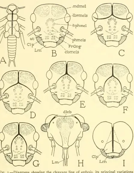
It is by no means a suture, and is here called the ecdysial cleavage line of the head. The cleavage line of the cuticle that opens to allow the insect to escape is therefore a. In some of the metabolic and hemimetabolic insects, the exuvial cleavage line of the head is completely or partially transferred to the adult (fig. 4A, B, D, CL).
The area of the face between and below the arms of the cleavage includes the areas commonly known as the forehead, klipen and lahrurn. If it represents such an important thing as the line of dorsal closure and junction of the head components, it is.
NO. 7 THE INSECT CRANIUM — SNODGRASS
8 SMITHSONIAN MISCELLANEOUS COLLECTIONS VOL. 10/
NO. 7 THE INSECT CRANIUM SNODGRASS 9
10 SMITHSONIAN MISCELLANEOUS COLLECTIONS VOL. IO7
NO. THE INSECT CRANIUM — SNODGRASS II
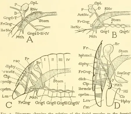
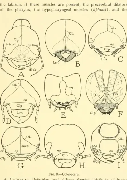
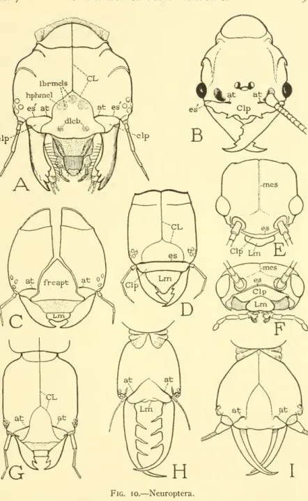
In the primitive state of the nervous system, accompanied by what may have been assumed to be the primitive musculature of the anterior part of the head (Fig. 2C), the frontal ganglion (FrGng) must lie between the compressive muscles of the clypeus (cprdp) and the postoral group of frontal muscles. In generalized pterygote insects, the frontal muscles of the hypopharynx are functionally the producers of this organ, but they are conserved in Neuroptera, Coleoptera and Hymenoptera, in which the hypopharynx inti-. The development of the insect hypopharynx (Fig. 2D, Hphy) from a metastomial lobe of the head creates a feeding passage (/w^) leading to the mouth between the inner wall of the labrum and the clypeus and the anterior wall of the hypopharynx.
At its inner end the alimentary passage is usually enlarged by a depression of the hypopharyngeal surface to form the alimentary pocket, thecibarium {Cb), below the clypeus. The pocket can therefore be expanded by the contraction of the compressor muscles of the clypeus, which thus become dilators of the cibarium (dlcb); it is compressible either by transverse muscles of the clypeal wall or by elasticity of the latter. With the greater development of the cibarium pump, the clypeal dilator muscles become considerably larger in size, and to accommodate these muscles the outer wall of the clypeus is correspondingly enlarged by an upward extension in the face.
Thus, there is always a close correlation between the size of the clypeus and the size of.
NO. 7 THE INSECT CRANIUM SNODGRASS 1
EXAMPLES OF THE ECDYSIAL CLEAVAGE LINE OF THE HEAD
14 SMITHSONIAN MISCELLANEOUS COLLECTIONS VOL. IO7
NO. 7 THE INSECT CRANIUM SNODGRASS 15
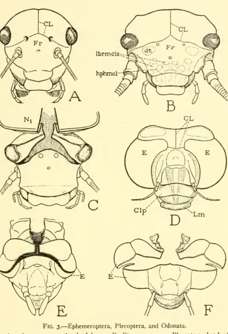
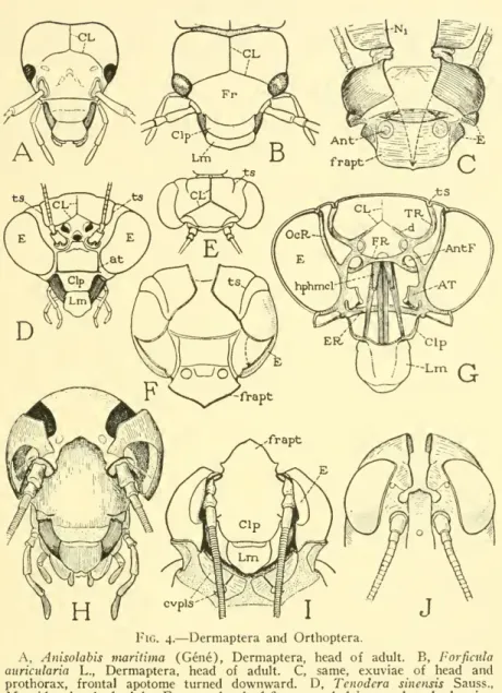
Orthoptcra.— Most of the Orthoptera retain at least part of the cuticular cleavage line in the adult, though in neither the imago nor the nymph dothearms always represent the full length of the exuvial cleavages which take place at ecdysis. In the Mantidae (Fig. 4D) the frontal arms of the cleavage line pass dorsally to the paired ocelli, and in the adult insect appear to be confluent with the temporal sulci {ts) returning from them over the top of the head. However, as already explained, this apparent continuity of the cleavage lines with the temporal sulci is due to the invasion of the former on the inner surface of the head wall (G) by spurs {d) from the temporal ridges (TR).
In the exuviae head of a nymphal mantis (E, F) it is seen that the temporal sulci (fs) do not reach the flanks of the fissure line. In the ecdysis of the mantid, the frontal slits extend into the compound eyes and cut deep into the cornea (F, £) as in Forficula (C). The lower halves of the cornea are then pulled down with the depression of the frontal apotome (F, frapt) and the mouthparts and antennae protrude posteriorly below the head capsule.
In Phasmatidae, Blattidae and Gryllidae, the arms of the cleavage line appear to terminate at the lateral ocelli. However, in a nymph of Periplaneta americana undergoing ecdysis (Fig. 4H), frontal clefts are seen to pass below the ocellar spots and proceed to the mesal angles of the antennal fossae, from where they turn upward along the dorsal margins of the fossae to points above the fossae. of the antennas. The parietal lobes of the head exuviae are then forced to either side of the new head in the new stage, producing deep folds through the corneas of the compound eyes, but not splitting them.
Finally, the facial region of the drag is turned flat ventrally (I) as in the mantid, forming deep folds of the eyes and the genae on each side, the edges of which partially overlap the bases of the antennae. In nymphs of Acrididae the line of the exuvial cleavage is scarcely perceptible, but in ecdysis the thoracic cleft extends over the top of the head and forks at the upper end of the frontal costa.
NO. 7 THE INSECT CRANIUM SNODGRASS 17
Although the anterior arms always pass in front of the eyes, and, when sufficiently extended, lateral to the antennae, the coronal stalk is of variable length and may be entirely absent. Many examples of the course of the "epicranial suture" in the Homoptera and the Heteroptera are given by Spooner (1938) in his comparative study of the head capsule of the Hemiptera. In some others, however, the coronal trunk is short and the forearms branch far back on the head, as illustrated by Spooner in Cimex lectiUarius (Cimicidae) and Ischnodemusfaliens (Lygaeidae), and as shown in the present article in Oncopeltus fasciatus (Lygaeidae) (Fig. 6 A) and in Cimex lectularius (B).
The ecdysial cleavages in Cimex follow the arms of the cleavage line and extend to the anterior dorsal corners of the eyes. The large frontoclypeal apotome of the exuviae (C, frcapt) is then turned forward and flat downwards, with the bill directed posteriorly, while the narrow parietal lobes {Prtl) which bear the eyes and the antennae spread outwards. In ecdysis of such species, the frontal fissure, which branches directly from the median fissure of the thorax and neck, splits (fig.
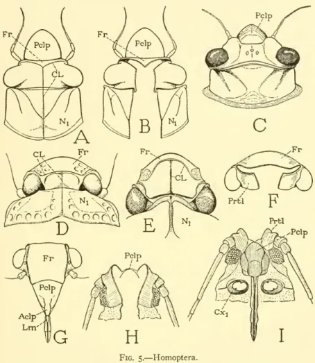
6 D), cut off small, lateral parietal triangles of the apex (Prtl) bearing. compound eyes, leaving the median part of the cranial margin intact. When the coronal stem is relatively long, the arms may diverge forward on top of the head, as in nymphalcicada (Fig. 5A), in which the frontal cleft is a small triangular front (B). 5G, has a large frontal area on the anterior surface of the head (Fig. 5G, Fr), the arms of the cleavage line are either transverse (D, F) or they curve posteriorly to points in front of the eyes (E).
20 SMITHSONIAN MISCELLANEOUS COLLECTIONS VOL. I07
NO. 7 THE INSECT CRANIUM SNODGRASS 21
In Pediculus hunianus corporis, the cleavage line is not visible on the head of younger nymphs; in later instars and in the adult it is. However, in mature nymphs, evidently just before the emergence of the imago, the cleavage line is a pale, distinctly double-edged part of the cuticle of the head (A), and it can now be observed that the frontal arms extend to the basal . membranes in the antennae. In ecdysis, the slits along the arms are further extended mesad of the antennae to points {x, x) on the sides of the conical anterior part of the head.
In the discarded exuviae, the frontal apotome (B, frapt) is directed anteriorly on the transverse axis between the tips of the frontal fissures {x, x), together with the snout-like cone of the head, while the parietal parts of the cuticle are removed . spread outwards, and in aventral view (B) they are seen to bear the eyes and the antennae. In the elongated head of Haeniatopinus suis, as shown by Stojanovich (Fig. 7D), the coronal trunk of the cleavage line is relatively long, but the arms extend just above the antennae to points in front of the antennae bases. The distribution of muscle attachments on the frontoclypeal region of the anoplurane head is shown by Stojanovich in several species, including Pediculus humanus (Fig. 7C) and Haeniatopinus suis (D).
It is seen that these muscles include, as in other insects, the cibarial dilators (dlcb) and the muscles of the labrum, hypopharynx, and pharynx, and in addition the antennal muscles. In Pedictdus (A, C) a weakly sclerotized band of the cuticle crosses the head between the antennal bases and appears to roughly separate the clypeal region from the frontal region, but it is doubtful whether.
NO. 7 THE INSECT CRANIUM SNODGRASS 23
24 SMITHSONIAN MISCELLANEOUS COLLECTIONS VOL. I07
NO. 7 THE INSECT CRANIUM SNODGRASS 25
26 SMITHSONIAN MISCELLANEOUS COLLECTIONS VOL. IO7
NO. 7 THE INSECT CRANIUM SNODGRASS 27
28 SMITHSONIAN MISCELLANEOUS COLLECTIONS VOL. IO7
NO. THE INSECT CRANIUM SNODGRASS 29
30 SMITHSONIAN MISCELLANEOUS COLLECTIONS VOL, 10/
NO. 7 THE INSECT CRANIUM SNODGRASS 3I
32 SMITHSONIAN MISCELLANEOUS COLLECTIONS VOL. 10/
NO. 7 THE INSECT CRANIUM SNODGRASS 33
34 SMITHSONIAN MISCELLANEOUS COLLECTIONS VOL. I07
NO. 7 THE INSECT CRANIUM SNODGRASS 35
36 SMITHSONIAN MISCELLANEOUS COLLECTIONS VOL. I07
NO. 7 THE INSECT CRANIUM— SNODGRASS 57
38 SMITHSONIAN MISCELLANEOUS COLLECTIONS VOL. IO7
NO. 7 THE INSECT CRANIUM — SNODGRASS 39
40 SMITHSONIAN MISCELLANEOUS COLLECTIONS \0L. IO7
NO. 7 THE INSECT CRANIUM SNODGRASS 4I
42 SMITHSONIAN MISCELLANEOUS COLLECTIONS VOL, IO7
NO. 7 THE INSECT CRANIUM SNODGRASS 43
44 SMITHSONIAN MISCELLANEOUS COLLECTIONS VOL. IO7
SUMMARY
NO. 7 THE INSECT CRANIUM SNODGRASS 45
46 SMITHSONIAN MISCELLANEOUS COLLECTIONS VOL. IO7
48 SMITHSONIAN MISCELLANEOUS COLLECTIONS VOL. 10/
50 SMITHSONIAN MISCELLANEOUS COLLECTIONS VOL. I07
NO. 7 THE INSECT CRANIUM SNODGRASS 5I
52 SMITHSONIAN MISCELLANEOUS COLLECTIONS VOL. 10/