The Intermuscular Bones and Ligaments of Teleostean Fishes
*
m
COLIN PATTERSON and
G. DAVID JOHNSON
I
I
SMITHSONIAN CONTRIBUTIONS TO ZOOLOGY • NUMBER 5 5 9
Emphasis upon publication as a means of "diffusing knowledge" was expressed by the first Secretary of the Smithsonian. In his formal plan for the institution, Joseph Henry outlined a program that included the following statement: "It is proposed to publish a series of reports, giving an account of the new discoveries in science, and of the changes made from year to year in all branches of knowledge." This theme of basic research has been adhered to through the years by thousands of titles issued in series publications under the Smithsonian imprint, commencing with Smithsonian Contributions to Knowledge in 1848 and continuing with the following active series:
Smithsonian Contributions to Anthropology Smithsonian Contributions to Botany Smithsonian Contributions to the Earth Sciences Smithsonian Contributions to the Marine Sciences
Smithsonian Contributions to Paleobiology Smithsonian Contributions to Zoology
Smithsonian Folklife Studies Smithsonian Studies in Air and Space Smithsonian Studies in History and Technology
In these series, the Institution publishes small papers and full-scale monographs that report the research and collections of its various museums and bureaux or of professional colleagues in the world of science and scholarship. The publications are distributed by mailing lists to libraries, universities, and similar institutions throughout the world.
Papers or monographs submitted for series publication are received by the Smithsonian Institution Press, subject to its own review for format and style, only through departments of the various Smithsonian museums or bureaux, where the manuscripts are given substantive review.
Press requirements for manuscript and art preparation are outlined on the inside back cover.
I. Michael Heyman Secretary
Smithsonian Institution
The Intermuscular Bones and Ligaments of Teleostean Fishes
Colin Patterson and G. David Johnson
SMITHSONIAN INSTITUTION PRESS Washington, D.C.
1995
Patterson, Colin, and G. David Johnson. The Intermuscular Bones and Ligaments of Teleostean Fishes. Smithsonian Contributions to Zoology, number 559, 85 pages, 16 figures, 2 plates, 8 tables, 1995.—Intermuscular bones are found only in teleostean fishes. They are segmental ossifications in the myosepta and generally are associated with ligaments. That association takes three forms: ontogenetic or structural continuity, when intermuscular bones ossify within ligament and/or are attached to the axial skeleton by ligament; serial homology, when a series of bones is continued rostrally or caudally by a series of ligaments; and homology, when a series of bones in one teleost is homologous with a series of ligaments in another. We recognize three series of intermusculars, epineurals, epicentrals, and epipleurals. Epineurals and epicentrals develop in a rostrocaudal gradient, whereas epipleurals develop rostrally and caudally from the region of the first caudal vertebra. We create a notation for recording the distribution and form of intermuscular bones and ligaments, and we map them in about 125 genera of teleosts from over 100 families, covering all major groups. The primitive state of Recent teleostean intermusculars is exemplified by Hiodon, in which all ossified epineurals are fused with the neural arches, and all epicentrals and epipleurals are ligaments. Some or all epineural bones are free (unfused) from the neural arches in other teleosts, and in many lower (nonacanthomorph) elopocephalans they develop an anteroventral branch so that they are forked proximally. Epineurals are primitively dorsolaterally directed, but the first one to three are deflected ventrally in a few nonacanthomorphs (argentinoids, some aulopiforms, Neoscopelus) and in lampridiform acanthomorphs. In Polymixia, the first epineural is displaced ventrally into the horizontal septum, and in all other acanthomorphs several or all epineurals are so displaced;
the bones generally called epipleurals in acanthomorphs are epineurals. Epicentrals lie in the horizontal septum and are primitively ligamentous. There are ossified epicentrals in Notopterus, Megalops, clupeiforms, gonorynchiforms, gymnotoids, Thymallus, and the aulopiforms Parasudis, Alepisaurus, and Omosudis. Epicentral ligaments sometimes include a cartilage rod distally (salmonoids, osmeroids, Maurolicus, Polymixia), and in many clupeoids the distal tip of each anterior epicentral bone is associated with a separate superficial chevron of cartilage.
Anterior epicentrals are lacking in some aulopiforms and among acanthomorphs in beryciforms, some zeiforms, and primitive percomorphs, so that the series of ligaments begins on the posterior abdominal vertebrae. All epicentrals are absent in some aulopiforms, in all examined paracanthopterygians and stephanoberyciforms, and in many percomorphs. A series of segmental, anterolaterally directed ligaments, "POTs," attaches to epicentrals in the horizontal septum of many teleosts. In percomorphs and zeiforms the anterior POTs acquire a new association, attaching to epineural bones secondarily positioned in the horizontal septum.
Epipleurals lie below the horizontal septum and are posteroventrally directed. There are ossified epipleurals in Heterotis, elopomorphs, clupeomorphs, esocoids, ostariophysans, argentinoids, stomiiforms, aulopiforms, myctophiforms, and Polymixia. Like the epineurals, in many lower (nonacanthomorph) elopocephalan teleosts the epipleural bones develop an anterodorsal branch so that they are forked proximally. Epipleurals are unossified in salmonoids and osmeroids (except Spirinchus and some galaxiines), and this, together with cartilaginous epicentrals in those groups, indicates that they are sister groups. Aulopiforms are uniquely characterized by attached epipleurals that extend forward to the first or second vertebra. In many aulopiforms, the anterior epipleurals are displaced dorsally into the horizontal septum, the reverse of the acanthomorph situation (epineurals displaced ventrally), and in most aulopiforms the primitive bidirectional pattern of epipleural development is replaced by a rostrocaudal gradient. The epipleural series is lost in all acanthomorphs except Polymixia and holocentrids. The distribution and structure of ribs and Baudelot's ligament also are mapped. The potential systematic value of the intermusculars is illustrated by a parsimony analysis of Aulopiformes, and intermuscular characters of many other groups are enumerated.
OFFICIAL PUBLICATION DATE is handstamped in a limited number of initial copies and is recorded in the Institution's annual report, Smithsonian Year. SERIES COVER DESIGN: The coral Montastrea cavernosa (Linnaeus).
Library of Congress Cataloging-in-Publication Data Patterson, Colin
The intermuscular bones and ligaments of teleostean fishes / Colin Patterson and G. David Johnson.
p. cm. — (Smithsonian contributions to zoology ; no. 559) Includes bibliographical references (p. 76).
1. Osteichthyes—Anatomy. 2. B o n e s . 3. Ligaments. 4. Muscles. I. Johnson, G. David. II. Title.
III. Title: Teleostean fishes. IV. Series.
Q L l . S 5 4 n o . 559 [QL639] 591 s—dc20 [597.50447] 94-10700
® The paper used in this publication meets the minimum requirements of the American National Standard for Permanence of Paper for Printed Library Materials Z39.48—1984.
Page
Introduction 1 Terminology and Material 1 A Note on the Tables 4 Acknowledgments 4 Intermusculars in Polymixia 4 Intermusculars in Nonacanthomorph Teleosts 6 Gonorhynchus 7 The Intermuscular Series and Associated Structures in Other Nonacanthomorph
Teleosts 11 Epineurals 11 Epipleurals 12 Epicentrals 14 Posterior Oblique Tendons 15 Myorhabdoi 16 Occiput, Cranial Ribs and Intermusculars, and Accessory Neural Arch 16 Baudelot's Ligament 17 Ribs 19 Comments on Individual Groups among Lower Teleosts 19 Osteoglossomorphs 20 Elopomorphs 21 Clupeomorphs 21 Ostariophysans 22 Esocoids 23 Salmonoids, Osmeroids, and Argentinoids 26 Aulopiforms 28 Intermusculars in Acanthomorph Teleosts 33 Holocentrids 34 Centropomids 35 Comparison between the Intermuscular Bones and Ligaments in Polymixia,
Holocentrids, and Centropomids 36 The Intermuscular Series and Associated Structures in Other Acanthomorph
Teleosts 38 Epineurals and Epicentrals 38 Posterior Oblique Ligaments 40 Myorhabdoi 41
"Neoneural" Ligaments and Bones 41 Occiput 42 Baudelot's Ligament 42 Ribs 42 Comments on Individual Groups among Acanthomorphs 42 Acanthomorpha 42 Lampridiforms 42 Paracanthopterygians 43 Stephanoberyciformes 43 Euacanthopterygii 44 Zeiformes 44
in
Beryciformes 44 Percomorpha (sensu Johnson and Patterson, 1993) 45 Smegmamorpha 46 Pleuronectifonnes 46 Conclusions and Summary of Systematic Consequences 47 Tables [Tables 1, 2, 4-8] 50 Appendix 1: Alphabetical List of Neopterygian Genera Cited in Text and
Tables 70 Appendix 2: Outline Classification of Neopterygians Including Genera Cited
in Text and Tables 73 Literature Cited 76 Plates 81 Table 3 85
The Intermuscular Bones and Ligaments of Teleostean Fishes
Colin Patterson and G. David Johnson
"In studying the skeleton, we generally give too little attention to the ligaments. The oversight is regrettable. When two organic systems have such close mutual relations as the bones and the ligaments, and especially when the elements of one system may happen to take on the characters of the other, to study one without taking account of the other will often create the most serious difficulties. In morphology there is no such thing as insignificant detail; each observation, no matter how trifling, may carry the germ of an explanation for others of much greater consequence."
Emile Baudelot (1868:84, our translation from the French)
Introduction
The work reported here began with an attempt by Patterson to understand the intermuscular bones and ligaments in Polymixia, the genus currently regarded as the sister taxon of all other Acanthomorpha (Rosen, 1985; Stiassny, 1986). Alone among Recent acanthomorphs, Polymixia has been described as having two sets of intermuscular bones, epineurals and epipleurals (Starks, 1904a; Patterson, 1964; Zehren, 1979).
Other Recent acanthomorphs have either a single series of bones, generally called epipleurals, or they lack intermuscular bones (bothid and samarid pleuronectiforms and aulostomid gasterosteiforms are exceptional in having extra autapomor- phous series of bones; Amaoka, 1969; Hensley and Ahlstrom, 1984; Jungersen, 1910:270; and below). The work expanded into the present collaboration after Johnson saw a draft manuscript by Patterson and thereupon pointed out that intermuscular ligaments are much more widely distributed than Patterson had realized. We began a wider survey of teleostean intermuscular bones and ligaments and soon found that interpreting and recording details of the form and distribution of those structures in cleared-and-stained specimens can be extremely difficult and is best undertaken by two people, Colin Patterson, The Natural History Museum, London, SW7 5BD, England. G. David Johnson, National Museum of Natural History, Smithsonian Institution, Washington, DC 20560.
Reviewers: Victor G. Springer, Smithsonian Institution; Gareth Nelson and Melanie J. Stiassny, The American Museum of Natural History; George Lauder, The University of California, Irvine.
alternating as observer and as critic and recorder. The plan of the paper is first to consider historical and current terminology, next to give a detailed description of conditions in Polymixia, and then to summarize our observations and interpretations in separate sections on lower (nonacanthomorph) and acantho- morph teleosts. We recognize that the results presented here do no more than scratch the surface of a very complex subject; we believe that the intermuscular bones and ligaments hold promise as a source of characters of systematic value, and we hope that others will pursue and realize that promise.
TERMINOLOGY AND MATERIAL
Intermuscular bones, which occur only in teleosts amongst Recent vertebrates, are segmental, serially homologous ossifi- cations in the myosepta. Their position in the myosepta distinguishes them from (pleural) ribs, which (with a few exceptions) are found not in myosepta but in the peritoneal membrane. In this paper we concentrate on the three principal series of intermusculars—epineurals, epicentrals, and epipleu- rals—that attach to the axial skeleton and which we show to be homologous throughout teleosts. In a few teleosts there are two or more additional series of intermusculars, the myorhabdoi (Chapman, 1944), which are commonest in the dorsal and ventral forward flexures of the myoseptum; however, myorhab- doi do not attach to the axial skeleton and are autapomorphous for those taxa in which they occur.
The terms "epineural," "epicentral," and "epipleural" were introduced by Richard Owen. "Epipleural" dates from 1846
(Owen, 1846:66), for bones "attached to, or near to, the heads of the ribs, [which] extend upward, outward, and backward, between the dorsal and lateral masses of muscles." "Epineural"
and "epicentral" were introduced in 1866 (Owen, 1866:43), and this division of the intermusculars into three series generally has been followed since. Owen's "type locality" for the three series was the herring, Clupea (Owen, 1866, fig. 37), which has epineurals above the horizontal septum, epipleurals below it, and epicentrals in it. Owen did not mention the horizontal septum as a criterion, but he named the three series "according to the vertebral element they may adhere to" (Owen, 1866:43), i.e., neural arch or spine for the epineural, centrum for the epicentral, and rib for the epipleural. But he acknowledged that this criterion is not definitive because "each may shift its place, rising or falling gradually along the series of vertebrae." Owen wrote that Esox and Thymallus have epineurals and epicentrals, Cyprinus has epineurals and epipleurals, Salmo has epineurals together with "gristly representatives of epipleurals," whereas Perca and Gadus have only epicentrals. We regard the first of those three statements as partially true (of Thymallus; Esox has ossified epineurals and epipleurals), the second {Cyprinus) as true, the third (Salmo) as false (Salmo has epineurals and
"gristly" (cartilaginous) epicentrals, not epipleurals), and the fourth (Perca, Gadus) as a pointer to one of the conclusions of this paper. We both initially thought Owen right, but we now think him wrong; the intermuscular bones of Perca (and other percomorphs) and Gadus (and other paracanthopterygians) are epineurals.
When all three series of intermuscular bones are present, as in Clupea, discriminating them is easy, and the horizontal septum (containing the epicentrals) is a useful criterion. When one (or two) series is missing and the remaining series
modified, naming becomes problematic; we postpone the question of defining criteria of the different series until the concluding section of the paper, after their modifications have been discussed.
Several other names have been applied to series of intermuscular bones in teleosts. In English, these include epimerals, hypomerals, and dorsal ribs. Monod (1963:271) gave a partial synonymy of these terms and others in French and German.
The epipleural series normally is easily distinguished from epicentrals and epineurals by an ontogenetic criterion. Whereas epineurals and epicentrals develop in a rostrocaudal gradient, first appearing and being most strongly developed on the anterior vertebrae, epipleurals develop both rostrally and caudally from about the level of the first caudal vertebra. The distribution of epipleurals in Polymixia (Figure 1) and in many lower teleosts (Tables 3-5) illustrates the restriction of the series to the middle of the body. Inference of rostral and caudal development is borne out by the developmental series of Albula in Table 3 and (to a lesser extent) by the series of Polymixia in Table 1.
One principal aim of this paper, implied by our epigraph, is to expand the concept of intermusculars to include ligaments as well as bones. We have found that teleostean intermuscular bones almost invariably are associated with ligaments (e.g., Figure 1, Plate lA). That association takes three forms. First, each intermuscular bone is generally an ossification within a ligament (e.g., Plate 2H), and intermuscular bones frequently are joined to the axial skeleton proximally by ligament (especially toward the caudal end of the series) and may be continued distally by ligament. Second, in a given teleostean fish each series of intermuscular bones (epineural, epicentral, epipleural) often is part of a more extensive series of ligaments;
FIGURE 1.—Intermuscular system of bones and ligaments in adult Polymixia lowei Giinther, based on BMNH 1987.12.7.1 (128 mm SL). The intermuscular bones are in solid black, the ligaments are shaded, and the epicentral cartilages are indicated by dots. (Scale bar in mm.)
the epineural, epicentral, and epipleural series of bones may be continued both rostrally and caudally by a series of ligaments (e.g., Plate 1 A). Third, where a teleost appears to lack one of the three series of intermuscular bones, that series (most commonly the epicentral series) may be represented by ligaments alone (e.g., Plate lB.C, Plate 2c,E,F). These three types of association between intermuscular bones and ligaments may be summa- rized as (1) ontogenetic or anatomical continuity; (2) serial homology; or (3) homology. Intermuscular ligaments occasion- ally are indicated in illustrations (e.g., Goodrich, 1909, fig. 336;
Kishinouye, 1923, pis. 13-15) or mentioned in descriptions (e.g., Phillips, 1942:489; Nursall, 1956:139; Taverne, 1978:209; Fink and Weitzman, 1982:61), but we found almost no account of them in the literature, even in the classics of detailed anatomy (e.g., Allis, 1903, 1909). In fact, the one account we found (through the help of Mark Westneat, FMNH;
also Westneat et al., 1993) is in Japanese (Kafuku, 1950) and deals only with ligaments in the horizontal septum, which contains the epicentral series in lower teleosts and the series of bones generally called epipleurals in acanthomorphs.
Kafuku studied the structure of the horizontal septum in representatives of 40 families of teleosts, including an elopomorph (Anguilla), three clupeomorphs (Clupea, Sardi- nella, Clupanodon), several ostariophysans (Chanos, three cyprinids, a cobitid) and other lower euteleosts {Oncorhynchus, Plecoglossus), an aulopiform (Saurida), and a range of acanthomorphs, including paracanthopts (Gadus, Ptero- phryne), smegmamorphs (a mugilid, an exocoetid, a scombre- socid), scorpaeniforms (representatives of four families), perciforms (representatives of about 20 families), a pleuronecti- form, and three tetraodontiforms. In all of these fishes he found that each vertebra gives origin to two tendons that lie in the horizontal septum, and he named them the anterior oblique and posterior oblique tendons (AOT, POT). Each AOT runs from the anteroventral part of the vertebral centrum out to the superficial lateralis muscle; according to Kafuku (1950:95) it often is cartilaginous distally, and sometimes it is called a
"dorsal rib," "intermuscular bone," or "accessory rib." Kafuku found that each POT originates on the posteroventral part of the vertebral centrum and also runs out to the superficial lateralis muscle, but the POT attaching to that muscle at the same point as a given AOT may come from the centrum located two {Scomber), three (Cyprinus), or up to eight (Katsuwonus, Thunnus) vertebrae posterior to that on which the given AOT originates; in general, AOTs extend directly posterolaterally to the superficial muscle, and POTs run more obliquely antero- laterally, penetrating through the AOTs on their way so that AOTs and POTs form an interconnected sliding latticework.
That latticework is particularly well developed in scombroids (Westneat et al., 1993) and is beautifully illustrated in pis.
13-15 of Kishinouye (1923). The number of POTs crossing a given AOT indicates the number of vertebrae separating the origin of that AOT from the origin of the POT inserting at its tip.
Kafuku (1950) made his observations of the ligaments in the
horizontal septum on parboiled formalin-fixed specimens. He gave no sizes for his specimens, but they were presumably large fishes. Westneat et al. (1993) also used large, dissected fishes. We worked mainly with cleared-and-stained specimens, most of them counterstained for cartilage and bone by the method of Dingerkus and Uhler (1977). In such specimens, we found POTs to be much more spotty in their distribution than the ubiquity documented by Kafuku (1950). We also found that when POTs occur, their distribution in a fish is more complex than the uniformity throughout the vertebral column implied by Kafuku's work. POTs certainly are easier to find in larger specimens than in the small individuals usually selected for clearing and staining, and differences between our observations and those of Kafuku and Westneat et al. may in part be due to different methods of investigation. However, in general we find that the system of intermuscular bones and ligaments is fully developed in quite small individuals of the species where we have studied ontogenetic variation or size ranges (examples may be found in Tables 1, 3, 8). Furthermore, we know of no instance where study of large specimens is necessary in order to find an intermuscular bone. The only probable exception is the development of cartilage rods within epicentral ligaments; we have found well-developed rods in sal monoids and osmeroids at 40-50 mm SL, but in Polymixia the rods do not become visible until about 100 mm SL, and they may develop in larger individuals of other taxa.
On nomenclature, Kafuku (1950) technically was correct to call the AOTs and POTs tendons because they connect muscle (superficial lateralis) and bone (vertebra). But when an intermuscular bone or cartilage lies in one of these tendons and is not directly attached to the axial skeleton, exactitude demands that one names the proximal portion (between the intermuscular bone or cartilage and the centrum; i.e., connect- ing bone or cartilage to bone) a ligament and the distal portion (connecting muscle to intermuscular bone or cartilage) a tendon. We avoid this and other problems of terminology by calling them all ligaments, whether or not an intermuscular bone or cartilage is involved. Kafuku (1950) dealt only with the horizontal septum, and in that septum intermuscular bones or cartilages develop only in his AOTs. Ligaments with a broadly similar posterolateral orientation to the AOTs may occur above and below the horizontal septum, in series with or including epineural and epipleural bones. We call these epineural and epipleural ligaments, and we call the AOTs epicentral ligaments. We refer to the proximal attachment of an intermuscular ligament, on the axial skeleton, as its origin, and we call its distal attachment, if any, its insertion. We comment on the POTs in our descriptive sections, but they never contain bone or cartilage and are not part of the intermuscular skeleton on which we concentrate.
As noted above, our material is mainly cleared-and-stained specimens and, where possible, is counterstained for cartilage and bone by the method of Dingerkus and Uhler (1977). We worked primarily by surveying the BMNH and USNM collections, borrowing material from other institutions where
necessary. (Institutional abbreviations follow Leviton et al.
(1985).) Rather than give a full list of material examined (which would run to many pages), in Appendices 1 and 2 we give alphabetical and systematic lists of all genera cited in the text and in Tables 1-5, 7, and 8. We examined specimens of many other genera beyond those listed in the Appendices. In addition to cleared-and-stained specimens, we studied some that were alizarin-stained without clearing before dissection, some dissected spirit specimens, and many dried skeletal preparations and radiographs of larger specimens. In Tables 3-5, 7, and 8, summarizing the intermusculars and associated structures in a wide variety of teleosts, we generally record structures on the left side of the specimen. Where there is noticeable variation between the two sides of a specimen, we record the state that appears normal, especially when we had access to more than one individual.
Although there can be little argument about the distribution of intermuscular bones in cleared-and-stained specimens, we cannot pretend that observing and recording intermuscular ligaments is an entirely objective procedure, particularly toward the caudal end of a series of ligaments, where they decrease in size and coherence. We have found that the condition of the specimen is important (how well digested and/or bleached), as is the lighting and the quality of the optical equipment. Incident light, in addition to or instead of transmitted light, often is useful in resolving the ligaments (e.g., Plate 2A-C), but too much reliance on incident light may overemphasize connective tissue sheets that do not deserve the term ligament. Transferring cleared-and-stained specimens to alcohol may render the ligaments more opaque, and so more visible. We have found that Leitz stereomicroscopes give the best resolution and may be essential in resolving details of ligaments in small specimens. In cases of doubt, we have changed illumination and observer until agreement was reached. Nevertheless, the observations of ligaments recorded in our tables include an unknown quantity of subjectivity, and we will be glad to see all or any of them checked by others. Our decisions, also recorded in the tables, on whether the pleural ribs of a specimen are preformed in cartilage or are membrane bone is based on the distinction between a rib tip that is an open, cartilage-capped cylinder and one that ends in a bony point. That distinction may be hard to draw in ribs that are very slender, or poorly stained, or decalcified, and there is an element of subjectivity.
A NOTE ON THE TABLES
In Tables 1-3, 7, and 8 we record features of the intermuscular bones and ligaments, and other aspects of the vertebral column, in about 125 genera of teleosts from over 100 families. The entries in those tables are, in effect, maps of the vertebral column or a form of illustration in which symbols stand in for objects. As with maps, some effort is necessary to learn the conventions. It will help readers who intend to study or use the tables, in conjunction with either our text or their own
specimens, to make a photocopy of the "Key to the Tables" (p.
50), a photocopy of Table 3, which is printed as a foldout, and photocopies of Tables 4, 5, 7, and 8 which may be cut and pasted to make the equivalent of foldouts of those tables.
ACKNOWLEDGMENTS
For the loan of specimens, we are most grateful to William D. Anderson Jr., Douglas P. Begle, Edward J. Crossman, M.
Norma Feinberg, Anthony C. Gill, Karsten E. Hartel, Dannie A.
Hensley, Robert K. Johnson, Gareth Nelson, Muneo Okiyama, James W. Orr, John R. Paxton, William J. Richards, Richard H.
Rosenblatt, Darrell J. Siebert, Melanie L.J. Stiassny, Kenneth J.
Sulak, Yoshiaki Tominaga, James C. Tyler, and Richard Winterbottom. For unpublished information we thank Lance Grande, Tony Harold, Gordon J. Howes, P. Humphry Greenwood, John E. Olney, Lynne R. Parenti, Darrell J.
Siebert, and Mark W. Westneat. For help in translating Japanese we are indebted to Louise and Seiji Arimatsu. We thank John Harshbarger for preparing histological sections. For reviewing the manuscript and providing many constructive suggestions for its improvement we are particularly grateful to George V. Lauder, Gareth Nelson, Victor G. Springer, Melanie J. Stiassny, and Richard L. Zusi. And for technical and logistic support of many kinds we are most obliged to Carole C.
Baldwin.
Intermusculars in Polymixia
The description that follows is based on four counterstained specimens of P. lowei Gunther: MCZ 64773, 12 mm SL; MCZ 95714, 28 mm SL; USNM 308378, 72 mm SL; and BMNH 1987.12.7.1, 128 mm SL; features of osteology also were checked in dried skeletons of P. japonica Gunther and P.
nobilis Lowe. Polymixia (Figure 1, Plate 2 A - D ) does not have two series of intermusculars (Starks, 1904a; Patterson, 1964;
Zehren, 1979), rather it has three: epineurals above the horizontal septum, epicentrals in that septum, and epipleurals below it. Ligaments occur in association with all three series.
In the adult, the entire upper, or epineural, series of bones and ligaments extends over about 24 vertebrae, from the first vertebra (VI) back to about the twelfth caudal (PU5). The epineural bone on VI differs from its successors in being larger and in lying in the horizontal septum, so that it is in series positionally with the epicentral ligaments behind it; we anticipate our discussion of the problem of its homology (p. 33) in describing it as an epineural. The first epineural is a stout bone, originating in a socket at the base of the (autogenous) first neural arch, at a level slightly ventral to the articulation of the epineural on the second neural arch (Rosen, 1985, fig. 18).
The bone extends posterolaterally in the horizontal septum and ends immediately beneath the lateral line nerve, at the level of V4. On V2 the epineural articulates at the base of the (fused) neural arch, but on V3-10 the epineurals originate on the centrum. On V3-8 the attachment is on the upper edge of the
Baudelot's ligament
Rib
FIGURE 2.—First five vertebrae and attached intermusculars in Polymixia lowei, based on BMNH 1987.12.7.1 (128 mm SL) and supplemented by USNM 308378 (72 mm SL). (Scale bar (1 mm) refers to the larger specimen.)
parapophysis, just above the head of the rib, and on the succeeding one or two vertebrae (V9-10) it is at or just above the midpoint of the anterior margin of the centrum. On V11 the epineural originates on the base of the neural arch, at about the same level as on V2, and on more posterior vertebrae the point of attachment of the epineural rises successively higher on the neural arch and spine. The last five to seven epineural bones are attached to the axial skeleton by ligaments that are progres- sively longer on more posterior vertebrae, and in the adult there are ligaments, with no included bone, on about four more neural spines, so that the entire series extends back to about PU5.
The lowermost, or epipleural, series of bones in Polymixia extends over some 10 vertebrae, from about V9 back to V19 (the seventh caudal). The first and the last one or two epipleural bones have no direct articulation with the axial skeleton but are attached by ligaments, and in front of the first ossified epipleural and behind the last there are from one to three ligaments with no included bone (Plate 2G). The remaining epipleurals attach near the heads of the last two or three pleural ribs and at successively lower points on the first five or six haemal spines (Figure 1).
Between the upper and lower intermuscular series of Polymixia there is a middle series, the epicentrals. As noted above, the first epineural lies in the horizontal septum. Behind and in series with this stout bone in the horizontal septum there is a row of epicentral ligaments (Plate 2B-D), one in each myocomma, extending from V2 back to the posterior caudal vertebrae (to about PU5 in the adult). Our epicentral ligaments
are the anterior oblique tendons (AOTs) of Kafuku's (1950) account of the teleostean horizontal septum. In Polymixia they are stout, cylindrical ligaments, whereas the posterior oblique tendons (POTs of Kafuku, 1950) are very feeble, each consisting of several strands. As shown in Figure 3, three POTs cross each epicentral ligament, and the POT inserting with a given epicentral ligament originates on the vertebra four behind that on which the epicentral ligament originates. The first epicentral ligament (Figure 2) originates immediately below the attachment of the epineural on V2. On V3 and succeeding abdominal vertebrae the ligament originates on the head of the rib, immediately below the origin of the epineural on the parapophysis. On anterior caudal vertebrae the ligament originates at the base of the haemal spine, and from about the sixth caudal vertebra onward it originates on the centrum. In the distal part of the second to sixteenth of these ligaments (V3-17) in our 128 mm SL specimen there is a series of cartilaginous rods (Plate 2D). We identify the tissue of these rods as cartilage by histology (in thin section and in squash preparations; it is cellular with much extracellular matrix), by comparing its reaction with alcian blue (deeply stained) with that of undoubted cartilage in the same specimen, and by comparison with similar rods in Salmo (Plate I D and below), both in thin section and in double-stained specimens, where cartilage previously has been verified histologically (Emeli- anov, 1935). Like the epineural bone on VI, each of these cartilaginous rods ends distally beneath the lateral line nerve.
They are most strongly chondrified distally, where their tips are often bifid, and they grow progressively shorter posteriorly.
7 8 9 10
POT
Rib Epicentral ligament
Epicentral cartilage
FIGURE 3.—Epicentral ligaments and posterior oblique ligaments (POTs) in horizontal septum in abdominal region of Polymixia lowei. The sketch, in dorsolateral view, shows the parapophyses and rib heads of V5-10 in BMNH 1987.12.7.1, with the epicentrals and POTs attaching to them. The cartilage in epicentral ligaments is shown by open circles. The vertebrae are numbered. (Scale bar in mm.)
Associated with the epicentral ligaments of Polymixia are the posterior oblique tendons (POT) of Kafuku (1950). The POTs lie in the horizontal septum and are clearly visible in our 128 mm specimen (Figure 3). Working back from the head, the first two POTs insert on the epicentral ligament of V5; one POT inserts about two-thirds of the way along that epicentral and another inserts at its tip where the epicentral ends in the lateral is muscle. These two POTs originate respectively on the head of the rib of V8 and the parapophysis of V9. Another slender POT originates on the head of the rib of V9 and inserts on the epicentral of V6. V10 gives origin to no POT. A POT originating on the parapophysis of V11 ends on the epicentral of V7. The POT originating on the parapophysis of VI2 (the last abdominal) inserts on the epicentral ligament of V8, and the POT originating on the haemal arch of V13 inserts on the epicentral of V9. This pattern is repeated back into the caudal region, with the POT inserting on a given epicentral ligament originating on the vertebra four behind. From VI1 back, the point of insertion of the POT is always at the tip of the epicentral ligament rather than some distance from its tip as it may be more anteriorly. Passing back along the column, the point of origin of the POTs rises from the haemal spine to the anteroventral margin of the centrum, and at V19-20 it shifts to the posterior part of the preceding centrum, so that V19 carries two POTs, one from the anteroventral and one from the posteroventral part of the centrum. From this point back to the last epicentral ligament (on V24) epicentrals and POTs show the pattern described by Kafuku (1950) as general in teleosts, with the epicentral originating at the front of a centrum and the POT at the rear. This pattern exists only on about five centra in Polymixia. The last visible POT originates on V25 (PU4). The POTs of Polymixia are very feeble in comparison with the epicentral ligaments. The epicentrals are stout cylinders, lying
on the dorsal surface of the horizontal septum, whereas the POTs are feeble, dorsoventrally compressed, strap-like liga- ments lying within the horizontal septum. The POTs pass ventral to the epicentral ligaments (although attached to the epicentral by connective tissue) and do not pass through or penetrate the epicentrals, which is the situation described by Kafuku (1950) in other teleosts. In the middle of the trunk, at approximately the first caudal vertebra, POTs are about half the size of the epicentral ligament originating on the same centrum.
As they pass forward, these POTs break up into two or three slender strap-like ligaments. Posteriorly, near the end of the series, each POT divides into five or six separate strap-like ligaments almost immediately after its origin from the centrum.
Anteriorly, at the front of the series, where the epicentral ligaments are more robust than posteriorly, the contrast between their size and the diminutive POTs is even more obvious (Figure 3). The anterior POTs divide into two or three branches, like those in the middle of the series. In our 72 mm SL specimen, POTs show much the same pattern as in the 128 mm SL specimen. We have not succeeded in seeing POTs in our two smaller specimens of Polymixia (12 and 28 mm SL).
The ribs of Polymixia extend from V3 to VI2. All are ossified in cartilage.
Table 1 introduces our method of recording intermuscular bones and ligaments in teleosts and shows the conditions in four specimens of Polymixia ranging from 11 to 128 mm SL.
Intermusculars in Nonacanthomorph Teleosts Polymixia has three sets of intermusculars that match Owen's (1866) topographic criteria for epineurals, epicentrals, and epipleurals. The situation in Polymixia is relatively uncomplicated because all the intermuscular bones are simple;
none shows the proximal or distal forking or branching that occurs in many lower teleosts, which may render their topography and relationships more difficult to decipher. In this section we first describe the intermusculars in the gonoryn- chiform Gonorynchus, as an example of a nonacanthomorph with the most extreme complications of that sort. (In keeping with our epigraph, the intricacies of the intermuscular bones and ligaments in Gonorynchus are not necessarily mere worthless detail for they may be phylogenetically informative.) We then review the structure and distribution of the three series of intermusculars and some associated structures in nonacanth- omorph teleosts, and finally we comment on the significance of variation in the intermusculars within and between some major groups of nonacanthomorphs.
Gonorhynchus
The description that follows is based on SIO 70-275, 146 mm SL (Figures 4, 6); we also include a drawing of a smaller double-stained specimen (Figure 5) made by one of us some years ago. Monod (1963) gave a full account of intermuscular bones in larger specimens (-300 mm SL). The situation in Gonorynchus is so complex that in the description we do not try to take account of variation among individuals (which certainly occurs, and may include interspecific variation, cf. Howes, 1985:280). We compare our observations with Monod's (1963) at the end of the description.
The epineural series in Gonorynchus ostensibly extends from the fourth vertebra (V4) back to about V47 (PU9), but as we will show, epineurals extend anterior to V4. In our 146 mm specimen there are ossified epineurals that attach to neural arches of V5-40 and unattached epineural bones on V41-47.
These epineural bones are all associated with or ossified within ligaments, and those on V5-40 are attached to the anterior part of the neural arch by ligament (Figures 5, 6). Those on V5-35 are forked proximally (Figures 4-6), with a primary anterome- dial branch attaching to the neural arch and a secondary anteroventral branch passing toward the transverse process (fused parapophysis) of the next vertebra in front. Passing back along the series, the point at which the epineural bifurcates becomes gradually more remote from its attachment to the neural arch, and at V36, in the region of the origin of the dorsal fin, the anteromedial branch ceases to join the distal part and anteroventral branch; beyond this point there are five or six short, simple epineurals attached to the neural arch by ligament, and lateral to them is a corresponding series of long bones that represent the distal portions and anteroventral branches of the posterior epineurals. This series of bones extends back to V47.
At the anterior end of the epineural series there is a similar separation of the epineurals into two parts. On V4 there is a short, simple epineural bone (En 4, Figure 4), which differs from its successors in attaching to the anterodorsal part of the centrum, not to the neural arch, and in articulating directly with the vertebra, with no intervening ligament. If one traces the distal parts of the epineurals forward in sequence, it is evident
that this short and simple epineural on V4 is merely the isolated anteromedial branch of an epineural, and that the distal part and anteroventral branch of this epineural exists as a long, separate bone (Vo 4, Figures 4, 5), extending down (like its successors) toward the region of the parapophysis of the next vertebra in front, V3. That vertebra bears the first rib, which is enlarged and articulates with a short transverse process. The tip of the epineural branch of V4 is directed toward the head of the rib rather than toward the transverse process. On V3 there is a ligament attaching to the centrum (En 3, Figure 4), a little lower than the attachment of the epineural bone on V4, and on the right side of our 146 mm specimen there is a diminutive epineural bone within the ligament. No epineural bone or ligament attaches to VI or V2. However, the epineurals of VI and V2, which appear to be missing when only vertebrae are considered, are represented by their distal parts and anteroven- tral branches, each directed proximally toward the axial skeleton of the segment in front. But on V I - 3 there is an additional complication in that the anterior ends of these three epineural branches are fused or coossified with the anterior members of the epicentral series; these epineural branches are described with the epicentral series, below.
The epicentral series of Gonorynchus extends from the cranium back to about V50, having about the same posterior extent as the epineurals. In our 146 mm SL specimen there are epicentral bones back to V40 and epicentral ligaments thereafter. Epicentral bones and ligaments attach to the tips of the transverse processes of abdominal vertebrae, to the base of the haemal arch of the first caudal vertebra (V43), and to the centrum on posterior caudal vertebrae. They lie in the horizontal septum, and all epicentral bones are simple, unbifurcated rods except for those on the occiput and V I - 2 , which are modified and associated with the anteroventral branches of the first few epineurals. On V4 the epicentral is a substantial bone (Ec 4, Figures 4,5) that attaches to the anterior margin of the large transverse process. On V3, which carries the enlarged first rib, there is apparently no epicentral. On V2 there is no rib, but there is a bifid transverse process. Attached by ligament to the posterior limb of the transverse process is a substantial intermuscular bone (Ec 2 + Vo 3, Figures 4,5). That bone bifurcates almost immediately into a posterodorsal branch and a posterolateral branch. The posterodorsal branch is in series posteriorly with the epineural bones, and it is the detached distal part and anteroventral branch of the epineural of V3. The posterolateral branch is in series posteriorly with the epicentrals, and it is the epicentral of V2. VI also has a bifid transverse process, like V2, and attached by ligament to the two limbs of the process are two large intermuscular bones. The proximal end of both may be bifid or trifid, and the bones are closely associated just beyond their attachment. The more posterior of the two bones (Ec 1 + Vo 2, Figure 5) splits into two branches, directed respectively posterodorsally and postero- laterally and agree with the epineural and epicentral branches of the intermuscular on V2 (this epineural branch on VI represents the epineural of V2). The more anterior intermuscu-
Ic limb of posttemporal
EnExo
Cranial ribs
Basioccipital
Glossopbaryngeal foramen Vagus foramen
Intercalar Enlc
Ec Exo + Vo 0 EcExo
Posttemporal Supracleithnim
Ec Boc + Vo 1
Rib 3
Ec4
Rib 5 Ec6
En 6
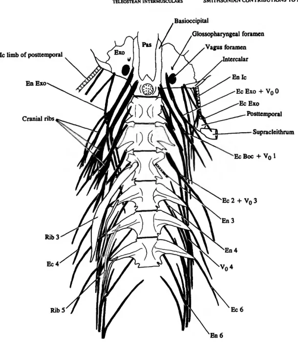
FIGURE 4.—Gonorynchus Igonorynchus (L,), SIO 70-275,146 mm SL. Ventral view of the first six vertebrae and the rear of the braincase, with the intermuscular bones in solid black. The ribs are removed on the left side (right side of figure). The left "cranial ribs" on the occiput and VI, and the epicentral (lower) branch of left compound intermuscular on VI are drawn as if they were cut through near their origin. The left posttemporal and supracleithnim are included, with the supracleithnim drawn as if it was cut through. The only ligaments shown are those representing the intercalar limb of the posttemporal and the epineural ligament of V3, which includes a diminutive bone on the right side. (Scale bar in mm.) (Abbreviations: Boc, basioccipital; Ec, epicentral; En, epineural; Exo, exoccipital; Ic, intercalar; Pas, parasphenoid; Vo, intermuscular representing unattached anteroventral limb and posterior body of an epineural. Plus signs between symbols indicate fusion. Intermusculars and ribs are numbered or labeled according to the vertebra (number) or bone (abbreviation) with which they are associated.)
Supraneurals Ec 1 + Vn 2
Enlc
En Exo
EcExo
Occipital cranial ribs
Ec Boc + Vn 1
Cranial rib 1
Ec 2 + Vn 3 Ec4
Rib 3
FIGURE 5.—Gonorynchus Igonorynchus. Anterior vertebrae and intermuscular bones in left dorsolateral view of RUSI 15048, 95 mm SL. The intermuscular bones are in solid black. The only ligament shown is that attaching the fifth epineural to the neural arch. (Scale bar 1 mm.) (Abbreviations: Boc, basioccipital; Ec, epicentral; En, epineural; Exo, exoccipital; Ic, intercalar, Vo, intennuscular representing unattached anteroventral limb and posterior body of an epineural. Plus signs between symbols indicate fusion. Intermusculars and ribs are numbered or labeled according to the vertebra (number) or bone (abbreviation) with which they are associated.)
Epineural bone
Epicentral bone
Rib
Epipleural bone
FIGURE 6.—Gonorynchus Igonorynchus, same specimen as Figure 4. Lateral view of vertebrae 25 and 26 (left) and of vertebrae 34 and 35 (right), with attached intermuscular bones and ligaments. The intermuscular bones are in solid Mack. (Scale bar in mm.) (Notation Do (as in Table 2) denotes unattached bones that are homologous with anterodorsal branch of an epipleural.)
lar on VI is directed posteroventrally in the peritoneal membrane, so that it lies in the plane of the ribs distally, where it breaks up into a brush-like termination, with about seven branches passing out into the musculature; we interpret this bone as the serial homologue of the two "cranial ribs" (below).
Anterior to these complex intermusculars on V1 there are seven more intermusculars, which insert on the cranium. These include the "cranial ribs" of previous descriptions of Gonoryn- chus (Chabanaud, 1931; Monod, 1963). The two most posterior of these bones insert close together on the basioccipital, just anterior to the lower margin of the occipital condyle. The more dorsal of the two (Ec Boc + Vo 1, Figures 4, 5) bifurcates into two branches, one posterodorsal and one posterolateral, respectively matching the epineural and epicentral branches of the succeeding bones (so that the epineural branch represents the epineural of VI). The more ventral bone is directed posteroventrally, breaks up into a brush-like termination in the peritoneal membrane, and is a cranial rib, matching the anterior intermuscular on VI in orientation and configuration. The next two intermusculars insert close together on the exoccipital, immediately beneath the vagus foramen. They show the same pattern as the two bones on the basioccipital, with the more dorsal bone (Ec Exo + Vo 0, Figure 4) bifurcating into epineural and epicentral branches and the more ventral ending in a bony brush in the peritoneal membrane in series with those of the two succeeding cranial ribs. Passing forward on the cranium, the next two intermusculars (Ec Exo, En Exo, Figures 4, 5) insert close together on the exoccipital just above and lateral to the vagus foramen. They are smaller than the bones behind them on the cranium, and together they match the upper of the two intermusculars on the exoccipital and on the basioccipital; the upper bone matches their epineural branch, and the lower matches their epicentral branch. The lower, epicentral, bone bifurcates and both limbs attach to the medial face of the supracleithrum. The last intermuscular on the cranium (En Ic, Figures 4, 5) is a slender bone attached to the intercalar, this intermuscular bifurcates distally, with an outer branch attach- ing to the medial face of the posttemporal and an inner dorsolateral branch (which bifurcates again) passing back into the musculature in series with the epineurals. Ridewood (1904:66) homologised this last intermuscular with the intercalar limb of the posttemporal, which he thought was otherwise absent in Gonorynchus, but we believe that the intercalar limb is represented by a stout, strap-like ligament that originates on the intercalar immediately beneath the point of origin of the epineural intermuscular and passes out to the posttemporal (Figure 4).
The epipleural series in our 146 mm SL Gonorynchus ostensibly extends from V11 back to V35 (the level of the pelvic-fin insertion), but, as we will show, it extends much further posteriorly. All the epipleurals to V35 are ossified, and there appears to be no rostral or caudal extension of the series in ligament. The bones are simple rods, attached by short ligaments to the tips of transverse processes (Figure 6). They are directed slightly posterodorsally, and most of them extend
back in the musculature for a distance equal to about one-and-one-half vertebrae. The first few and last few bones are shorter than those in the middle of the series and are attached to the transverse process by longer ligaments. Beginning at V30, and extending posteriorly to V52, there is another series of epipleural bones that clearly mirror the detached anteroventral branches of the posterior epineural bones. These bones (Do
Figure 6) lie unattached (free) in the lateral musculature above the epipleurals and lateral to the middle part of the epicentrals;
they are oriented obliquely ventrolaterally, and, in the anterior part of the series, each begins close to the middle part of the epicentral bone of the preceding vertebra. The first few bones are short (about the length of a vertebra), but they lengthen posteriorly (up to about two vertebrae long) and become oriented more horizontally.
We have not found any posterior oblique tendons (POTs) in Gonorynchus, and Baudelot's ligament also is absent.
Ribs of Gonorynchus extend from V3 to V34. The first is enlarged and is preformed in cartilage. The remainder are slender and ossified as membrane bone.
Table 2 illustrates our method of recording intermuscular bones and ligaments as applied in Gonorynchus.
Monod (1963) gave an excellent account of intermuscular bones in Gonorynchus based on dissected specimens two or three times as large as ours (-300 mm SL). His observations match our own in most respects. Monod's specimen has 62 preural vertebrae, whereas our 146 mm specimen has 55. He used an alphabetical notation (a 1-50, b 1-50, etc.) for the different series, but he concluded that his a series comprised epineurals, his b series epicentrals, his c series ribs, and his d and e series epipleurals. Monod's e series extends from V33 into the caudal region, and posteriorly from V49 onward each e bone is fused with the tip of an epipleural (Monod's d), so that his e bones correspond to the anterodorsal branch of the epipleural (Do in Table 2 and Figure 6). In our smaller specimen (146 mm SL), principal epipleurals (attached to the axial skeleton) extend only to V35, overlap the e series (beginning at V30) for only a few segments, and are nowhere fused with them. Fusion between the two may occur in larger individuals, as the series of principal epipleurals extends to more posterior vertebrae. Monod found that the principal (anteromedial) and distal (anteroventral branch + posterior body of the bone; notation Vo in Table 2 and Figure 6) branches of epineurals did not separate until about V51, and that the medial branch did not disappear until V54 (cf. V36 and V41 in our specimen). On the back of the cranium and the first few vertebrae, where the intermusculars of Gonorynchus are most intricate, Monod's observations and interpretations agree with our own in most details.
Monod included three "cranial ribs" in his c series, two originating on the cranium and one on V1. We can find no other interpretation for these three bones, which (as noted above) appear to lie in the peritoneal membrane. We have not found cranial ribs in any other teleost (the supposed "cranial ribs" of Chanos and other gonorynchiforms are epicentrals, Table 4)
and infer that they are autapomorphous for gonorynchids. We do not believe that they are homologous with "true" ribs, and we use a different symbol for them in Table 2.
Monod failed to notice that the anteroventral (secondary) limb of each epineural is directed toward the transverse process of the vertebra in front of that to which the primary (anteromedial) limb attaches. He also referred to the primary limb, attaching to the neural arch, as the accessory branch, and he took the anteroventral limb and the posterior part of each epineural to represent the principal part of the bone. He found no intermusculars on V2 and sought to explain this by a phylogenetic shift of the intermusculars from the posterior part of one centrum to the anterior part of its successor. Intermuscu- lars exist on V2 in our specimens, so no such explanation is necessary. The absence of an epicentral on V3 seems more to demand explanation, and we return to it below, in the section on ostariophysans.
Gonorynchus has an intermuscular system more intricate and challenging than that of any other teleost in our sample.
THE INTERMUSCULAR SERIES AND ASSOCIATED STRUCTURES IN OTHER NONACANTHOMORPH TELEOSTS
Tables 3-5 record the distribution and form of intermuscular bones and ligaments in a range of nonacanthomorphs. In this section we comment on the three series of intermusculars in those fishes and on their POTs, ribs, and certain other features recorded in the tables.
EPINEURALS.—The primitive teleostean condition with regard to intermuscular bones is to have epineurals alone (Patterson and Rosen, 1977:129; Schaeffer and Patterson, 1984:61). Actinopterygian epineurals are primitively devel- oped as short, broad outgrowths of the anterior neural arches that are preformed in cartilage and ossify perichondrally (in cartilage bone) so that in fossil actinopterygians they are hollow and open at the tip, which was cartilage-capped in life.
Short epineural processes of this type occur on the first few vertebrae in Devonian lungfishes (Rosen et al., 1981, fig. 54) and palaeoniscoids (Gardiner, 1984, figs. 121-124) and may well be primitive for osteichthyans. Short, cartilage-tipped epineural processes occur on about the first 20 vertebrae in the Jurassic neopterygian Hulettia (Schaeffer and Patterson, 1984, figs. 15, 17C). It is synapomorphous for teleosts to have much longer epineurals, equal in length to several vertebrae (e.g., the early Jurassic Pholidophorus bechei, Schaeffer and Patterson, 1984, fig. 17D). These long epineurals were still hollow and cartilage-tipped in Pholidophorus, a condition that we ob- served in P. bechei (the type species), the early Jurassic P.
germanicus, and two late Jurassic species, "P." macrocephalus and the species described by Patterson (1975) as "the Callovian Pholidophorus sp." The cartilaginous anlagen of the epineurals evidently have regressed in all Recent teleosts, as have those of some or all the pleural ribs in many teleosts (Emelianov, 1935;
Tables 2-5, 7, 8). We have seen no sign of cartilage in epineurals of any Recent teleost (including the developmental
series of some taxa), and Emelianov (1935) found no cartilage in epineural bones of the teleosts that he investigated, which included Salmo and a clupeoid. It is therefore synapomorphous for Recent teleosts to have solid, membrane bone epineurals, and this derived condition also characterizes some Jurassic stem-group teleosts. We observed solid, membrane bone epineurals, without cartilage cores or tips, in the late Jurassic
"leptolepids" Ascalabos, Tharsis, and the species described by Patterson (1975) as "the Callovian Leptolepis sp.," as well as in the late Jurassic ichthyodectiform Allothrissops and the late Jurassic elopocephalan Anaethalion. However, in the early Jurassic Leptolepis coryphaenoides (type species of the genus), the epineurals are hollow and evidently had a cartilage core in life, but their tips are fully ossified, so that there was no cartilage cap. In the early Jurassic Proleptolepis, the epineurals seem to show the same condition: they are hollow, but the tips of at least the most posterior ones (our material is incomplete) are fully ossified. Solid, membrane bone epineurals, therefore, seem to characterize the node above Leptolepis coryphaenoides in the cladogram in Patterson and Rosen (1977, fig. 54), or ichthyodectiforms, Ascalabos, Tharsis, and all higher teleosts, and epineurals with a cartilage core but without a cartilage tip characterize a node above Pholidophorus bechei and below Leptolepis coryphaenoides in that cladogram. Those two taxa are separated by Pholidolepis dorsetensis in the cladogram. We examined the available material of that species, and although we can confirm that the epineurals had a wide cartilaginous core, we could see no well-preserved epineural tips; the lack of distal tapering of the epineurals is suggestive of the condition in Pholidophorus (cartilage-tipped) rather than that in Leptolepis (solid tips).
Primitive continuity between epineurals and neural arches persists in all epineurals of Pholidophorus bechei and of Leptolepis coryphaenoides. In both genera, epineurals are confined to the abdominal region. In the derived late Jurassic
"Pholidophorus" macrocephalus, only about the first eight epineurals are continuous with the neural arch, and the remainder (they extend back to about V22, and the first caudal is V27) are free, articulating with a socket on the neural arch. In the ichthyodectiforms Thrissops (Taverne, 1977a) and Al- lothrissops, all epineurals are continuous with the neural arch;
they extend back to the third or fourth caudal vertebra. In Tharsis, epineurals extend back to about the first caudal vertebra, and there appear to be two or three free epineurals posteriorly, articulating with the neural spine. In Ascalabos, epineurals extend to about the fifth caudal vertebra, and, as in Tharsis, the last two or three are free and articulate with the neural spine. As in the above stem-group teleosts, primitive continuity between anterior epineurals and neural arches persists in various more-derived fossil and living teleosts (e.g., the Jurassic Anaethalion and Recent Elops, Arratia, 1987, figs.
3, 31; Hiodon, Taverne, 1977b, figs. 22, 23; Pantodon, Taverne, 1978, figs. 49-53; Esox, Figure 8; Alepocephalus, Gosline, 1969, fig. 11; Clupea, Figure 7A, and Ramanujam.
1929:389; stomiiforms, Weitzman, 1974, fig. 85; Fink and
Weitzman, 1982, fig. 5). In all these forms (except the osteoglossomorphs Hiodon, where all epineurals are fused to the neural arch (Table 3), and Pantodon, where all but the first are so fused (Taverne, 1978)), the more posterior epineurals articulate with the neural arch or spine, and the most posterior ones are either free or connected with the neural spine by ligament. In more-derived teleosts, anterior epineurals are separate, either articulating with the vertebra (e.g., Rosen, 1985, figs. 9, 11, 13, 15-17) or attaching to it by ligament, and posterior epineurals are free from the vertebral column and often are forked proximally (e.g., Figures 4, 5, 8, Plate lE.F Weitzman, 1962, fig. 14; Jollie, 1962, fig. 6.40, "epimerals"), a condition that we take to be synapomorphous for elopo- cephalans. When epineurals are forked anteriorly (as in Gonorynchus, above), there is an anteromedial branch that represents the primitive connection with the neural arch and an anteroventral branch that passes forward in the myoseptum, roughly parallel to the vertebral column, into the epaxial forward flexure of the myomere (Nursall, 1956, fig. 8).
Epineurals are virtually universal in nonacanthomorph teleosts, occurring in all "leptolepids," ichthyodectiforms, osteoglossomorphs, elopomorphs, and clupeomorphs, in ostariophyans (except siluroids), in esocoids, argentinoids, osmeroids (osmeroids sensu Rosen, 1974, were said by Rosen to lack intermusculars, but all osmeroids that we have examined have epineurals), salmonoids, stomiiforms, aulopi- forms, and myctophiforms (cf. Tables 3-5).
The only nonacanthomorphs lacking epineural bones among those recorded in Tables 3-5 are the southern osmeroids Stokellia and Lepidogalaxias, the salangid Salangichthys, and the umbrid Novumbra (they also are lacking in Dallia). In Stokellia, Novumbra, Dallia, and salangids, the epineural series is represented by a series of ligaments with the same distribution as epineural bones in other lower euteleosts. We found the same condition in Retropinna, and it may be a synapomorphy corroborating the sister-group relationship between that genus and Stokellia (Begle, 1991), but McDowall (1971, 1976) reported that ossified epineurals also are absent in Aplochiton, Lovettia, and Prototroctes. In Lepidogalaxias, there are no epineural bones or ligaments, a unique condition.
In other osmeroids (Osmerus, Hypomesus, Table 4) and various other lower euteleosts (e.g., the stomiiform Diplophos and the aulopiforrns Ahliesaurus, Scopelosaurus, Scopelarchoides, Synodus, and Trachinocephalus, Table 5), the series of epineural bones is extended caudally by a series of ligaments, without included bone. In the evermanellid aulopiform Coc- corella, there is only one epineural bone (true of all evermanellids according to Johnson, 1982:55), and the epineu- ral series is continued caudally as ligament.
Epineurals are posterodorsally inclined in the epaxial backward flexure of the myoseptum (Nursall, 1956, fig. 8), and their distal tips are normally (and primitively) all aligned at the same level. But in Polymixia (Figure 2), the bone that we interpret as the first epineural is out of alignment with the rest of the series and lies in the horizontal septum. In a few
nonacanthomorphs, the distal part of the first one or more epineurals also is displaced ventrally relative to their succes- sors, although, in contrast to Polymixia, they are always above the horizontal septum. We have observed this ventral displace- ment in alepocephaloids, argentinoids, several aulopiforms, and the neoscopelid Neoscopelus (notation b, or f {in Tables 4, 5). In alepocephaloids and argentinoids, the tips of the first three (Glossanodon, Leptoderma, Searsia) or four {Argentina) epineurals are displaced, a unique condition that we take to be synapomorphous, corroborating the sister-group relationship of these two taxa (Greenwood and Rosen, 1971; Begle, 1991). In aulopiforms, the tips of the first one to three epineurals may be displaced, and in Neoscopelus only that of the first is displaced (none is displaced in the other neoscopelid genera, Scopelengys and Solivomer).
In Polymixia, the proximal articulation of the epineurals on V3-10 is not on the neural arch but on the centrum (V9-10) or parapophysis (V3-8). The proximal ends of some anterior epineurals also are displaced ventrally in a few nonacantho- morphs. Among osteoglossomorphs, most epineurals originate on the centrum in mormyrids and in the notopterids Xeno- mystus (Table 3) and Papyrocranus. Among the elopomorphs in our sample, a few anterior epineurals originate on the centrum in Notacanthus, and among clupeomorphs about 15 do so in Anchoa (notation B2 in Table 3). In euteleosts, Esox has 20 or more epineurals originating on the centrum, from about V10 onward (Table 4), and the aulopiforms Scopelosaurus, Ahliesaurus (both Notosudidae), Scopelarchoides (Scopelarchidae), Coccorella (Evermanellidae), Bathysaurus, and Gigantura have a number of epineurals originating on the centrum or (in Scopelosaurus) on the parapophysis (Table 5).
EPIPLEURALS.—As noted above, the epipleural series gener- ally is easily distinguished from epicentrals and epineurals by ontogeny and distribution; epineurals and epicentrals develop in a rostrocaudal gradient, first appearing and being most strongly developed on anterior vertebrae, whereas epipleurals develop both rostrally and caudally from about the level of the first caudal vertebra.
Epipleural bones are absent in the most primitive teleosts, such as Mesozoic pholidophorids, early Jurassic leptolepids, and the Mesozoic ichthyodectiforms. When epipleurals first appear in the geological record, in late Jurassic "leptolepids,"
such as Ascalabos, Tharsis, and Todiltia, they are confined to the last few abdominal and first few caudal vertebrae. In small Todiltia (37 mm SL), for example, there are five pairs of epipleurals in that region, and in the largest specimens (60-80 mm SL) there are seven or eight (Schaeffer and Patterson, 1984:54). Ascalabos and Tharsis have about six epipleurals in that region, Anaethalion has about 12, and Elops has up to about 40 (Taveme, 1974). Osteoglossomorphs generally lack epipleurals, but in Heterotis they occur on up to 10 vertebrae (V26-36, Taverne, 1977b; cf. Table 3), and notopterids have been described as having a long series of them (Taverne, 1978).
However, we interpret notopterid "epipleurals" differently; we consider them to be myorhabdoi (see section on osteoglosso-


![The significance mythical creatures and their symbols in the first six series of Harry Potter by J.K. Rowling toward the storyline [abstrak dan teks sedang dalam proses] - USD Repository](data:image/gif;base64,R0lGODlhAQABAIAAAP///wAAACH5BAEAAAAALAAAAAABAAEAAAICRAEAOw==)