Gregory and Conrad (1943) made extensive comparisons of the skeleton of Luvarus with that of a "typical". Most of the osteological description in this paper is based on two cleaned and counterstained specimens, 79.2 and 301 mm SL. An early draft of the manuscript was improved by the persuasive and constructive criticism of R.P.
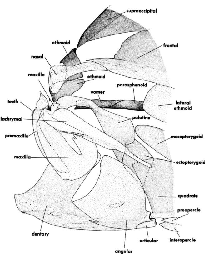
Cressey (USNM) identified copepod parasites from one of the specimens and advised us of their potential significance in relation to the relationships of Luvarus. The posterior end is forked where it articulates by cartilage and slight interdigitation with the basioccipital, forming the floor of the shallow posterior myodome. Ethmoid (= Mesethmoid): A huge, rectangular ethmoid cartilage (not shown) forms the largest mass of the neurocranium in front of the orbit.
Ventrally, the posteromedial edge of the sphenotic door cartilage articulates with the anterolateral region of the dorsal head of the hyomandibula (forms part of hyomandibular fossa).
The bases of the medial four rays overlap only the cartilaginous border of the hypopural plate. The first (unbranched) main ray of the caudal fin above and below has the greatest degree of overlap with the skeleton of the caudal fin. The first spine has a single set of serrations (not shown in the figures) along the proximal half of the length of each posterolateral wing.
The slender ventral shaft articulates posteroventrally with the anterodorsal edge of the neural arch of the second vertebra. The vomer and lateral ethmoid ossifications are larger, and the surfaces of the latter are extremely spongy. The posterior end of the ceratohyal is more strongly sutured with the anterior end of the epihyal than in the smaller specimen.
Similar in configuration to that of the 79.2mm sample, but most elements are more robust. The lateral or anterior margin of the pelvic spine is more strongly toothed and rugose than in the smaller specimen. More of the main rays are branched and segmented than in the smaller specimen, and the bases of the rays overlap the hypural plate more extensively.
There is a very small notch at the midline of the posterior margin of the hypural plate. Similar to that of the 301 mm specimen (Figure 19), except most of the legs are stronger, the gills are heavier, shorter and less spiny, and the basihyal more rounded than in the smaller specimens (Figures 9 and 19). Caudal notches on the dorsal and ventral parts of the tail peduncle found on some other specimens are not seen in this individual.
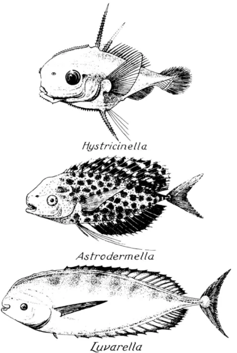
A shallow groove is present from the snout to the upper edge of the operculum above the eye. The bases of most of the caudal-fin rays are deeply robust and broadly overlap the hypural plate. Previous classifications have most often treated acanthuroids as members of the older Squamipinna (sensu Cuvier, 1817).
In all other squamipinnes and most percoids, the surfaces of the skull bones are relatively smooth.
Pmx D
Parietals are absent in scatophagids (Figure 40) and siganids but are present in Luvarus (only in the
If parietals were absent in the common ancestor of scatophagids and acanthuroids, they must have reappeared in the common ancestor of Luvarus, Zanclus, and Acanthuridae. An equally simple hypothesis is that parietals were lost independently in scatophagids and siganids. A third possibility, that the loss of parietals is a synapomorphy of scatophagids and siganids, is ruled out by the many synapomorphies described below that place siganids as the sister group of other acanthuroids.
At least one derived character supports the hypothesis that ephippidids are the sister group of acanthoroids.
In the absence of information about the importance of this innovative specialization in relation to the two (and possibly four) reductive synapomorphies of scatophagids and acanthuroids, we assume that Scatophagidae is the sister group of Acanthuroidei and postulate that a specialized gill filament arrangement has either arisen independently in epipidids and acanthuroids, or that it has been lost secondarily, are scatophagids. For most characters, change in the hypothetical phyletic sequence of the first two outgroups will not affect the polarity assessment because the primitive state characterizes them both and usually also other squamipins. ADULT MORPHOLOGY.—The following 11 synapomorphies confirm the monophyly of Acanthuroidei, which includes Siganidae, Luvaridae, Zanclidae, and Acanthuridae.
In addition, the connection point is at the posteroventral corner of the lacrimal, so the main body of the lacrimal lies above the projected path of the infraorbital ring (Figure 43). In both outgroups, other squamipinnes, and most percoids, there is a more intimate association between the lacrimal and second infraorbital, and the main body of the lacrimal lies below the path of the infraorbital ring. In cyanids it lies between the open neural arch of the first centrum; in all other acanthuroids it is anterior to the neural spine of that centrum, fully within the first internal space.
In both outgroups, other squamipinnes and most percoids (see Johnson, 1984) it is located more posteriorly, in the second or third internal space. In both outgroups, other squamipinnes, and most percoids, the posttemporal canal joins the main trunk lateral line through a short bony canal at the dorsal end of the supracleithrum. Movement of the upper jaw is mainly limited to rotation, the variously developed rostral cartilage acts as a pivot against the vomer, protrusion is extremely limited or impossible.
It is equally parsimonious to assume independent acquisition of the relatively shorter articular in siganids. In both outgroups, other squamipins, and most percoids, the ventral extension of the supraoccipital (spina occipitalis of Allis, 1909) is embraced laterally by dorsal extensions of the exoccipitals that are closely attached to the supraoccipital on either side of the extension. In both outgroups and other squamipins, the supraoccipital crest forms a large, triangular crest with a thickened anterior margin.
Both outgroups, other squamipines, and most percoids have one or more predorsal bones (see Smith and Bailey, 1961, and Johnson, 1984). In both outgroups, other squamipinnes (some pomacanthids are exceptional), most percoids and Luvarus, the fourth pharyngeal tooth plate is oriented transversely, so the long axis and tooth rows (if the teeth are arranged in rows) are perpendicular to the body axis.
In other acanthuroids, the dorsal soft rays range from 19 to 41, and the anal soft rays from 19 to 35. Cycloid scales are found among squamipines only in the ephippidids Ephippus and Rhinoprenes; they apparently evolved multiple times among percoids, where ctenoid scales are most common and presumably primitive (Johnson, 1984). It is bony in all other acanthuroids, both outgroups, and in other squamipines where it occurs.
Because the parasphenoid is a dermal bone, this cartilaginous apophysis in siganids may not be homologous to the bony apophysis of other groups. The anterior uroneural pair is reduced to two small bony nodes (Figure 29) lying free in the space between the urostyle and the two anterior epurals. In other acanthuroids, such as the outgroups, other squamipines, and most percoids, this uroneural pair is much longer and the anterior part forms an embracing saddle.
In other acanthuroids, both outgroups, and most percoids, the first pleural rib is on the third vertebra. The anterior surface of the ethmoid is vertical and forms a shallow, concave facet that receives the broad base of the large conical rostral cartilage. See character 42 for a description of the ethmoid in other acanthuroids and the general outgroup discussion for a description of its configuration in Drepane, chaetodontids, and pomacanthids.
The medial of these (described as two separate cartilages by Mok and Shen, 1983) articulates anteriorly with the second pharyngobranchial; the lateral one lies freely next to the medial one. A median accessory cartilage is associated with the dorsal gill arches in some acanthurids, but to our knowledge paired cartilages like those of siganids do not occur elsewhere among perciforms. A small, autogenous, triangular cartilage is present lateral to the fourth pharyngobranchial tooth plate, between the anterior cartilaginous tip of the third epibranchial and the posterior cartilaginous tip of it.

- The configuration of the fourth pharyngeal tooth- plate is unique in having a T-shaped dorsal process; the
- Replacement teeth on the jaws and pharyngobran- chials are not enclosed in bone, but instead lie free in
- There is a thin, elongate rod of cartilage between the haemal spines of the second and third preural centra
- There is an overlapping articulation between the proximal-medial and distal radials in the spinous dorsal
- The first dorsal pterygiophore inserts fully in the first interneural space, and the anterior portion of its
- Three epurals are present in the larvae, but these eventually consolidate into a single element that fuses to
- Hypurals 1-4 fuse to form a single hypural plate
- Hypural 5 remains separate from hypural 4 but fuses to the uroneural pair which in turn fuses to the
- There are only two dorsal spines and no anal spines. Among other acanthuroids the number of dorsal
- The first dorsal (first three in Zanclus, see character 47) and first anal pterygiophores bear a
- There is a conical, spine-like protuberance on each frontal near the anter odor sal margin of the orbit
- The second (third in Zanclus,) dorsal and pelvic spines are the first fin rays to form and they rapidly
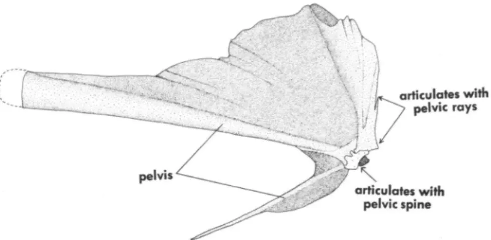
- The pelvic girdle has an unique configuration, being essentially T-shaped and so oriented that the
- A thin, slightly curved spine projects ventrally from the posteroventral corner of the angular (= retro-
- The single postcleithrum (on each side) ossifies very early (3 mm) and forms a strong, almost vertically
- The posterior margin of the preopercle develops two minute spines near the angle, but these are lost by 5
- There is a vertically oriented, serrate ridge on the lateral surface of the opercle. A comparable ridge is
- The second anal spine is notably elongate and the first anal pterygiophore is an enormous, columnar strut,
- The scales are extremely long and narrow (the long axis is vertically oriented). Naso larvae have ovoid
- Scales on the head and cheek and most of those on the body anterior to the postcleithra lack the central
In other acanthoroids, both outgroups, other squamipinnes, and most percoids, there is a fourth pharyngobranchial cartilage firmly attached to the dorsal (lateral in Zanclus and acanthurids) surface of the fourth pharyngeal tooth plate. The palatine lies well anterior to the lateral ethmoid and has no articulation with it. In siganids, both outgroups, other squamipines, and most percoids, the posterior part of the palatine has a condylar articulation with the lateral ethmoid.
The extensive interposition of the proximal radials in Luvarus does not occur elsewhere among acanthuroids. It is notched anteriorly and the ethmoid cartilage continues anteriorly over the parasphenoid and vomer to the tip of the snout. The anterior end of the ethmoid is always a simple, flat, horizontal or slightly oblique plate.
See Johnson and Washington (1987) for a discussion of the serial homology of dorsal spines in Zanclus and acanthurids. First supernumerary spine on first dorsal and anal pterygiophore reduced and lacking pointed distal tip; lies beneath the surface of the skin. Some or all of the specialized scales, where present in larvae (absent in Paracanthurus and Zebrasoma), are resorbed and some or all of the adult scales are formed anew, rather than being transformed directly from existing larval scales (see characters 71 and 81) .
LARVAE MORPHOLOGY - Acanthuroid larvae have a complex morphology that differs greatly from that of adults. Spines on the ascending processes of the premaxilla are unique to these groups among larval perciforms. This spine gradually reduces with growth in Luvarus and eventually fuses with the dorsal surface of the pterygiophore (fused in our 301 mm SL specimen).
This flange resembles those of the first dorsal and anal pterygiophores, but unlike them is only present in larvae.