S M I T H S O N I A N C O N T R I B U T I O N S T O B O T A N Y 0 N U M B E R 3 7
Pollen Morphology and the Relationship of the
Plumbaginaceae, Polygonaceae, and Prirnulaceae to the
Order Centrospermae
Joan W. Nowicke
and John 3. Skvnrln
SMITHSONIAN I N S T I T U T I O N PRESS City of M7ashington
1977
Nowicke, Joan W., and John J. Sklarla. Pollen Morphology and the Relation- ship of the Plumbaginaceae, Polygonaceae, and Primulaceae to the Order Centro- spermae. Siiiz1hsoniun Contl-zbzitions to L’olany, number 37, 64 pages, 200 figures, 3 tables, 197’i.-Three families, Plumbaginaceae, Poljgonaceae, a n d Pi imulaceae, are consiclei ed to be related to or dei i\ ed from the 0 1 der Centrospei mae by 1 ari- ous authors. These three families hai e anthocyanin pigments i n contrast to the betalains found in all but t u o families in the Centrospermae. I n addition, all three are known to h a l e staich-tjpe sieie-tube plastids in contrast to the piotein type found in all centiospermous families. Examination of the pollen of 134 species by SEN, T E N , and light micioscopy reiealed g.reat dii ersity, especially in the Polygonaceae, but not the spinulose and tubuliferous/punctate ektexine, which characterizes the i ast majoritj of the centrospermous taxa. Recent ei idence argues against a close relationship of the Plumbaginaceae, Pol) gonaceae, and Primulaceae with the Centrospei mae, and the absence of any pollen tjpes com- mon to the three families further suggests that they are not closely related to each other.
OFFICIAL PVBLICATIOS DATE is handstamped in a limited n u m b e r of initial copies and is recorded in the Institution’s a n n u a l report, Smithsoninn Year. SERIFS COVER D E S I G N : Leaf clearing from the katsura tree C e r c i d i p h y l l t c m japoniczcm Siebold a n d Zuccarini.
Library of Congress Cataloging i n Publication Data Sorvicke, Joan LV.
Pollen morphology a n d t h e relationship of t h e Plumbaginaceae, Polygonaceae, a n d Primulaceae (Smithsonian contributions to botany ; no. 37)
to the o r d e r Centrosperrnae.
Bibliography: p.
1. Plumbaginaceae. 2. Pol>-gonaceae. 3 . Primulaceae. 4 . Centrospermae. 5 . Palynotaxonomy.
6. Pollen morpholorry. I. Sktarla, Tohn T., ioint a u t h o r . 11. T i t l e . 111. Series: Sinithsonian Institution. SkithsoGian contributioGs to 6ota”uy ; no. 37.
QKl.S2747 no. 37 [QK495.A12] 581’.08s [583’.672] 77-608070
Contents
Page
Introduction
...
1Materials and Methods
...
3Results of Pollen Analyses
...
4CENTROSPERMAE
...
4PLUMBAGINACEAE
...
5POLYGONACEAE
...
8PRIMULACEAE
...
1 1 Discussion and Conclusions...
12Literature Cited
...
15Tables
...
16Figures
...
23111
...
Pollen Morphology and the Relationship of the
Plumbaginaceae, Polygonaceae, and Primulaceae to the
Order Centrospermae
Joan W. Nowicke and John 3. Skvarla
Introduction
T h e Centrospermae (Caryophyllales) represents one of the most controversial orders in the angio- sperms. This group of at least 10 families, which in the past has been cited as one of the few natural orders based mostly on morphological characteris- tics, has unique N-containing pigments, the beta- lains, and a distinctive structure in the sieve-tube plastids. Both Cronquist (1968) and Takhtajan (1969) unite the betalain families, Aizoaceae, Ama- ranthaceae, Basellaceae, Cactaceae, Chenopodiaceae, Didiereaceae, Nyctaginaceae, Phytolaccaceae, and Portulacaceae, as well as two anthocyanin families, Caryophyllaceae and Molluginaceae, in the order Caryophyllales ( k Centrospermae). Both authors include Halophytum ameghinoi Spegazzini (a beta- lain taxon) and the Gyrostemonaceae in this order:
Cronquist (1968) treats Halophytum as a monotypic genus in the Chenopodiaceae while Takhtajan (1969) gives it family status; Cronquist regards the
Gyrostemonaceae as part of the Phytolaccaceae, and Takhtajan separates it as a distinct family. Takh- tajan recognizes the Tetragoniaceae as a separate family while Cronquist includes it in the Aizoaceae.
Dysphania is treated as a member of the Chenopo- diaceae by both authors; and finally Takhtajan also places the Hectorellaceae and the Bataceae in this order.
Thorne (1968) in a synopsis of angiosperm classi- fication has placed the betalain families in a sub- order, Chenopodiinae, one of two constituting the order Chenopodiales. He recognized the Gyrostemo- naceae and the Halophytaceae as distinct families but treated the Molluginaceae as a subfamily in the Aizoaceae, the Achatocarpaceae as a subfamily in the Phytolaccaceae, and Dysphania as a member of the Chenopodiaceae. T h e other suborder, Caryo- phyllinae, consists of two families, the Caryophylla- ceae and the Polygonaceae. T h e next order, Bati- dales, consists of only the Batidaceae, but Thorne apparently regards this family as somewhat related t i t h e Chenopodiales since both orders are united under a superorder, Chenopodiiflorae.
Joan W . Nowicke, Department of Botany, National Museum
of Natural History, Smithsonian Institution, Washington, D . C . 20560. John J . Skvarla, Department of Botany and Micro- biology, University of Oklahoma, Norman, Oklahoma 73019.
Mabry, and Turner and Behnke
and Turner (1971) have treated the betalain fami- 1
lies as a distinct group, separate from, yet closely allied to, the two anthocyanin families, Caryo- phyllaceae and Molluginaceae.
Evidence from ultrastructural research on sieve- tube plastids (Behnke and Turner, 1971; Behnke, 1976) and pollen morphology (Nowicke, 1975;
Skvarla and Nowicke, 1976) has reinforced the close tie between the betalain families and the Caryo- phyllaceae a n d the Molluginaceae. I n a n investiga- tion of these plastids (colorless leucoplasts found in the sieve-tubes) of the above-mentioned families (Behnke and Turner, 1971) and the Magnoliidae and Ranunculidae (Behnke, 1971), two main types were found: plastids accumulating only starch and designated as the S-type; and plastids accumulating at least some protein, variously deposited, and desig- nated as the P-type. T h e betalain families and the Caryophyllaceae a n d the Molluginaceae all had the P-type plastid i n which proteinaceous filaments formed a peripheral ring usually around a crystal- loid. IVithin the above-mentioned families, Behnke (1976) was able to distinguish three groups based on the crystalloid: globular, the most common; polyg- onal, found i n the Caryophyllaceae and i n two other genera, L i m e u m (Molluginaceae) a n d Stegno- sperma (Phytolaccaceae); and crystalloid-free, hav- ing only the ring, found in two families, the Amaranthaceae and the Chenopodiaceae.
Examination of the pollen of 177 species (No- wicke, 1975) by light microscopy and SEM revealed that 85% had a similar type of ektexine pattern or sculpture, which was described as spinulose and the tectum perforate, the openings described as tubules or punctae. This was the predominant ektexine pat- tern in every betalain family and i n the Caryo- phyllaceae and the Molluginaceae; in some families, i.e., the Phytolaccaceae and the Molluginaceae, this pattern was the only one found. Examination of selected taxa by T E M (Skvarla and Nowicke, 1976) revealed that the predominant pollen-type i n the betalain families and i n the Caryophyllaceae has a similar, sometimes identical, wall structure.
Pollen of the Achatocarpaceae, Bataceae, Gyro- stemonaceae, and Theligonaceae was also examined, but none of the species i n these small families has the spinulose and tubuliferous/punctate ektexine described above. T h e Bataceae, Gyrostemonaceae, and Theligonaceae all have the starch-type plastid and lack the betalain pigments. T h u s the evidence
from palynology, pigmentation, and plastid struc- ture indicates that these three families are not closely related to the Centrospermae. T h e relation- ship of the Achatocarpaceae, a family unknown with regard to pigmentation, to the Centrospermae is more obscure-the pollen morphology does not, in Nowicke’s opinion (1975) support a close tie, b u t on the other hand the two genera that comprise the family, Achatocarpus and Phaulothamnus, do have the P-type of plastid (Behnke, 1976).
T h e Gyrostemonaceae have in fact been the sub- ject of a multidisciplinary study (Goldblatt, et al., 1976) i n which all lines of evidence, including cyto- logical, argue strongly against any relationship of this family to the Centrospermae.
Although there is almost universal agreement on treating the betalain families as a closely related group, the precise definition of the order Centro- spermae, i.e., to include or exclude the Caryo- phyllaceae and Molluginaceae, may never be re- solved to everyone’s satisfaction. Of more interest in view of the accumulating data on the Centro- spermae (sensu lato) is the question of the deriva- tion of the families Plumbaginaceae, Polygonaceae, and Primulaceae from this order.
Takhtajan (1969) considers the Polygonales, con- taining only the Polygonaceae, as near the Caryo- phyllales, especially the Portulacaceae and Basella- ceae, and probably derived from the same stock as the Caryophyllales (Centrospermae). His views o n the Plumbaginaceae are similar: nearest to Portula- caceae and Basellaceae and probably derived from the same stock as Caryophyllales. H e does admit that the pollen morphology of the Plumbaginaceae is different-apparently from that of Portulacaceae and Basellaceae.
A4ccording to Cronquist (1968: 185), “both the Polygonaceae and Plumbaginaceae are pretty clearly related to the Caryophyllales. Both have a single, basal, bitegmic, crassinucellate ovule i n a com- pound, unilocular ovary, and both have trinucleate pollen. These characters are not known to occur i n combination outside the subclass Caryophyllidae.
T h e Polygonaceae are further bound to the Caryo- phyllales by similarities in the pollen and by a more or less transitional group of genera which are vari- ously referred to the Caryophyllaceae or treated as a separate family, Illecebraceae. T h e Plumbagina-
NUMBER 31 3 ceae are somewhat more isolated but may also be
derived from the Caryophyllaceae.”
I n one respect the system of Thorne (1968) paral- lels those of Cronquist (1968) and Takhtajan (1969) i n that all three authors consider the Polygonaceae as related to the Caryophyllaceae. Although Thorne treats the Primulales and Plumbaginales as adjacent orders indicative of some relationship, they are far removed from the Chenopodiales, at least in his linear sequence.
Philipson (1975) in a review paper on evolution- ary lines in the dicotyledons states that there is general agreement to exclude the Primulaceae from the subclass Caryophyllidae (sensu Cronquist, 1968).
“Almost as universal is the acceptance of the Poly- gonaceae as at least peripheral to the group. T h e position of the Plumbaginaceae remains more de- batable” (Philipson, 1975:74).
Hutchinson (1959: 117) regarded the Caryophyl- lales (Elatinaceae, Molluginaceae, Caryophyllaceae, Ficoidaceae, and Portulacaceae) as “a prolific herba- ceous group which has given rise to apetalous orders such as Polygonales, Chenopodiales, and perigynous petaliferous families as Lythrales, besides sympeta- lous groups as Gentianales and Primulales.” T h e Illecebraceae are given family status and included as the only other family in the Polygonales. I t is noteworthy that Hutchinson included H e m iayia, Paronychia, and Scleranthus, as well as rllecebrzirn, among the genera listed at the end of the family description. He united the Primulaceae and Plum- baginaceae as the order Primulales, closely related to the Caryophyllaceae and perhaps the Saxifraga- ceae. T h e Primulaceae, Polygonaceae, and Plumba- ginaceae contain the anthocyanin pigments, and the last two, at least, are known to have the starch-type of plastid (Behnke and Turner, 1971) and appar- ently the Primulaceae also has this type (Behnke, pers. comm.).
I n an effort to resolve the limits and relationships of the Centrospermae, pollen of Plumbaginaceae, Polygonaceae, and Primulaceae were examined by light microscopy, SEM, and TEM. For purposes of comparison and the reader’s convenience, we have included a brief discussion and a SEM and T E M of a species from several betalain families, and from the Caryophyllaceae and Molluginaceae.
ACKNOWLEDGMENTS.-In a n extensive study like this numerous persons have contributed in one way
or another, but Janice Bittner at the Smithsonian Institution and William Chissoe at the University of Oklahoma deserve special thanks for their fine technical assistance and helpful suggestions. T h e scanning electron photographs were prepared by the Scanning Electron Microscope Laboratory at the Smithsonian Institution. We also wish to thank Aaron Goldberg and Stanwyn G. Shetler for their critical review and Clara Ann Simmons for her assistance in the preparation of the manuscript.
This research was supported in part by BMS grant 75-19846 to John J. Skvarla.
Materials and Methods
Pollen of 20 species in the Plumbaginaceae (Table 3), 85 in the Polygonaceae (Table 4) and 29 in the Primulaceae (Table 5) was examined by light microscopy, scanning electron microscopy (SEM), and transmission electron microscopy (TEM). Also examined and included are 38 additional taxa from the centrospermous families (Table 2). Pollen sam- ples were removed from herbarium material and acetolyzed according to procedure outlined in Erdt- man (1966). Samples for the SEM were vacuum- coated with gold, and examined and photographed with a Cambridge Stereoscan M K IIA microscope.
Samples for the T E M were incorporated into agar, dehydrated through increased concentrations of ethyl alcohol, and subsequently embedded in araldite-epon resins (Skvarla, 1973). Some pollen samples were stained in 0.125y0 OsO, in 0.1 M so- dium cacodylate buffer for two hours prior to agar incorporation. Thin-sections were made with dia- mond knives, collected on uncoated grids and stained with uranyl acetate and then lead citrate.
Electron microscope observations were made with a Philips model-200 transmission electron micro- scope. Slides of all samples are deposited at the Palynological Laboratory, Department of Botany, Smithsonian Institution.
This study is a comparison of the pollen mor- phology of three families, Plumbaginaceae, Poly- gonaceae, and Primulaceae, with the pollen of the Centrospermae, and detailed measurements and/or descriptions are not given. T h e species examined are listed in Tables 2-5; the names were taken from the herbarium labels or the most recent annota- tions. T h e identifications would have to be mis-
taken at the level of family to be significantly mis- leading i n this type of study, but all vouchers and geographical locations are given i n Tables 2-5.
Results of Pollen Analyses
Pollen analysis of the Plumbaginaceae, Polygona- ceae, and Primulaceae revealed diverse forms within each family, especially the Polygonaceae, but n o dis- tinctive pollen type(s) was common to all three families. For this reason each family will be dis- cussed separately following a review of the Centro- spermae.
CENTROSPERMAE
FIGURES 1-28
T h e common pollen types in the Centrospermae are illustrated i n Figures 1-6: 3-colpate, pantopo- rate, and pantocolpate, all with a spinulose and tubuliferous/punctate ektexine. With the exception of some taxa i n the Caryophyllaceae which are 3-colporoidate, the apertures in the Centrospermae pollen are simple. T h e most wriable class of aper- tures is that of pantoporate. Anredera scandens (Figure 3) is usually 6-porate, with one pore on each plane of the roughly cube-shaped grain. Gymnocar- pos frziticosum pollen (Figure 4), with large, sunken pores in a geometric pattern, contrasts sharply with that of Chenopodizrm ambrosioides (Figure 5), which has numerous small pores.
Figures 7 through 12 illustrate at high magnifica- tion ( X 7500) the ektexine surface, spinulose, and tubuliferous/punctate, which characterizes the T ast majority (85%) of the centrospermous taxa. Hama- tocactus septispinus pollen (Figure 12) has the largest perforations among the species examined by SEM.
Figures 13 through 18 illustrate specialized or un- usual forms, but at least one species, Cal-dioizerna ramosissima (Figure 16), has the typical ektexine.
T w o other taxa, Psilotrichtim amplum (Figure 13), considered unusual because of the stellate aperture plates, and Herniaria glabra (Figure 15), in which the grains have a rare tetrahedral shape, also have the common ektexine pattern but a modified ~ e r - sion of it-the spines and perforations are much reduced.
T h e internal structure of the exine in various centrospermous pollen types is illustrated i n Fig- ures 19-28. Characteristically i n the centrospermous taxa, the ektexine is well developed while the en- dexine, with few exceptions (e.g., Mesembryanthe- mum variabile, Figure 19; Herniaria glabra, Figure 24), is developed only in the region of the aperture.
Apertures with spine-shaped flecks of ektexine are common in the Centrospermae and in this study are illustrated by Opuntia (Figure 22), Limeuni (Fig- ure 26), and Gymnocarpos (Figure 23). Representa- tive pollen from nine families of the Centrospermae were examined by TEM. Comparison of these data with those of earlier T E M work in the Centro- spermae (Skvarla and Nowicke, 1976) allows us to expand our knowledge of this order, and a brief summary for each family is presented below.
AIZoAcEAE.-Mesem bryanthemum variabile pol- len (Figure 19) is considerably different from that of M . crystallinum (Skvarla and Nowicke, 1976, fig.
13) in possessing a thinner tectum and columellae, as well as an extremely thick foot layer and a nar- row but consistent endexine. Mesembrganthemum, however, is a large and unsatisfactory genus and structural yariation is not unexpected. T h e exine stratification of M . varia bile bears a striking simi- larity to that of Boerhavia erecta of the Nyctagina- ceae (Sklai-la and Nowicke, 1976, fig. 30).
been shown with SEhI and T E M to be highly pleo- morphic (Nowicke, 1975; Skvarla and Nowicke, 1976). T h e exine structure of Psilotrichurn a m p l u m (Figure 20) extends this pleomorphism by indicat- ing a complete absence of endexine.
BASELLACEAE.-some pollen in this family is unique in having a cuboidal shape (Nowicke, 1975;
Skvarla and Nowicke, 1976). T h e structure of Anl-edera scandens pollen (Figure 21), is quite dif- ferent from that of Basella alba (Skvarla and Kowicke, 1976, fig. 19) but similar to that of Bozissingniiltia gracilis (Roland, 1968, pl. 8: fig. 4), a more typical member of the family.
CACTACEAE.-Tlle structure of Opzintia Zindhei- meri pollen (Figure 2 2 ) is consistent with other ex- amples of the Cactaceae. Comparison of this micro- graph with one of Allztaz~clia (Skvarla and Nowicke, 1976, fig. 28) again underscores the close relation- ship of the Cactaceae and Didieriaceae.
AMARANTHACEAE.-The pollen of this family has
NUMBER 37 5 CARYOPHYLLACEAE.-The two taxa examined,
G y m n o c a r p o s f r u t i c o s u m (Figure 23) and Herniaria glabra (Figure 24), correspond in exine structure to that previously noted for the family (Skvarla and Nowicke, 1976, figs. 41-48). Additional comments on H . glabra will be given later.
n o p o d i u m ambrosioides (Figure 25) agrees with other examples in the family: thick tectum with few spines and a thin foot layer.
MoLLUGINACEAE.-The exine structure of this family has not been previously examined. Pollen of L i m e u m viscoszim (Figure 26) appears similar to the pollen of C o m e t e s surattensis of the Caryophyl- laceae (Skvarla and Nowicke, 1976, fig. 41), as well as to M e s e m b r y a n t h e m u m crystallinurn of the Aizoaceae (Skvarla and Nowicke, 1976, fig. 13).
These relationships are of interest in the context of the introductory remarks concerning the Mol- luginaceae.
NYCTAGINACEAE.-~ bronia angzistifolia (Figure 27) pollen morphology differs from that of previous taxa examined by lacking an endexine. As indicated earlier (Skvarla and Nowicke, 1976), however, the Nyctaginaceae display a spectrum of exine mor- phology.
N a i o c r e n e pal-vifolia (Figure 28) is similar to that of other taxa examined in the family, most notably C a l y p t r i d i u m u m b e l l a t u m (Skvarla and Nowicke, 1976, fig. 38).
Several taxa whose placement or affinities are in dispute were also examined. Geocarpon m i n i m z i m (Figure 9), the single species of this monotypic genus, has an ektexine pattern that is characteristic of the order Centrospermae, but the presence of anthocyanins would seem to restrict the family placement to Caryophyllaceae or Molluginaceae.
T w o more monotypic genera, Hectorella and Lyallia, have been united as a separate family, Hectorellaceae (Philipson and Skipworth, 196l), or included in the Caryophyllaceae (Eckhardt, 1964).
Material of Hectorella was not available, and a pollen sample of Lyallia kerguelensis yielded only 10-12 grains, all of which were 3-colpate with a spinulose and sparsely punctate ektexine. As with Geocarpon, the pollen morphology of Lyallia ap- pears characteristic of the Centrospermae, but in- conclusive with regard to status or family affinity CHENOPGDIACEAE.-The exine structure Of C h e -
PORTL'LACACEAE.-The exine morphology Of
since these types are of wide occurrence in the order. T h e evidence from plastid structure is more definitive and supports the treatment of Hectorella and presumably also Lyallia as a separate family, or at least argues for their exclusion from the Caryophyllaceae. Hectorella has the P-type plastid but lacks the polygonal central crystalloid that char- acterizes the Caryophyllaceae (Behnke, 1975).
T h e pollen types in Figures 15 and 16 are rep- resentative of a small group of genera that have been treated as a subtribe, the Jllecebrinae, in the Caryophyllaceae (Pax and Hoffman, 1934) or given family status, the Illecebraceae (Hutchinson, 1959).
Pax and Hoffman regarded the subtribe as consist- ing of four genera, I l l e c e b r u m , H a y a , Cardionema, and Chaetonychia, while Hutchinson's concept of the Illecebraceae includes several additional genera, among them, Herniaria. Pax and Hoffman placed this particular genus in the first subtribe, Parony- chiinae, with the Illecebrinae as the second. Cron- quist (1968:185) regards these genera as a transi- tional group between the Caryophyllaceae and the Polygonaceae. Figure 15 is a single grain of Her- niaria glabra and is extremely similar to those of I l l e c e b r u m verticillatum in scanning electron mi- croscopy and light microscopy. Herniaria glabra is illustrated because the material of I l l e c e b r u m was insufficient for examination by TEM. Figure 16 is C a r d i o n e m a ramosissima; material of H a y a and Chaetonychia was not available.
For a detailed discussion of pollen morphology in the order Centrospermae the reader is advised to consult Nowicke (1975) and Skvarla and Nowicke (1976).
PLUMB ACIN ACEAE FIGURES 29-81
Twenty species and one variety, representing nine genera (Table 3), were examined by light microscopy and SEM, and eleven of these species were sectioned and examined by T E M . I n all taxa, the apertures appeared to be simple, mostly 3-zonocolpate, rarely 4- (or 5-)zonocolpate, and in one collection of Ceratostigma w i l l m o t t i a n z i m (Fig- ure 59) the grains were pantocolpate. T h e ektexine was either reticulate or verrucose, two sharply dis- tinct morphologies with no intermediate forms.
These correspond to the two pollen types noted for
the family by Erdtman (1966:325); the Plumbago type, which is verrucose, and the Armeria type, which is reticulate. T h e Plumbago type (Figures 2 9 4 0 , 59-64) has a well-defined foot layer, highly irregular columellae and a continuous tectum. Ex- tending from the latter is another set of columellae, designated as verrucae, thicker than those below the tectum, with the terminal sculpturing appearing as fine bristles in T E N . T h e Armeria type (T’g ‘ 1 ures 41-58) has a foot layer supporting straight, regular columellae, which are fused distally into an incom- plete tectum of the reticulate configuration.
Heterostyly, frequently associated with dimorphic stigmas and dimorphic pollen grains, is a well- documented phenomenon i n the Plumbaginaceae (Baker, 1948, 1953, 1966; Dulberger, 1975; Philipp, 1974). Although the collections at the U S . h’ational Herbarium are too limited and valuable to permit a study of heterostyly per se, and this is not o u r in- tention, the survey of the Plumbaginaceae revealed seemingly controversial results. Neither author claims taxonomic expertise in the Plumbaginaceae, but the collection sampled i n each case was similar to or within the range of variation of the remaining collections of the particular species as identified.
Few specimens had sufficient numbers of open flowers to permit a designation of short- or long- styled with any degree of confidence, nor could we follow Baker’s (1966) scheme of labeling the two Armeria forms “A” or “B” since this too is based on style length. However, the results i n two species with the Plumbago type reinforce each other, and the pollen forms found in four species with the Armeria type reinforce each other. T h e following discussions apply only to the particular collections listed in Table 3 for each species, and within each major type the different grains are referred to as forms.
What appears to be the two forms in the Plum- bago type pollen can be illustrated by two collec- tions of Ceratostigma g r i f i t h i i from China (Figures 29-32) and two collections of P l u m bag0 europaea (Figures 35-38). I n each species there is a form with pointed verrucae (Figures 32, 38) and a form with more rounded verrucae (Figures 30, 36). TZ’hether these two forms actually represent dimorphic pollen and are associated with heterostyly remains to be answered, but our results agree, for the most part, with those of Erdtman (1970).
Baker (1948, 1966) regarded Ceratostigma and P l u m b a g o as having monomorphic pollen, but the similarity of the two forms makes them difficult to distinguish using only light microscopy. As a result only those species examined also by SEAM have been designated as having pointed verrucae or rounded verrucae in Table 3. Collections having the pointed verrucae were found in P l u m bag0 rosea (Figures 33, 34) and in Ceratostigma w i l l m o t t i a n u m (Figures 59-62). A collection identified as P l u m b a g o auri- culata (Figures 39, 40) had the more rounded verrucae.
T h e above reservation does not apply to the Armeria type in which the two forms are strikingly distinct even in light microscopy. T h e Armeria forms can be illustrated by two collections each of Armeria maritima (Figures 43-46), L i m o n i u m uiilgare (Figures 51, 5 2 ) , and Statice sinziata (Figures 55-58) and one collection of Statice tenella (Figures 47-50) with several unattached branches, presum- ably from different plants. I n each of the above species there is a prominently (or coarsely) reticu- late form (Figures 45-48, 51, 55, 56) and a finely reticulate form (Figures 43, 44, 49, 50, 5 2 , 57, 58).
ilihile each form exhibits subtle interspecific differ- ences, i.e., the coarsely reticulate grain of A r m e r i a maritima may be slightly different from the coarsely reticulate grain in Statice sinuata, the classification of grains as to coarsely or finely reticulate could have been made without knowledge of the alternate form. T h e remaining Armeria types illustrated by S E N are G o n i o l i n u m collinum (Figures 53, 54) and L i m o n i u m viciosoi (Figures 41, 42). I n both species the collection sampled had the coarsely reticulate form.
TEA1 observations corroborate the dimorphism shown by SEM. T h e verrucose type is represented by Ceratostigma grifithii (Figure 69): the fine bristle-like columellae heads correspond to the rounded verrucae illustrated in Figure 30 by SEM;
i n Figure 70, the verrucae have thicker bristles, and those with perfect longitudinal section illustrate the large blunt protrusion of the pointed verrucae shown in Figure 32 by SEM. I n A r m e i i a maritima (Figures 67, 68) and Statice sinziata (Figures 77-80), representative of the reticulate morphology, T E N reinforces the differences in diameter and length of the columellae between the finely and coarsely reticulate forms, and also the existence of a fine
NUMBER 37 7 network of sporopollinen around the muri of the
coarsely reticulate forms (Figure 67), which was not readily apparent in the finely reticulate forms. Our observations of A . maritima agree with the T E M investigations of Erdtman and Dunbar (1966).
These workers designated the coarsely reticulate exines as “A-line” and the more finely reticulate exines as “B-line.” Direct correlations in exine struc- ture between our TEMs (Figures 67, 68) and those of Erdtman and Dunbar (1966, figs. 1, 2) are ob- vious.
I n all samples examined the endexine pollen wall unit that characteristically occurs beneath the foot layer in the majority of angiosperm poIlen grains is highly reduced in the Plumbaginaceae. We have noted its presence only in the aperture region, and, therefore, it is depicted in just a few of the trans- mission electron micrographs included in this report (Figures 65, 66, 68, 71, 73, 74, 80). Internal foramina in the columellae and foot-layer (Figures 65-67, 75) are also found in the Plumbaginaceae. This feature has been described in the large family Compositae by Skvarla and Larson (1965). These holes are of sporadic distribution and are most commonly noted in the Armeria pollen type.
I n Ceratostigma willmottianurn, a collection from India had 3-colpate grains (Figures 61, 62), and a collection from China had pantocolpate grains (Fig- ures 59, 60). T h e pantocolpate grain has larger clus- ters of verrucae, but at high magnification, x 7500 (Figures 60, 62), the tips of the verrucae in both forms are very similar in the terminal structure.
These results do not correlate with those of Cera- tostigma grifithii and Plumbago europaea, but di- morphic pollen grains based on aperture structure are not unknown (Kohler, 1976). Additional sam- ples are needed to confirm the alternate form as 3-colpate.
As stated above, this study was not intended to document or identify the species of Plurnbaginaceae with dimorphic pollen. We are aware that a de- tailed investigation of each of the species listed in Table 3 might well reveal that a majority of these have dimorphic grains. Also, most pollen samples in this family consist of a single flower with the five anthers. This sampling technique limits damage to the specimen, but does not yield large numbers of grains.
If, especially in the Armeria types, 15 or 20 an-
thers from each collection had been examined, the larger sample might have produced results more compatible with those of previous workers. I n the case of L i m o n i u m vuZgare, examination of four col- lections (Table 3) revealed three with the coarsely reticulate form (Figure 51) and one with the finely reticulate form (Figure 52). Our coarsely reticulate form corresponds exactly with the “A” grain of Baker (1966, fig. I), but we did not find the form which he illustrated as the alternate or “B” (1966, fig. 2) and described as bearing a pattern of small spines on the surface of the grain. In fact, the closest similarity to his “B” grain is Plumbago rosea (Fig- ures 33, 34). It seems unlikely that both major pollen types, which are morphologically and struc- turally very different, would be found in one species, but our results do not exclude this possibility.
Another problematical taxon is Armeria maritima var. sibiricu (Turczaninow) Lawrence. This particu- lar variety, sometimes treated as a subspecies or even as a species, has been investigated by a number of workers, with differing, but not necessarily mutually exclusive, results. Both Baker (1966:355-356) and Philipp (1974:41) regard “sibirica” as monomorphic, having the papillate stigma and Type A pollen (coarsely reticulate form). However, in a study of collections made between longitudes 30°W and 60°E, Praglowski and Erdtman (1969) described six pollen forms, and found as many as four in a single anther. These incIude an “A” and “B” that cor- respond to our coarsely and finely reticulate forms.
Of all the Plumbaginaceae examined in our study, 39 collections representing 20 species, only Armeria maritima var. sibirica had both forms within a single sample. T h e holdings of var. sibirica at the United States National Herbarium are mostly from Greenland and Canada, with only two from Norway. Pollen was removed from a single inflores- cence on each of six collections (Table 3 ) . T h e SEhIs, taken at a range of low magnifications, illus- trate mixtures of the finely and coarsely reticulate Armeria forms. Unfortunately this variety was ex- amined late in the study and the micrographs could not be included here. However, two observations seem worthy of note: the coarsely reticulate form appears to be predominant, and, secondly, the dis- tinction between the two forms is not as striking as in the other dimorphic Armeria taxa. Philipp (1974, fig. 1) illustrates grains from dimorphic
Danish A r m e r i a m a r i t i m a (1974, fig. IA,B) which agree very well with our Figures 4 3 4 6 , b u t more importantly the two S E W S from monomorphic Greenland plants (1974, fig. Ic,D) also show a pos- sible dilution of the distinction: the mesh of what could be the finely reticulate form (fig. l c ) is larger, and the mesh of what could be the coarsely reticu- late form (fig. 1 ~ ) is slightly smaller.
Philipp (1974:49) also cites the existence of large and small pollen grains in the hybrids between the dimorphic subspecies, A . maritima ssp. mari- t i m a X ssp. elongata. T h e proportion of the grains varies widely; i n one cyme the percentage of large grains i n open flowers ranged from 45 to 90. I n one plant the maximum variation between the cymes was from 24y0 to 90% large grains. T h e variation i n size is not exactly comparable to the structural variation i n the exine, but her results, those of Praglowski and Erdtman (1 969), and ours, indicate that one plant, or even one inflorescence, is capable of producing variable or dimorphic grains.
T o summarize the results of pollen analysis in this family-the striking difference i n the two types designated as “Armeria” and “Plumbago,” the dis- tinction of the Plumbago type from all other pollen examined to date i n these studies, the existence of the two Armeria forms within a single inflorescence raise more questions than they answer. T h e exist- ence of heterostyly and dimorphic pollen grains in the Plumbaginaceae, however, i n n o way detracts from the conclusion of this study: None of the pollen examined i n the Plumbaginaceae is simi- lar or related to the common type found in the Centrospermae.
POLYCONACEAE
F I C U R E ~ 82-173
T h i s is a much larger family than either the Plumbaginaceae or Primulaceae. T h e possibility of a relationship with the Centrospermae is more widely held and thus was examined in greater depth. T h e 85 species (Table 4) representing 36 genera were examined by light microscopy and SERI; 32 of these species were sectioned and ex- amined by T E M .
T h e Polygonaceae are one of the most palyno- logically diverse families i n the dicotyledons, and
this extensive variation may have great taxonomic potential at all levels, particularly that of generic definition.
T h e aperture structure is more complex and vari- able than in the Primulaceae and much more so than in the Centrospermae or Plumbaginaceae.
IVhile the most common type in the Polygonaceae is 3- (rarely 4-) colporate, the endoaperture, readily delimited i n light microscopy, is variable and in- cludes some zonorate types. Also found i n the family are taxa with pantoporate (Figures 113, 114, 134) and pantocolpate (Figures 112, 118, 120, 133) apertures. Some collections of 3-colporate taxa had occasional grains that were syncolporate.
T h e surface of the ektexine varies widely and in- cludes tectate and nontectate (reticulate) forms. T h e tectate forms can be punctate (Figures 82-85);
punctate-striate (Figures 94-99): prominently spinu- lose (Figures 133, 134); or perforate and small- spinulose (Figures 136, 138); and there are some unique types that are difficult to describe accurately i n text (Figures 98, 106, 108, 110). T h e nontectate forms are finely to coarsely reticulate (Figures 112-
123). I n some taxa the grains had one type of sur- face outlining the colpi, and another on the poles and mesocolpial ridges (Figures 130, 140).
T h e most common surface pattern is punctate.
T h e size and distribution alone of these punctae vary widely, but a n additional characteristic is pres- ent in many taxa-the punctae are connected by grooves or striae which are also variable in depth, width, and placement (Figures 89, 91, 93, 95, 97, 99).
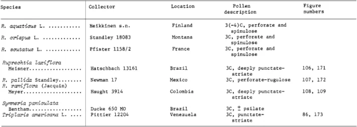
I n R u p r e c h t i a laxiflora (Figure 106), the tectum be- tween the striae is variously upraised, producing a n uneven surface. I n R u p r e c h t i a ramiflora (Figures 108, log), the intergroove tectum is more promi- nently upraised. I n A t r a p h a x i s buxifolia (Figures 98, 99), the connecting striae are larger, tend to be parallel, and result in, at least superficially, a striate- like surface pattern. These distinctions are not as conspicuous in TERI ( A t r a p h a x i s , Figure 143;
R u p r e c h t i a laxiflora, Figure 171; R . pallida, Figure 172), but this, of course, is frequently the case when cross-sections are compared with surfaces having irregular variations.
Within the broad category of punctate ektexines, the following six subtypes with some representative species can be recognized:
1. Ektexine finely punctate: Chorizanthe breweri,
NUMBER 31 9 Eriogonum correllii, and Nemacaulis denudata
(Figure 82).
2. Ektexine with clustered punctae: Centrostegia thurberi, Chorizanthe fimbriata (Figure 84), C. pani- culata (Figure 85), Eriogonum parishii, and E. race- mosum.
3. Ektexine punctate, the punctae connected by small striae: Eriogonum inflatum, E . marifolium (Figures 90, 91), E. thomasii, Mucronea californica (Figures 88, 89), Oxytheca trilobata (Figure 83).
4. Ektexine more prominently punctate-striate:
A n t i g o n u m guatemalense, Calligonum comosum (Figures 94, 95), G y m n o p o d i u m antigonoides (Fig- ures 92, 93), Harfordia macroptera (Figures 96, 97), Muehlenbeckia chilensis (Figures 102, 103), M . poly- botryar, N e o m illspaugh ia panicu Eata, and T r i p laris americana (Figure 86).
5. Ektexine with prominent sunken punctae:
Fagopyrum esculentum (Figure 87), M u e h l e n beckia tamnifolia (Figures 104, 105).
6. Ektexine microreticulate: O x y g o n u m drege- a n u m , 0 . zeyheri, and Podopterus mexicanus (Fig.
ure 132).
T h e subtypes listed above should be treated with reserve since the distinction between consecutive ones is not very great, particularly between subtypes 3 and 4. Also, variation within a sample made classification of some taxa arbitrary or impossible.
Observations of exine wall with T E M indicate that the six subtypes established by SEM have con- siderable structural diversity as well. Sixteen of these taxa were examined in sections: Chorizanthe breweri (Figure 145) and Nemacaulis denudata (Fig- ure 157) of subtype 1, Chorizanthe paniculata (Fig- ure 146) and Eriogonum parishii (Figure 149) of subtype 2, Eriogonum marifolium (Figure 148) and Oxytheca trilobata (Figure 158) of subtype 3, A n tigo n u m guatema lense (Figure 142), Ca 11 igon um comosum (Figure 144), Harfordia macroptera (Fig- ure 152) and Muehlenbeckia polybotryar (Figure 156) of subtype 4, and Muehlenbeckia tamnifolia (Figure 154) and Fagopyrum esculentum (Figure 150) of subtype 5 are all essentially similar in pos- sessing a moderate to thick tectum, long, narrow columellae (Calligonum comosum, Figure 144, ex- cepted), a very reduced (absent?) foot-layer, and a distinguishable endexine. T h e TEM’s of some of the above taxa revealed a n unusual characteristic that may be of phylogenetic significance: Chori-
zanthe spp. (Figures 145, 146), Eriogonum parishii (Figure 149), Nemacaulis (Figure 157), and Oxytheca trilobata (Figure 158) all had a tectum in which the inner face was conspicuously granular.
T w o of these 16 taxa examined by TEM, M u e h - Eenbeckia chilensis (Figure 153) and Podopterus mexicanus (Figure 160), are distinguished from the preceeding ones by a prominently developed foot layer and an endexine well developed only in the region of the aperture. G y m n o p o d i u m (Figure 151) and Triplaris (Figure 173), with a thin foot-layer and endexine, appear intermediate between the above groups.
T h e second most common surface pattern is a reticulum formed by larger and fewer columellae, arranged in a network and distally fused, thus pro- ducing incomplete tectum. Differences in the size and placement of the primary columellae and in the surface of the lumina produce wide varieties and some recognizable pollen types. Grains with this ektexine were 3-colpate (colporate?) (Figures 116, 122), pantocolpate (Figures 112, 118, 120), or pantoporate (Figures 113, 114). I n Polygonurn capi- t a t u m (Figures 122, 123) and P. dielsii (Figures 116, 117) the columellae are massive, and a high, con- spicuous reticulum is the result. T h e lumina are larger in P. dielsii, but in both species they are almost free of any columella material. I n Poly- g o n u m acuminatum (Figure 113) and P. orientale (Figures 114, 115), both pantoporate, the muri alter- nate with large lumina filled with smaller, free columellae. In three pantocolpate taxa, Persicaria coccinea (Figure 112), Polygonurn a m p h i b i u m (Fig- ures 120, 121), and P. virginianum (Figures 118, 119), the columellae which form the reticulum are smaller in length and diameter, closely placed, and a finer reticulum results. I n all three the lumina are filled with small, free columellae.
Thin-sections of Polygonum orientale (Figure 159), P. dielsii (Figure 166), P. a m p h i b i u m (Figures 162, 163), and P. virginianum (Figure 169) empha- size the SEM observations.
T h e formation of the reticulum in the above Polygonaceae is unusual: the ridges or muri are formed by distal fusion along the midline of two rows of columellae, which are opposite or sometimes alternate, producing a “staggered” effect best illus- trated in a “planar” view (Figures 115, 117).
Four species of Polygonum, P . a f i n e (Figure 124),
P. bistorta (Figures 128, 129), P. vaccinifolium (Fig- ures 126, 127) and P. uiviparum (Figure 125), which belong, among others, to the Bistorta complex (Hedberg, 1947), and an Australian species of Muehlenbeckia, M . cunninghamii (Figures 100, 101), have a n ektexine pattern that could be de- scribed as sparsely spinulose and sparsely punctate.
Grains of all five taxa were 3-colporate, the endo- aperture well defined. I n thin-section, Mziehlen- beckia cunninghumii, (Figure 155) is very similar to the other species of Muehlenbeckia examined by T E N (Figures 153, 154, 156).
It is difficult to reconcile the T E M of P. bistorta (Figure 164) with the sparsely spinulose and sparsely punctate ektexine depicted in SEM (Figures 128, 129). T h e lack of a continuous tectum may be due i n part to an oblique section a n d perhaps to the densely packed columellae. T h e SEM’s of P. vac- cinifolium (Figures 126, 127), with the larger per- forations in the tectum, are more compatible with the cross-section illustrated in T E M (Figure 168).
Koenigia islandica, Polygonum cyanandyium (Figures 134, 135), and P. forrestii (Figure 133) had grains that were either pantoporate or pantocolpate with a prominently spinulose ektexine. Comparison of these grains (Figures 133-135) with the common type in the Centrospermae (Figures 1-12) indicates that the above species could not be related paly- nologically to that order. Polygonzim f orrestii (Fig- ure 167) was the most distinctive Polygonum of those examined by T E M . T h e exine consists of a thin endexine and an ektexine containing abun- dant holes or internal foramina. T h e surface of the tectum was composed of very large, solid spines with numerous, small projections i n between.
Hedberg (1947), i n a classic study of pollen morphology in the genus Polygonum, sensu lato, recognized ten pollen types for which he gave descriptions and listed species. H e segregated Poly- gonzim convolvultis (Figures 140, 141), P. cristatunz, and P. dumetorum as slightly aberrant forms of his Tiniaria type. These three species have almost identical pollen grains: 3-colporate with a zonorate endoaperture, the ektexine echinate near the colpi and psilate at the poles and the mesocolpial re- gions. T h e above characteristics make the grains distinct, not only from the more typical Tiniaria pollen, but from the remaining species examined i n the present study. T h i s particular morphology is
paralled by that of Polygonella fimbriata (Figure 130) a n d closely related species (Horton, 1963.
181-183): both groups have grains with similar apertures and a dimorphic ektexine i n which the two surface patterns have the same distribution.
Examination of P. convolvulus in T E M (Figure 165) revealed a remarkably uniform tectum (throughout the psilate area of the ektexine) and foot layer, underlain by a prominent endexine.
T h i s rare combination of well-developed foot layer and endexine was also found in Polygonella (Fig- ure 161), which reinforces the parallel exomor- phologies. Polygonzim cilinode, P. czispidatum, and P. mzilfiporiim, which Hedberg lists as typical Tiniaria, have 3-colporate grains and a uniform ektexine, punctate-striate or prominently punctate.
Four species of R u m e x were examined: R.
aqziaticzis, R . acetosa (Figures 138, 139), R . aceto- sella, and R . sczitafus, all of which had a perforate and very finely and evenly spinulose ektexine (Fig- ure 139). T w o species of Emex had an ektexine (Figure 136) similar to that of Rumex, but with a distinctive aperture structure. T h e ectoaperture is very reduced in length, and its polar margins almost coincide with those of the endoaperture.
T h e close similarity of the ektexine i n E m e x and Rzimex and its restricted occurrence reinforce Dam- mer’s (1893) consecutive placement of the two genera. Comparison of the ektexine of R u m e x (Figure 139) with the common Centrospermae type (Figures 7-12) indicates that there is not a close relationship between the two groups. T h e section of Riimex acetosn (Figure 170) reveals a thin exine in which the columellae and especially the foot- layer-endexine unit are greatly reduced.
T h e ektexine of seven species i n the large New TVorld genus Coccoloba illustrates a continuous variation. ,411 taxa are 3-colporate, and Coccoloba cordafn (Figures 110, 111) has an ektexine best de- scribed as columnar-pyramidal o n a base of ran- domly oriented small rods; C. bm-beyuna has an ektexine similar to Riiprechtia ramifEora (Figures 108, 109); C . diven~ifolin is similar to Calligonzim comosiim (Figures 94, 95); Coccoloba uenosa and C. obovata are prominently punctate; and C. @ri- mensis has a microreticulate ektexine similar to Podopfertis mexicanzis (Figure 132) and Polygonella polygama (Figure 131), b u t with slightly smaller lumina.
NUMBER 97 11
T h e internal morphology of Coccoloba cordata pollen (Figure 147) also appears unique for the family. T h e ektexine surface consists of prominent irregular protuberances supported by a moderateIy thick tectum underlain by notably reduced colu- mellae, greatly thickened foot-layer, and an ex- tremely thin endexine, if any.
T h e genus Lastarriaea has been regarded as con- sisting of two endemic species, L. chilensis Remy in Chile, and L. coriacea (Goodman) Hoover in Baja and Southern California. T h e California taxon has been treated as L. chilensis ssp. californica Gross, or as a species of Chorizanthe, C. coriacea Good- man, who had to select another specific epithet since “californica” was already applied. T h e pollen of Lastarriaea chilensis (Figure 137), L. coriacea, and Chorizanthe species (Figures 84, 85) are all very similar, 3-colporate with a punctate ektexine. This is the most common morphology in the family, however, and the similarity does not necessarily indicate that Lastarriaea is congeneric with Chori- zanthe or is closely related. For the same reason, the close similarity of the pollen in the two species of Lastarriaea does not, a priori, indicate a single species. Careful observation of the floral morphol- ogy when sampling for pollen, however, revealed that the flowers of the plants from Chile and from California are nearly identical in structure. Good- man (1934), in a revision of the North American species of Chorizanthe that included the California taxon, described the genus as having nine stamens, rarely six or three. T h e California and Chilean species of Lastarriaea have only three stamens.
Within limits of the taxa investigated (Table 4), a number of pollen types were found in only one species, and such cases are discussed separately here.
Polygonella fimbriata (Figure 130). Grains 3- colporate, the endoaperture zonorate; the tectum variable: finely punctate in the region of the col- pus, conspicuously reticulate at the mesocolpial ridges and more so at the poles. T h e TEM also indicates that P. fimbriata is distinctive for the family. This conclusion is based on the presence of a well-developed foot-layer and a thickened en- dexine (Figure 161), two characteristices that were rarely combined. See also the earlier discussion of Polygonum convolvulus and allied species.
Polygonella polygama (Figure 131). Grains 3- colporate, the endoaperture zonorate, the nexine
thickened at the margins; the ektexine
-+
micro- reticulate.Comparison of the major subdivisions of the Polygonaceae (Dammer, 1893; Roberty and Vau- tier, 1964; Reveal and Howell, 1976) with pollen morphology reveals little correlation. This lack of correlation is due primarily to two complementary phenomena: the wide distribution of the punctate- striate pollen type, which cuts across subfamily and tribal lines, and the wide array of pollen types within one genus, Polygonum. Thus far, the pollen morphology would support Reveal’s concept (Reveal and Howell, 1976) of the subfamily Eriogonoideae.
Twelve of the 13 genera that he assigned to this subfamily, have been examined by light microscopy and/or SEM, and all are 3-colporate with either punctate or punctate-striate ektexines, the most common pollen type in the Polygonaceae. However, an unusual characteristic illustrated by TEM, a granular inner surface of the tectum, is known thus far to be restricted to the Eriogonoideae: Chori- zanthe (Figures 145, 146); Eriogonum (Figures 148, 149); Nemacaulis (Figure 157); Oxytheca (Figure 158). Another genus in this subfamily, Harfordia (Figure 152), is slightly granular. Examination of additional genera by TEM might well reinforce the value of this characteristic and also the validity of the subfamily.
PRIMULACEAE
FIGURES 174-200
Twenty-nine species (Table 5) representing 22 genera were examined by light microscopy and SEhI, and 9 of these were sectioned and examined by TEM.
T h e structure of the aperture in the Primulaceae is more complex than in the Plumbaginaceae and in the Centrospermae. If the number is three or four, the apertures are generally compound, and the endoaperture is easily seen in light microscopy.
A number of taxa with 3-colporate grains had a bridge over the endoaperture formed by the exten- sion of the two lateral margins of the colpus (Fig- ures 175, 180-184). One species, Lysimachiopsis hillebrandia, had 4-colporate grains in which each side of an endoaperture frequently terminated in a V-shaped process. Sometimes the V from one endo-
aperture and that of the adjacent one formed the outline of a diamond. Grains with 5-8 apertures, e.g., some Primula species, are generally zonocol- pate (Figure 186). I n some taxa, eg., Cortusa m a t t h i o l i L. (Figure 188), the colpi fuse at the poles to form a triangular apocolpial field.
Different surface patterns were due largely to variation in the perforation of the tectum. I n some grains the surface was finely punctate (Lysimachia hy b ri d a , Figure 175; Douglasia m o n t a n a , Figure 182), in others prominently punctate (Coris m o n - speliensis, Figure 174; Cyclamen n e o p o l i t a n u m , Figure 181), irregularly perforate (Naum bergia t h r y s i f i o ~ ~ , Figure 176), or microreticulate (Hot- tonia palustris, Figure 185; Primula veris, Figures 186, 187; O m p h a l o g r a m m a vinciflorum, Figure 191). Still others (Lysimachia terrestris, Figure 177;
Stimpsonia chamaedryoides, Figures 178, 179) might be described as finely reticulate. Additionally, in some grains the tectal perforations were most pro- nounced in the mesocolpial regions (Coris, Figure 174; Nazimbergia, Figure 176; Lysimachia terrestris, Figure 177; Anagallis, Figure 183).
Transmission electron microscopy indicates a variability of exine structure within the family.
Most taxa examined have a well-developed foot- layer and tectum, e.g., Anagallis (Figure 192), Glaztx (Figure 194), Naiimbergia (Figure 196), Onzphalog?-amma (Figure 197), k short columellae, e.g., Glntix (Figure 194), N a u m b e r g i a (Figure 196), and an endexine that is massive in the region of the colpus and thinner but still prominent in the meso- colpial regions, e.g., Anagallis (Figure 192), Glaux (Figure 194), Nazimbergia (Figure 196), Onzphalo- g r a m m a (Figure 197), Stimpsonia (Figure 199). I n Lysimachia hy brida (Figure 195) the irregular tec- tuin, incomplete columellae, and possibly the exten- sive endexine, are due to oblique section. T w o species of Primzila, P. officinalir (Figure 198) and P. ueris (Figure 200), are exceptional, the foot-layer seemingly absent.
Heterostyly and dimorphic pollen grains are known to occur in the Primulaceae. Punt, et el.
(1974) acknowledge this condition in some species of Primiila and in H o t t o n i a palust?-is, as well as dimorpliic pollen grains in T r i e n t a l i s europaea, due apparently to polyploidy. In Glaux m a r i t i m a the collection of Redfield s.n. from Maine was dis- tinct enough from that illustrated by Punt, et al.
(1974, pl. 14: figs. 1, 2) that the identification of Redfield s.n. was rechecked and verified, but still a second sample, H a a k a n a s.n. from Finland, was examined, and these grains were more similar to those illustrated by Punt, et al. (1974). T h e differ- ences in Glaux pollen may be due to heterostyly, not reported in the literature to our knowledge, or the pollen grains of this species may be simply rather variable. T h e r e are also some differences between our illustration of H o t t o n i a palustris (Fig- ure 185) and that of Punt, et al. (1974, pl. 4: figs.
11, 12; pl. 5: figs. 1-6), but the distinctly prolate shape shown in Figure 185 may be an artifact of preparation, i.e., collapse. JVe acknowledge the existence of heterostyly in the Primulaceae and dimorphic pollen (as in the Plumbaginaceae) b u t did not want the present study to be diluted by a detailed examination of this phenomenon. JVhether i t is the long- or short-styled form that is illustrated for Hottonia, Primula, and other Primulaceae, both authors feel strongly that the alternate form will not be shown to have the ektexine characteristic of so many of the centrospermous taxa (Figures 1-12).
For detailed descriptions and measurements, the reader is advised to consult the article by Punt, et al. (1974).
Discussion and Conclusions
Of the three families examined i n this study, the Pollgonaceae is the most frequently considered as being related to the Centrospermae. Of the families that comprise this order, the Caryophyllaceae ap- pears to be the most likely family from which the Po1)gonaceae could be derived or related to, since both have the anthocjanin pigments. T h e data from plastic1 structure, however, does not support a relationship between the Polygonaceae and Caryo- phjllaceae; the Caryoph)llaceae not only have the protein or P-type plastid characteristic of the Centrospermae, but the plastids have a central crys- talloid with a distinctive polygonal shape Behnke, 1976:42); the Polygonaceae have the more common starch or S-t) pe plastid, but admittedlj this char- acteriration is based o n a small number of species.
T h e data presented in this palynological study indicate that the Polygonaceae and Car)ophyllaceae are not related. Although the Polygonaceae is a large and paljnologically diverse family, examina-
NUMBER 37 18
tion of 85 species representing 36 genera (Figures 82-173), revealed no pollen types similar to those in the Caryophyllaceae. (For additional information o n Caryophyllaceae, see Nowicke, 1975; Skvarla and Nowicke, 1976). T h e pollen data do not support a relationship between the subtribe Illecebrinae (Caryophyllaceae) or the larger Illecebraceae and any of the Polygonaceae examined in this study.
T h e grains of Illecebrum verticillatum are almost identical to those of Herniaria glabra (Figure 15):
in both species the pollen has a distinctive tetra- hedal shape with a large aperture on each of the three (four?) faces. Cardionema ramosissima (Figure 16) has pantoporate grains, roughly cube-shaped and with a spinulose and tubuIiferous/punctate ektexine. T h e unusual grains of Illecebrum verti- cillatum and Herniaria glabra have not been found in any other taxa examined to date, including those remaining in the order Centrospermae, as well as the Plumbaginaceae, Polygonaceae, and Primu- laceae. These results would support Hutchinson’s (1959) inclusion of Herniaria in the Illecebraceae, but not necessarily the family status since the re- maining species examined, including another species of Herniaria, H . hirsuta, all have a common panto- porate grain.
An argument could be raised that the pollen of the Bistorta complex in Polygonum (Figures 124- 129) and that of Muehlenbeckia cunninghamii (Fig- ures 100, 101) provide some evidence for a rela- tionship to the Centrospermae. However, we think it much more likely that the occurrence of this ektexine surface in a limited number of species is a reflection of the enormous palynological diversity in the Polygonaceae: ektexines that are 3- psilate, punctate, punctate-striate, conspicuously spinulose, perforate with small spines, microreticulate, finely reticulate to coarsely reticulate; apertures that are porate, pantocolpate or colporate with variable endoapertures; and grains that are probably unique (limited to the Polygonaceae), such as those of Poly- gonella fimbriata (Figure 130) and related species, Polygonum convolvulus (Figures 140, 141) and re- lated species, or Coccoloba cordata (Figures 110, I t is also clear that a diversity of structural forms exist in the Polygonaceae. These observations par- allel the sculpturing pleomorphism illustrated by SEM. T h e T E M reveals wide variation in the de- 11 1).
velopment of two layers-the endexine and foot- layer. T h e endexine can be absent (apparently) as in Podopterus (Figure 160), thin but recognizable as in Chorizanthe paniculata (Figure 146), well de- veloped as in Atraphaxis (Figure 143) and N e m a - caulis (Figure 157), and greatly enhanced as in Eriogonum spp. (Figures 148, 149). T h e foot-layer has a similar range of variation: from apparently absent in Chorizanthe breweri (Figure 145) and Nemacaulis (Figure 157) to prominently thickened in Coccoloba cordata (Figure 147) and Podopterus (Figure 160), with many intermediate stages repre- sented in other taxa.
Another unusual characteristic is the granular inner surface of the tectum found in a number of closely related genera: Chorizanthe spp. (Figures 145, 146), Eriogonzirn (Figure 149), Nemacaulis (Figure 157), and Oxytheca (Figure 158).
While the tectum tends to be thin in the above taxa, in others, Fagopyrum (Figure 150) and Tri- plaris (Figure 173), it is much thicker.
T h e structure of the exine in the Bistorta com- plex of Polygonurn (Figures 164, 168), densely packed columellae, is unique in all the taxa exam- ined with T E M in this study or previously (Skvarla and Nowicke, 1976) and thus diminishes the pos- sibility that the Centrospermae and the Poly- gonaceae could be linked by this group. I n fact, i t seems quite notable that in spite of the diverse exine structures in this family, there is very little similarity to the Centrospermae.
T h e spinulose and punctate ektexine surface found in the Polygonaceae or in the Centrospermae, for that matter, is simple enough to have arisen independently in any of these taxa. However, the significance attached to this ektexine in the Cen- trospermae is that in every betalain family, as well as in the Caryophyllaceae and Molluginaceae, the overwhelming majority of the species examined had pollen grains with a spinulose and tubuliferous,’
punctate ektexine, which, moreover, were some- times identical between families. Insofar as pollen structure is concerned, a more defensible relation- ship could be proposed between the Polygonaceae and any dicot family with a preponderance of punctate ektexines. Pollen grains with a promi- nently reticulate ektexine are widely (and ran- domly?) distributed among dictoyledon families, but this may be due to parallel evolution and does
not necessarily mean that these families are related to each other or to the Polygonaceae.
This study reinforces current opinion (Cronquist, 1968; Philipson, 1975:74; Takhtajan, 1969:220) that the Primulaceae are not related to nor derived from the Centrospermae. According to Cronquist (1968:177, 223) the only special feature in common between the Caryophyllales (Centrospermae) and the Primulales is the free-central placentation. T h e results of pollen analysis indicate that the ancestors a n d / o r relatives of the Primulaceae are not in the centrospermous families. Although the pollen of the Primulaceae exhibits some variation (Figures 174- 200), none of the taxa examined had grains with a counterpart in the Centrospermae.
Unlike the Primulaceae, the Plumbaginaceae are still regarded as being related to the Cen