maharani roxy <[email protected]>
Re: Online Paper Submission
2 pesan
Rohan JCF Corp <[email protected]> 7 Juli 2022 19.50
Kepada: [email protected] Dear Dr. Maharani,
Thank you for submitting your valuable work to our Journal. There is an author fee USD 1000 associated with the accepted articles.
Please confirm and submit your article by using the link below to start the review process.
Link: http://rds.manuscriptmanager.net Kindly get back to us for further queries.
Have a great day.!
Regards, Rohan Reddy RDS
On Thu, Jul 7, 2022 at 3:53 PM Info SBDR <[email protected]> wrote:
---Ursprüngliche Nachricht---
Von: [email protected] [mailto:[email protected]]
Gesendet: Donnerstag, 7. Juli 2022 00:17 An: [email protected]
Betreff: Online Paper Submission
First Name : Maharani Middle Initial : Laillyza Last Name : Apriasari Title : Dr
Institution : University of Lambung Mangkurat Department : Oral Medicine
Address 1 : Veteran street no. 128B Address 2 :
City : Banjarmasin State :
ZIP/Postal Code : 70239 Country : Indonesia Date of Birth (MM/DD/YYYY):
Phone Number :
E-mail : [email protected] Cover Letter:
---
Filename : COVER LETTER-Review of Diabetic Studies.docx Manuscript File(s):
---
File No. 1 : Manuscript- Anti inflamatory DM Neovascular.docx File No. 2 : Figure legend.docx File No. 3 : Table 1.docx
--
This email has been checked for viruses by AVG.
https://www.avg.com
Gmail - Re: Online Paper Submission https://mail.google.com/mail/u/0/?ik=bfd8cef53a&view=pt&search=...
1 of 2 10/10/2022, 11:22 AM
Rohan JCF Corp <[email protected]> 8 Juli 2022 10.45 Kepada: [email protected]
Dear Dr. Maharani,
Thank you for submitting your valuable work to our Journal. There is an author fee USD 1000 associated with the accepted articles.
Please confirm and submit your article by using the link below to start the review process.
Link: http://rds.manuscriptmanager.net Kindly get back to us for further queries.
Have a great day.!
Regards, Rohan Reddy RDS
[Kutipan teks disembunyikan]
Gmail - Re: Online Paper Submission https://mail.google.com/mail/u/0/?ik=bfd8cef53a&view=pt&search=...
2 of 2 10/10/2022, 11:22 AM
maharani roxy <[email protected]>
Follow-Up: Online Paper Submission
1 pesan
Rohan JCF Corp <[email protected]> 8 Juli 2022 20.39
Kepada: [email protected]
On Fri, Jul 8, 2022 at 8:15 AM Rohan JCF Corp <[email protected]> wrote:
Dear Dr. Maharani,
Thank you for submitting your valuable work to our Journal. There is an author fee USD 1000 associated with the accepted articles.
Please confirm and submit your article by using the link below to start the review process.
Link: http://rds.manuscriptmanager.net Kindly get back to us for further queries.
Have a great day.!
Regards, Rohan Reddy RDS
On Thu, Jul 7, 2022 at 5:20 PM Rohan JCF Corp <[email protected]> wrote:
Dear Dr. Maharani,
Thank you for submitting your valuable work to our Journal. There is an author fee USD 1000 associated with the accepted articles.
Please confirm and submit your article by using the link below to start the review process.
Link: http://rds.manuscriptmanager.net Kindly get back to us for further queries.
Have a great day.!
Regards, Rohan Reddy RDS
On Thu, Jul 7, 2022 at 3:53 PM Info SBDR <[email protected]> wrote:
---Ursprüngliche Nachricht---
Von: [email protected] [mailto:[email protected]]
Gesendet: Donnerstag, 7. Juli 2022 00:17 An: [email protected]
Betreff: Online Paper Submission
First Name : Maharani Middle Initial : Laillyza Last Name : Apriasari Title : Dr
Institution : University of Lambung Mangkurat Department : Oral Medicine
Address 1 : Veteran street no. 128B Address 2 :
Gmail - Follow-Up: Online Paper Submission https://mail.google.com/mail/u/0/?ik=bfd8cef53a&view=pt&search=...
1 of 2 10/10/2022, 11:23 AM
City : Banjarmasin State :
ZIP/Postal Code : 70239 Country : Indonesia Date of Birth (MM/DD/YYYY):
Phone Number :
E-mail : [email protected] Cover Letter:
---
Filename : COVER LETTER-Review of Diabetic Studies.docx Manuscript File(s):
---
File No. 1 : Manuscript- Anti inflamatory DM Neovascular.docx File No. 2 : Figure legend.docx File No. 3 : Table 1.docx
--
This email has been checked for viruses by AVG.
https://www.avg.com
Gmail - Follow-Up: Online Paper Submission https://mail.google.com/mail/u/0/?ik=bfd8cef53a&view=pt&search=...
2 of 2 10/10/2022, 11:23 AM
maharani roxy <[email protected]>
RDS-2022-7-2 Manuscript submission confirmation
1 pesan
Review of Diabetic Studies <[email protected]> 9 Juli 2022 05.57 Balas Ke: Rohan Reddy <[email protected]>
Kepada: [email protected]
Manuscript: RDS-2022-7-2 - Anti Inflammatory Effect of Channa micropeltes Extract in Angiogenesis of Diabetes Wound Healing
Authors: Maharani Laillyza Apriasari (Corresponding Author), Dewi Puspitasari (Co-author), Juliyatin Putri Utami (Co-author)
Date submitted: 2022-07-08 Dear Dr. Apriasari
Thank you very much for submitting the above manuscript. Please refer to the manuscript number in all correspondence concerning the manuscript as listed above.
The manuscript will now be forwarded to our Editors and reviewers and we shall inform you as soon as a decision has been made by the editorial board.
The progress of your manuscript can be followed in the progress report that can be accessed from your account overview.
Sincerely,
The Editorial Office
YOUR SIGN IN INFORMATION
Website: https://www.manuscriptmanager.net/rds Email: [email protected]
Forgot password or not signed in before?
Click the URL below to create/reset your password.
https://www.manuscriptmanager.net/sLib/v4/retrieve_pw.php?paramScreen=
EbCodUIbZQ1eMrxls4WJboyyjOPBDdQrMAlq9/SXGTc=
Gmail - RDS-2022-7-2 Manuscript submission confirmation https://mail.google.com/mail/u/0/?ik=bfd8cef53a&view=pt&search=...
1 of 1 10/10/2022, 11:24 AM
maharani roxy <[email protected]>
Manuscript Received and Peer-review Acceptance - RDS-2022-7-2
3 pesan
Rohan JCF Corp <[email protected]> 9 Juli 2022 10.29 Kepada: [email protected], [email protected], [email protected]
Dear Dr. Maharani Laillyza Apriasari,
Thank you for submitting your valuable work to our Journal.
Kindly confirm the publication charges 1000 USD upon the article acceptance. So that we can initiate the review process.
Awaiting your reply.
Regards, Rohan Reddy RDS
On Sat, Jul 9, 2022 at 3:27 AM Review of Diabetic Studies <[email protected]>
wrote:
Manuscript: RDS-2022-7-2 - Anti Inflammatory Effect of Channa micropeltes Extract in Angiogenesis of Diabetes Wound Healing
Authors: Maharani Laillyza Apriasari (Corresponding Author), Dewi Puspitasari (Co- author), Juliyatin Putri Utami (Co-author)
Date submitted: 2022-07-08 Automatic notification:
Dear Rohan Reddy
The above manuscript has been submitted to the Editorial Office. Please login and process it at your earliest convenience.
YOUR SIGN IN INFORMATION
Website: https://www.manuscriptmanager.net/rds Email: [email protected]
Forgot password or not signed in before?
Click the URL below to create/reset your password.
https://www.manuscriptmanager.net/sLib/v4/retrieve_pw.php?paramScreen=
Mj31lVrpFhRlnbWMkdsC7tyz1QhMZg+yD917nU/76kc=
Gmail - Manuscript Received and Peer-review Acceptance - RDS-2022-7-2 https://mail.google.com/mail/u/0/?ik=bfd8cef53a&view=pt&search=...
1 of 2 10/10/2022, 11:27 AM
maharani roxy <[email protected]> 9 Juli 2022 13.01 Kepada: Rohan JCF Corp <[email protected]>
Cc: [email protected], [email protected] Dear Rohan,
Thank you for the information. Yes, i agree to pay it.
Best regards,
Maharani L. Apriasari
[Kutipan teks disembunyikan]
Rohan JCF Corp <[email protected]> 9 Juli 2022 13.06 Kepada: maharani roxy <[email protected]>
Cc: [email protected], [email protected] Dear Dr. Maharani,
Thank you for confirming the publication charges.
Soon, we will get back to you with the review comments.
Regards, Rohan Reddy RDS
[Kutipan teks disembunyikan]
Gmail - Manuscript Received and Peer-review Acceptance - RDS-2022-7-2 https://mail.google.com/mail/u/0/?ik=bfd8cef53a&view=pt&search=...
2 of 2 10/10/2022, 11:27 AM
maharani roxy <[email protected]>
Manuscript Accepted for Publication with Minor Revision - RDS-2022-7-2
1 pesan
Rohan JCF Corp <[email protected]> 12 Juli 2022 10.53 Kepada: maharani roxy <[email protected]>
Cc: [email protected], [email protected] Dear Dr. Maharani,
Greetings,
We have received comments for your article entitled "Anti Inflammatory Effect of Channa micropeltes Extract in Angiogenesis of Diabetes Wound Healing" Our decision is to accept the article with minor revision.
Review comments:
I went through the manuscript and found it to have a very good concept with basic laboratory experimental research, however it needs to be revised based on the comments and
suggestions I put as Review points on the corresponding points in the manuscript.
Please find the attached file. We request you to kindly follow the review suggestions and send us the revised manuscript by 20 July 2022. So, we will forward the article to our English
editing team.
Meanwhile, we request you to confirm your mode of payment 1) Credit Card or 2) Bank Transfer. An invoice can be raised accordingly.
Feel free to contact us for further queries.
Regards, Rohan Reddy RDS
On Sat, Jul 9, 2022 at 10:36 AM Rohan JCF Corp <[email protected]> wrote:
Dear Dr. Maharani,
Thank you for confirming the publication charges.
Soon, we will get back to you with the review comments.
Regards, Rohan Reddy RDS
On Sat, Jul 9, 2022 at 10:31 AM maharani roxy <[email protected]> wrote:
Dear Rohan,
Thank you for the information. Yes, i agree to pay it.
Gmail - Manuscript Accepted for Publication with Minor Revision -... https://mail.google.com/mail/u/0/?ik=bfd8cef53a&view=pt&search=...
1 of 2 10/10/2022, 11:28 AM
Best regards,
Maharani L. Apriasari
Pada tanggal Sab, 9 Jul 2022 10.29, Rohan JCF Corp <[email protected]> menulis:
Dear Dr. Maharani Laillyza Apriasari,
Thank you for submitting your valuable work to our Journal.
Kindly confirm the publication charges 1000 USD upon the article acceptance. So that we can initiate the review process.
Awaiting your reply.
Regards, Rohan Reddy RDS
On Sat, Jul 9, 2022 at 3:27 AM Review of Diabetic Studies
<[email protected]> wrote:
Manuscript: RDS-2022-7-2 - Anti Inflammatory Effect of Channa micropeltes Extract in Angiogenesis of Diabetes Wound Healing
Authors: Maharani Laillyza Apriasari (Corresponding Author), Dewi Puspitasari (Co- author), Juliyatin Putri Utami (Co-author)
Date submitted: 2022-07-08 Automatic notification:
Dear Rohan Reddy
The above manuscript has been submitted to the Editorial Office. Please login and process it at your earliest convenience.
YOUR SIGN IN INFORMATION
Website: https://www.manuscriptmanager.net/rds Email: [email protected]
Forgot password or not signed in before?
Click the URL below to create/reset your password.
https://www.manuscriptmanager.net/sLib/v4/retrieve_pw.php?paramScreen=
Mj31lVrpFhRlnbWMkdsC7tyz1QhMZg+yD917nU/76kc=
Manuscript 7-2 (3).docx 35K
Gmail - Manuscript Accepted for Publication with Minor Revision -... https://mail.google.com/mail/u/0/?ik=bfd8cef53a&view=pt&search=...
2 of 2 10/10/2022, 11:28 AM
1. Introduction
Indonesia ranks sixth among countries with a high prevalence of diabetes mellitus (DM). In an uncontrolled state, DM will result in various oral problems such as xerostomia, candidiasis, stomatitis, gingivitis and periodontitis. The healing of such conditions is often complicated due to hyperglycemia that initiates chronic inflammation [1-3]. Previous study revealed that Channa micropeltes ointment at 20% concentration or Channa striata ointment at 10% concentration applied topically can accelerate wound healing in a DM rat model. Both species are categorized in the same genus and contain albumin as well as omega-6 that acts as an antioxidant and anti- inflammatory [3,4]. As an antioxidant, Channa micropeltes extract elevates superoxide dismutase (SOD) activity and lowers malondialdehid (MDA) level on Day 7 [2]. As an anti- inflammatory, the topical application of Channa micropeltes at 20% concentration on the back skin of a diabetic rat model can increase the number of macrophage and lymphocyte cells on Day 8 and gradually reduce it on Day 14 [1].
Macrophage is the key inflammatory process in wound healing. Reduction in macrophage number at the end of inflammatory stage demonstrates tissue recovery by producing growth factors and cytokines and inducing as well as terminating angiogenesis [5,6]. Macrophage is also produced Nuclear Factor kappa B (NF-κB) that regulates the inflammatory response of metabolic disease such as DM. A state of hyperglycemia in DM will increase reactive oxygen species (ROS) and advanced glycation end products (AGEs) that elevate chronic inflammation through the activation of NF-κB . This will change vascular endothelial growth factor (VEGF) expression that will generate the damage to blood vessels in the angiogenesis process [7,8].
The extract of Channa micropeltes has shown to promote wound healing on the skin of diabetic rat model by reducing macrophage number at the end of inflammatory stage [3]. There has been no study that explores the effect of Channa micropeltes application on NF-κB , VEGF, and neovascular cells that are pivotal components in the DM wound healing process. Based on that, a study to analyze the expression of NF-κB , VEGF and neovascular cells number at the inflammatory stage of the DM wound healing process was warranted.
2. Methods
This study was an experimental laboratory research study incorporating a post-test only control
group design. It was approved by Ethical Clearance Committee, Faculty of Dentistry, Universitas
Lambung Mangkurat, Banjarmasin, South Kalimantan, Indonesia, with number 111/KEPKG- FKGULM/EC/III/2020.
2.1 Manufacturing Channa micropeltes and Channa striata extract
Preparing both Channa micropeltes and Channa striata extract used fresh fish weighing 600- 1000 grams. Each extract was later steamed in a pan for 25-35 minutes at a temperature of 60
oCelsius. The flesh was enclosed with flannelette and pressed in a hydraulic device. Furthermore, Channa micropeltes and Channa striata were centrifuged for 15 minutes at a speed of 6000 rpm.
Each extract was kept inside a dark glass bottle and then covered with aluminum foil and clean pack.
2.2 Formulation of Channa micropeltes and Channa striata ointment
Adeps lanae (Asian chemicals, Semarang) in a weight of 16.875 grams and vaselin flavum (PT.
Brataco, 1295578) in a weight of 23.125 grams were used in the formulation of the Channa micropeltes ointment. Meanwhile, a combination of 16.875 grams adeps lanae and 28.125 grams vaselin flavum were combined in the formulation of Channa striata ointment. Adeps lanae was initially poured into different tubes for each extract and later added gradually with either Channa micropeltes at 20% concentration or Channa striata at 10% concentration. After the extract was fully absorbed by adeps lanae, the mixture was then mashed to obtain a homogenous consistency. Subsequently, the composition was further mixed with vaselin flavum and mashed again until homogenous.
2.3 In vivo study
This study included 2-3 months old male Wistar (Rattus novergicus) rats (weight, 250-300
grams) obtained from an animal laboratory at the Faculty of Medicine, University of Lambung
Mangkurat. The 24 rat specimens were kept in cages, and the temperature and humidity were set
within ±25 °C and 60%, respectively. They were fed standard BR-II, and they had access to
boiled water ad libitum. Rats with hyperglycemia were obtained by injecting streptozotocin
(STZ) at 35 mg/kg dosage until the blood glucose level was over 126 mg/dL-1; non-diabetic rats
were ones without intervention. All animals were divided into 3 treatment groups consisting of
20% Channa micropeltes extract ointment, 10% Channa striata extract ointment, and placebo
ointment as control. Each substance was applied topically 3 times daily (every 6-8 hours). An incisional wound was made on the back of the rats with 1 cm length and 1 mm depth using sterile scalpel under inhaled anesthesia of 5 ml diethyl ether.
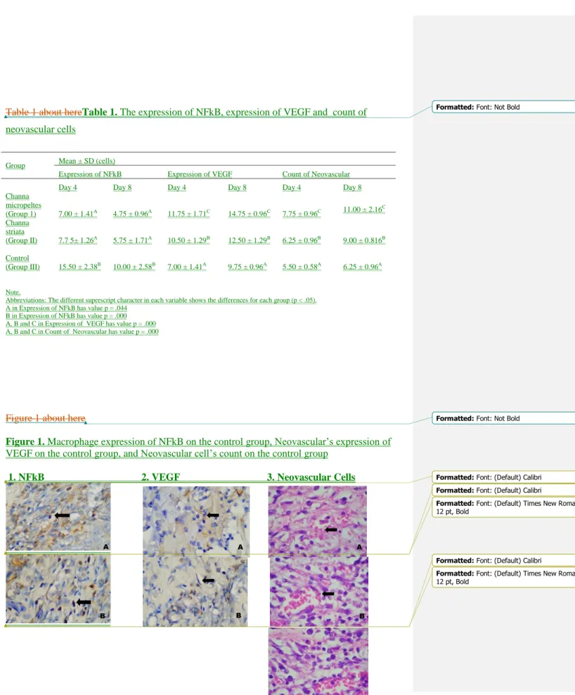
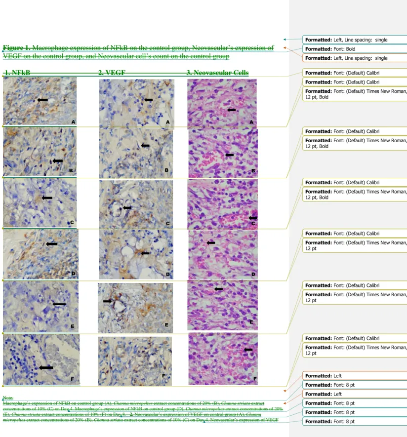
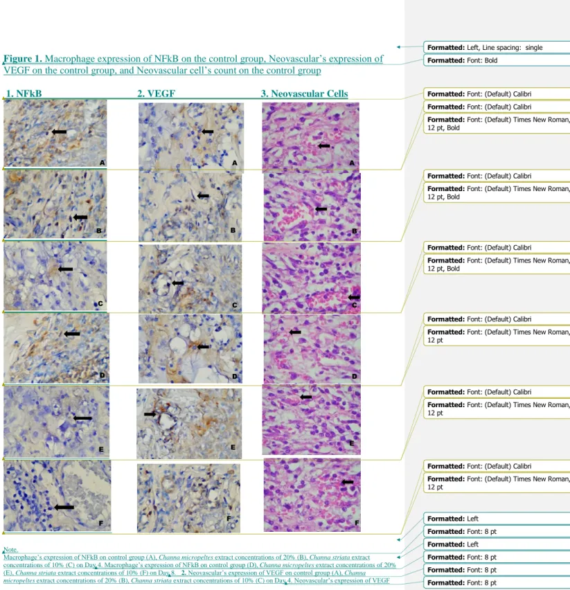
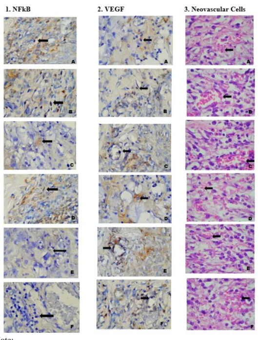
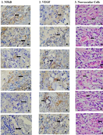
After the 4th and 8th day of application, rats were euthanized by inhaling a lethal dosage of diethyl ether. The back skin was then biopsied for histopathology examination using hematoxylin eosin (HE) to evaluate macrophages and neovascular cells, and immunohistochemistry (IHC) to evaluate NF-κB and VEGF. The number of macrophages and neovascular cells were calculated in 3 different field locations using a light microscope (Olympus, WA) at 400 magnifications and subsequently calculated for its average. IHC staining was performed using anti-mouse NF-kB monoclonal antibody (Santa Cruz Biotechnology Inc, Santa Cruz, CA, NF-kB p65 (F-6): sc 8008) and anti-mouse VEGF monoclonal antibody (Santa Cruz Biotechnology Inc, Santa Cruz, CA, VEGF (C1): sc 7269). Positivity NF-κB expression was defined as only distinct nuclear immunostaining, which is considered as activated NF-κB in the studied field at 100 magnifications.
2.4 Data analysis
The results were analyzed using a 2-way analysis of variance parametric test based on the Shapiro-Wilk normality test and Levene’s variance homogeneity test. The results showed normal data distribution and homogenous data variances. Consequently, further analysis by means of a post hoc Bonferroni test was conducted with a statistical significance of p < 0.05.
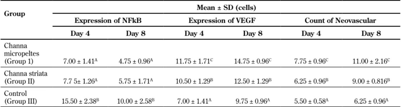
3. Results
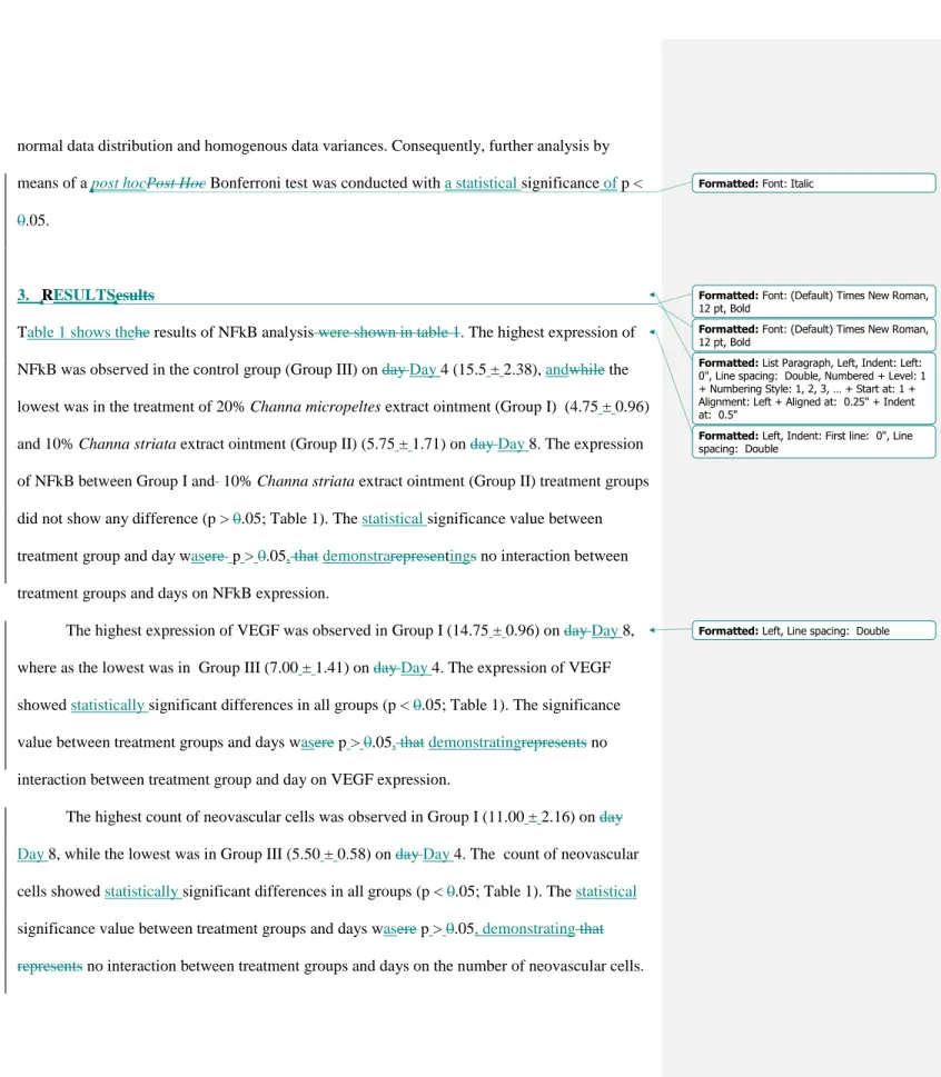
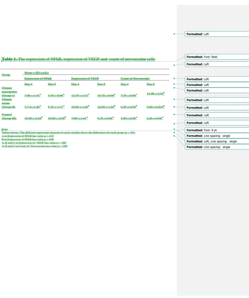
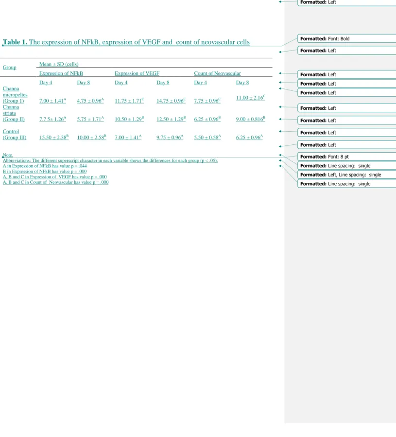
Table 1 shows the results of NF-κB analysis. The highest expression of NF-κB was observed in the control group (Group III) on Day 4 (15.5 ± 2.38), and the lowest was in the treatment of 20%
Channa micropeltes extract ointment (Group I) (4.75 ± 0.96) and 10% Channa striata extract ointment (Group II) (5.75 ± 1.71) on Day 8. The expression of NF-κB between Group I and 10% Channa striata extract ointment (Group II) treatment groups did not show any difference (p
> .05; Table 1). The statistical significance value between treatment group and day was p > 0.05, demonstrating no interaction between treatment groups and days on NF-κB expression.
The highest expression of VEGF was observed in Group I (14.75 ± 0.96) on Day 8, whereas the
lowest was in Group III (7.00 ± 1.41) on Day 4. The expression of VEGF showed statistically
significant differences in all groups (p < 0.05; Table 1). The significance value between treatment groups and days was p > 0.05, demonstrating no interaction between treatment group and day on VEGF expression.
The highest count of neovascular cells was observed in Group I (11.00 ± 2.16) on Day 8, while the lowest was in Group III (5.50 ± 0.58) on Day 4. The count of neovascular cells showed statistically significant differences in all groups (p < 0.05; Table 1). The statistical significance value between treatment groups and days was p > 0.05, demonstrating no interaction between treatment groups and days on the number of neovascular cells.
4. Discussion
DM is characterized with an increase in blood glucose level that induces glycation reaction. This process will result in amadory production to formulate toxic proteins (AGEs). Interaction between AGEs and a receptor advanced glycation end product (RAGE) will increase the signal for nicotinamide adenine dinucleotide phosphate (NADPH) oxidase which produces superoxide anion. This process elevates the production of reactive oxygen species (ROS) which are the key for molecular signaling as well as the development of inflammatory disorders such as DM.
Excessive production of ROS will complicate the healing process of wounds in DM [2,9].
Channa micropeltes contains albumin and omega-6 fatty acid. Albumin can decrease ROS by cutting chained oxidative reaction in the ROS formation’s process. Albumin can bind metal ions and also catch oxygen that processing hydrogen peroxide into non radical compound. Omega-6 fatty acid, especially arachnodic acid are the keys to anti-inflammatory processes. It plays the role in stimulating macrophages to release growth factors, such as VEGF. Arachidonic acid will be metabolized through an enzymatic mechanism such as the 5-lipoxygenase and cyclo- oxygenase pathways that produce leukotrienes, prostaglandins, and thromboxane A2. These can stimulate cell migration and new local vascularization in the wound healing process of DM [4,10].
In previous studies, Channa micropeltes extract ointment at 20% concentration and Channa
striata extract ointment at 10% concentration were shown to promote the wound healing process
in DM. Channa micropeltes and Channa striata are categorized in the same genus. Both species
contain albumin, the secondary antioxidant that can bind metal ion in ROS formation [1,2]. ROS
induces an inflammatory response through the activation of Nuclear Factor kappa B (NF-κB ).
NF-κB signal is the main key of chronic inflammation in DM [7,8]. This is demonstrated by the study result on Day 4 that reveals the highest expression of NF-κB in control group, while the result for 20% Channa micropeltes extract application was comparable to 10% Channa striata extract application.
Our study result on Day 8 demonstrates the reduction of NF-κB expression in both Channa micropeltes extract ointment at 20% concentration and Channa striata extract ointment at 10%
concentration when compared to the control. Topical application of Channa micropeltes extract at 20% concentration or Channa striata extract at 10% concentration may reduce excessive ROS, thereby suppressing NF-κB expression. Both extracts possess potential as natural substances that may inhibit the expression of NF-κB . Previous studies reveal that the impediment of pro- inflammatory NF-κB from therapeutical application of several natural and synthetic ingredients will be a good target to manage vascular complication in DM [7]. A prolonged inflammatory response can be resolved by the inhibition of NF-κB. As an anti-inflammatory substance, Channa micropeltes will reduce inducible nitric oxide synthase (iNOS) and cyclooxygenase 2 (COX2) that suppress the NF-κB gene regulator. This will prevent prolonged inflammation in the wound healing process of DM [11,12].
Excessive activation of NF-κB will cause abnormal DNA transcription which includes various gene expression of vascular complications occurring in VEGF, Platelet Derived Growth Factor (PDGF), Endothelin-1 (ET-1) and Transforming Growth Factor beta (TGF-β) that cause vascular cell damage [7]. VEGF as pro-angiogenic modulators encounter down regulation in DM, disturbing the angiogenesis process [13,14]. Prior studies demonstrate Vascular Endothelial Growth Factor A (VEGF-A) protein and messenger ribonucleic acid (mRNA) level in the wound of a diabetic rat model which shows reduction when compared to the group with a normal wound. DM leads to the decrease of angiogenesis in wound healing, so that it lessens vascular and capillary density [13].
The angiogenic effect is initiated by VEGF-A binding to Vascular Endothelial Growth Factor
Receptor-2 (VEGFR-2). Angiogenesis stimulation through PI3K-Akt-eNOS will cause
endothelial cell to migrate, proliferate, and differentiate. Molecular signals will be commenced
by phosphatidylinositol 3 kinase from serine/threonine kinase Akt/protein kinase B. Akt/PKB
through the phosphorylation of endothelial nitric oxide synthesis on Ser 1177, and will stimulate
NO production, vasodilatation, and endothelial cell migration [15,16]. This result presents that
Channa micropeltes extract ointment at 20% concentration increases the highest expression of VEGF when compared to Channa striata extract ointment at 10% concentration or the control on Day 8. This exhibits the potential of Channa micropeltes extract ointment at 20% concentration to promote angiogenesis on the DM wound healing process.
Previous study by Carabelly et al. [1] reveals that the application of Channa micropeltes extract ointment at 20% concentration may elevate macrophage number on Day 8 and reduce them on Day 14. Macrophage regulates angiogenesis signals in neovascular along the formation of granulation tissue process [17]. This concept is in accordance with this study as the number of macrophages was observed the highest on Day 8 and followed by the increase of neovascular cell number by the application of Channa micropeltes extract ointment at 20% concentration on Day 8. The highest number of neovascular cells also was observed in the application of Channa micropeltes extract ointment at 20% concentration when compared to Channa striata extract ointment at 10% concentration or the control in Day 8. This study did not continue to further days to limit the parameters.
We can conclude that the application of Channa micropeltes extract ointment at 20%
concentration on the wound of a diabetic rat model can reduce the expression of NF-κB. The anti-inflammatory effect of Channa micropeltes can elevate the expression of VEGF and the number of neovascular cells in angiogenesis process of diabetic wound healing. Our findings require further research using different parameters, adding time of evaluation, and using larger experimental animals.
Acknowledgements: This study was financed by the Faculty of Dentistry, University of Lambung Mangkurat. The authors express their gratitude.
Conflicts of interest statement: The authors have no conflicts of interest to report.
Ethical approval: Approval for this study was provided by the Ethical Clearance Committee,
Faculty of Dentistry, Universitas Lambung Mangkurat, Banjarmasin, South Kalimantan,
Indonesia (#111/KEPKG-FKGULM/EC/III/2020).
maharani roxy <[email protected]>
Re: Follow-Up: Manuscript Accepted for Publication with Minor Revision - RDS-2022-7-2
3 pesan
Rohan JCF Corp <[email protected]> 12 Juli 2022 21.20
Kepada: maharani roxy <[email protected]>
Cc: [email protected], [email protected]
On Tue, Jul 12, 2022 at 8:23 AM Rohan JCF Corp <[email protected]> wrote:
Dear Dr. Maharani, Greetings,
We have received comments for your article entitled "Anti Inflammatory Effect of Channa micropeltes Extract in Angiogenesis of Diabetes Wound Healing" Our decision is to accept the article with minor revision.
Review comments:
I went through the manuscript and found it to have a very good concept with basic laboratory experimental research, however it needs to be revised based on the comments and suggestions I put as Review points on the corresponding points in the manuscript.
Please find the attached file. We request you to kindly follow the review suggestions and send us the revised manuscript by 20 July 2022. So, we will forward the article to our English editing team.
Meanwhile, we request you to confirm your mode of payment 1) Credit Card or 2) Bank Transfer. An invoice can be raised accordingly.
Feel free to contact us for further queries.
Regards, Rohan Reddy RDS
On Sat, Jul 9, 2022 at 10:36 AM Rohan JCF Corp <[email protected]> wrote:
Dear Dr. Maharani,
Thank you for confirming the publication charges.
Soon, we will get back to you with the review comments.
Regards, Rohan Reddy RDS
On Sat, Jul 9, 2022 at 10:31 AM maharani roxy <[email protected]> wrote:
Dear Rohan,
Thank you for the information. Yes, i agree to pay it.
Best regards, Maharani L. Apriasari
Pada tanggal Sab, 9 Jul 2022 10.29, Rohan JCF Corp <[email protected]> menulis:
Dear Dr. Maharani Laillyza Apriasari,
Thank you for submitting your valuable work to our Journal.
Kindly confirm the publication charges 1000 USD upon the article acceptance. So that we can initiate the review process.
Awaiting your reply.
Regards, Rohan Reddy RDS
Gmail - Re: Follow-Up: Manuscript Accepted for Publication with Mi... https://mail.google.com/mail/u/0/?ik=bfd8cef53a&view=pt&search=...
1 of 2 10/10/2022, 11:29 AM
On Sat, Jul 9, 2022 at 3:27 AM Review of Diabetic Studies <[email protected]> wrote:
Manuscript: RDS-2022-7-2 - Anti Inflammatory Effect of Channa micropeltes Extract in Angiogenesis of Diabetes Wound Healing
Authors: Maharani Laillyza Apriasari (Corresponding Author), Dewi Puspitasari (Co-author), Juliyatin Putri Utami (Co-author)
Date submitted: 2022-07-08 Automatic notification:
Dear Rohan Reddy
The above manuscript has been submitted to the Editorial Office. Please login and process it at your earliest convenience.
YOUR SIGN IN INFORMATION
Website: https://www.manuscriptmanager.net/rds Email: [email protected]
Forgot password or not signed in before?
Click the URL below to create/reset your password.
https://www.manuscriptmanager.net/sLib/v4/retrieve_pw.php?paramScreen=Mj31lVrpFhRlnbWMkdsC7tyz1QhMZg +yD917nU/76kc=
maharani roxy <[email protected]> 13 Juli 2022 08.05
Kepada: Rohan JCF Corp <[email protected]>
Dear Rohan Reddy,
Thank you for the fast reponse. I have revised the manuscript, and i hope it is in accordance to the reviewer. i will pay by Bank Transfer.
Best Regards,
Maharani Laillyza Apriasari
[Kutipan teks disembunyikan]
Manuscript 7-2 (3)-Revised.docx 41K
Rohan JCF Corp <[email protected]> 13 Juli 2022 11.34
Kepada: maharani roxy <[email protected]>
Dear Dr. Maharani, Greetings,
Thank you for sending us the revised manuscript.
Please find the attached wire transfer invoice. We request you to kindly process the payment by 18 July 2022, and send us the payment receipt for our reference. So, we can proceed further.
Awaiting your reply.
Regards, Rohan Reddy RDS
[Kutipan teks disembunyikan]
RDS-7-2 Wire Invoice.pdf 71K
Gmail - Re: Follow-Up: Manuscript Accepted for Publication with Mi... https://mail.google.com/mail/u/0/?ik=bfd8cef53a&view=pt&search=...
2 of 2 10/10/2022, 11:29 AM
ABSTRACT
Background: Channa micropeltes extract contains albumin and Omega-6 which possess anti- oxidant and anti-inflammatory agent that can promote macrophages in wound healing process of diabetes mellitus (DM). Objective: to analyze Nuclear Factor kappa B (NFkB) and Vascular Endothelial Growth Factor (VEGF) expression as well as neovascular cells in the inflammatory stage of DM wound healing. Materials and methods: The twenty-four males Rattus novergicus were divided into three groups that were 20% Channa micropeltes ointment (Group I), 10%
Channa striata extract ointment (Group II), and placebo ointment as control (Group III).
Ointments were applied three times daily. Results: The highest expression of NFkB was observed in Group III on day 4 (15.50 ± 2.38), while the lowest was in treatment of Group I and Group II on day 8 (4.75±0.96). The highest expression of VEGF was observed in Group I on day 8(14.75±0.96), while the lowest was Group III on day 4 (7.00±1.41). The highest count of neovascular cells was observed in Group I on day 8 (11.00±2.16), while the lowest was in Group III on day 4 (5.50±0.58). Conclusion: Channa micropeltes have an anti-inflammatory effect by regulating NFkB expression and elevating VEGF expression in angiogenesis process of DM wound healing.
Keywords: Channa micropeltes, nfkb, vegf, neovascular, wound healing, diabetes mellitus
Introduction
Indonesia is ranked sixth among the countries with high prevalence of Diabetes mellitus in the world. In uncontrolled state, Diabetes mellitus will result in the variance of oral problems such as xerostomia, candidiasis, stomatitis, gingivitis and periodontitis. The healing of such conditions is often complicated due to hyperglycemia that initiates chronic inflammation.1,2,3 Previous study revealed that Channa micropeltes ointment at 20% concentration or Channa striata ointment at 10% concentration that was applied topically can accelerate wound healing in diabetes mellitus rat model. Both species are categorized in the same genus and contain albumin as well as omega 6 that act as anti-oxidant and anti-inflammation.3,4 As antioxidant, Channa micropeltes extract elevate Superoxide dismutase (SOD) activity and lower Malondialdehid (MDA) level on day 7.2 As anti-inflammation, the topical application of Channa micropeltes at
20% concentration on the back skin of diabetic rat model could increase the number of macrophage and lymphocyte cells on day 8 and gradually reduce it on day 14.1
Macrophage is the key of inflammatory process in wound healing. Reduction in macrophage number at the end of inflammatory stage demonstrates tissue recovery by producing growth factors, cytokines, and induce as well as terminate angiogenesis.5,6 Macrophage is also produced Nuclear Factor kappa B (NFkB) that regulates inflammatory response of metabolic disease such as diabetes mellitus. Hyperglycemia state in diabetes mellitus will increase Reactive Oxygen Species (ROS) and Advanced Glycation End Products (AGEs) that elevate chronic inflammation through the activation of NFkB. This will change Vascular Endothelial Growth Factor (VEGF) expression that will generate the damage of blood vessel cell in angiogenesis process.7,8
The extract of Channa micropeltes has been proven to promote wound healing on the skin of diabetic rat model by reducing macrophage number at the end of inflammatory stage.3 There has been no study that explore the effect of Channa micropeltes application on NFkB, VEGF, and neovascular cells that are pivotal components in diabetes mellitus wound healing process. Based on that, a study to analyze the expression of NFkB, VEGF and neovascular cells number at the inflammatory stage of diabetes mellitus wound healing process should be conducted.
Materials and Methods
This study was an experimental laboratory research incorporating post-test only control group design. It was approved by Ethical Clearence Commitee, Faculty of Dentistry, Universitas Lambung Mangkurat, Banjarmasin, South Kalimantan, Indonesia, with number 111/KEPKG- FKGULM/EC/III/2020.
Manufacturing Channa micropeltes and Channa striata extract
Preparing both Channa micropeltes and Channa striata extract used fresh fish weighed 600-1000 g. Each of it was later steamed in a pan for 25-35 minutes under 60o temperature. The flesh were enclosed with flannelette and pressed in hydraulic device. Further, Channa micropeltes and Channa striata were centrifuged for 15 minutes within 6000 rpm speed. Each extract was kept inside dark glass bottle and then covered with aluminum foil and clean pack.
Formulation of Channa micropeltes and Channa striata ointment
Adeps lanae (Asian chemicals, Semarang) in a weight of 16.875 g and vaselin flavum (PT.
Brataco, 1295578) in a weight of 23.125 g were used in the formulation of Channa micropeltes ointment. Meanwhile, a combination of 16.875 g adeps lanae and 28.125 g vaselin flavum were utilized in the formulation of Channa striata ointment. Adeps lanae was initially poured into different tubes for each extract and later added gradually with either Channa micropeltes at 20%
concentration or Channa striata at 10% concentration. After the extract was fully absorbed by adeps lanae, the mixture was then mashed to obtain homogenous consistency. Subsequently, the composition was further mixed with vaselin flavum and mashed again until homogenous.
In Vivo Study
This study included 2-3 months-old male Wistar (Rattus novergicus) rats (weight, 250–
300 g) obtained from animal laboratory, Faculty of Medicine, University of Lambung Mangkurat. The total of 24 rats were kept in cages, and the temperature and humidity were set within ±25 °C and 60%, respectively. They were fed standard BR-II, and they had access to boil water ad libitum. Rats with hyperglycemia was obtained by injecting Streptozotosin (STZ) at 35 mg/kg dosage until the blood glucose level was over 126 mg/dL-1, while non Diabetic Rats is rats without intervention. All animals were divided into 3 treatment groups consisting of 20%
Channa micropeltes extract ointment, 10% Channa striata extract ointment, and placebo ointment as control. Each substance was applied topically three times daily (every 6-8 hours).
Incisional wound was made on the back of the rats with 1 cm length and 1 mm depth using sterile scalpel under inhaled anesthesia of 5 ml diethyl ether.
After the 4th and 8th day of application, rats were euthanized by inhaling lethal dosage of diethyl ether. The back skin was then biopsied for histopathology examination using Haematoxyllin Eosin (HE) to evaluate macrophages and neovascular cells, and Immunohistochemistry (IHC) to evaluate NF-κB and VEGF. The number of macrophages and neovascular cells were calculated in three different field locations using a light microscope that were counted using light microscope (Olympus, United States) at 400 magnifications and subsequently calculated for its average. Immunohistochemistry (IHC) staining was performed using anti-mouse NF-kB monoclonal antibody (Santa Cruz
Comment [Ma1]: Both products to mentioned trade mark, source and date if related
Biotechnology Inc. NF-kB p65 (F-6) : sc 8008) and anti-mouse VEGF monoclonal antibody (Santa Cruz Biotechnology Inc. VEGF (C1) : sc 7269). Positivity NF-κB expression was defined as only distinct nuclear immunostaining, which is considered as activated NF-κB in the studied field at100 magnification.
Statistical Analysis
The results were analyzed using two-way analysis of variance parametric test based on Shapiro Wilk normality test and Levene’s variance homogeneity test. The results showed normal data distribution and homogenous data variances. Consequently, further analysis by means of a Post Hoc Bonferroni test was conducted with significance p < 0.05.
Results
The results of NFkB analysis were shown in table 1. The highest expression of NFkB was observed in the control group (Group III) on day 4 (15.5±2.38), while the lowest was in the treatment of 20% Channa micropeltes extract ointment (Group I) (4.75±0.96) and 10% Channa striata extract ointment (Group II) (5.75±1.71) on day 8. The expression of NFkB between Group I and 10% Channa striata extract ointment (Group II) treatment groups did not show any difference (p > 0.05; Table 1). The significance value between treatment group and day were p>0.05 that represents no interaction between treatment groups and days on NFkB expression.
The highest expression of VEGF was observed in Group I (14.75±0.96) on day 8, where as the lowest was in Group III (7.00±1.41) on day 4. The expression of VEGF showed significant differences in all groups (p < 0.05; Table 1). The significance value between treatment groups and days were p>0.05 that represents no interaction between treatment group and day on VEGF expression.
The highest count of neovascular cells was observed in Group I (11.00±2.16) on day 8, while the lowest was in Group III (5.50±0.58) on day 4. The count of neovascular cells showed significant differences in all groups (p < 0.05; Table 1). The significance value between treatment groups and days were p>0.05 that represents no interaction between treatment groups and days on the number of neovascular cells.
Discussion
Diabetes mellitus is characterized with an increase in blood glucose level that induces glication reaction. This process will result in amadory production to formulate toxic protein called Advanced Glication End Products (AGEs). Interaction between AGE and Receptor Advanced Glication End Product (RAGE) will increase the signal for nicotinamide adenine dinucleotide phosphate (NADPH) oxidase which produces superoxide anion. This process elevates the production of Reactive Oxygen Species (ROS) which are the key for molecular signaling as well as the development of inflammatory disorders such as diabetes mellitus.
Excessive production of ROS will complicate the healing process of wound in diabetes mellitus.2,9
Channa micropeltes contains albumin and omega-6 fatty acid. Albumin can decrease ROS by cutting chained oxidative reaction in the ROS formation’s process. Albumin can bind metal ions and also catch oxygen that processing hydrogen peroxide into non radical compound.
Omega-6 fatty acid, especially arachnodic acid are the key of anti inflammatory. It plays the role in stimulating machropages to release growth factors, such as VEGF. Arachinodic acid will be metabolized through an enzymatic mechanism such as the 5-lipoxygenase and cyclo-oxygenase pathways that produce leukotrienes, prostaglandins, and thromboxane A2. These can stimulates the cell migration and new local vascularization in wound healing process of diabetes mellitus.4,10
In previous studies, Channa micropeltes extract oinment at 20% concentration and Channa striata extract ointment at 10% concentration were proven to promote wound healing process in diabetes mellitus. Channa micropeltes and Channa striata are categorized in same genus. Both species contain albumin, the secondary antioxidant that can bind metal ion in ROS formation.1,2 ROS induce inflammatory response through the activation of Nuclear Factor kappa B (NFkB). NFkB signal is the main key of chronic inflammation in diabetes mellitus.7,8 This is proven by this study result on Day 4 that reveals the highest expression of NFkB in control group, while the result for 20% Channa micropeltes extract application was comparable to 10%
Channa striata extract application.
Study result on day 8 demonstrates the reduction of NFkB expression in both Channa micropeltes extract ointment at 20% concentration and Channa striata extract ointment at 10%
concentration when compared to control. Topical application of Channa micropeltes extract at 20% concentration or Channa striata extract at 10% concentration may reduce excessive ROS,
thus suppressing NFkB expression. Both extract possess potential as natural substance that may inhibit the expression of NFkB. Previous study reveals that the impediment of proinflammatory NFkB from therapeutical application of several natural and synthetic ingredients will be a good target to manage vascular complication in diabetes mellitus.7Prolonged inflammatory response can be resolved by the inhibition of NFkB. As anti-inflammatory substance, Channa micropeltes will reduce inducible Nitric Oxide Synthase (iNOS) and Cyclooxygenase 2 (COX2) that suppress NFkB gene regulator. This will prevent prolonged inflammation in wound healing process of diabetes mellitus.11,12
Excessive activation of NFkB will cause abnormal DNA transcription which include various gene expression of vascular complication occurring in VEGF, Platelet Derived Growth Factor (PDGF), Endothelin-1 (ET-1) and Transforming Growth Factor beta (TGF-b) that cause vascular cell damage.7 VEGF as pro-angiogenic modulators encounter down regulation in diabetes mellitus, thus disturbing the angiogenesis process.13,14 Prior study demonstrates Vascular Endothelial Growth Factor A (VEGF-A) protein and messenger Ribonucleic Acid (mRNA) level in the wound of diabetic rat model which shows reduction when compared to the group with normal wound. Diabetes mellitus leads to the decrease of angiogenesis in wound healing, so that it lessens vascular and capillary density.13
The angiogenic effect is initiated by VEGF-A binding to Vascular Endothelial Growth Factor Receptor-2 (VEGFR-2). Angiogenesis stimulation through PI3K-Akt-eNOS will cause endothelial cell to migrate, proliferate and differentiate. Molecular signal will be commenced by Phosphatidylinositol 3 kinase from serine/threonine kinase Akt/protein kinase B. Akt/PKB through the phosphorylation of endothelial Nitric Oxide synthesis on Ser 1177 will stimulate NO production, vasodilatation, and endothelial cell migration.15,16 This result presents that Channa micropeltes extract ointment at 20% concentration increase the highest expression of VEGF when compared to Channa striata extract ointment at 10% concentration or control on day 8.
This exhibits the potential of Channa micropeltes extract ointment at 20% concentration to promote angiogenesis on diabetes mellitus wound healing process.
Previous study by Carabelly (2019) reveals that the application of Channa micropeltes extract ointment at 20% concentration may elevate macrophage number on Day 8 and reduce them on Day 14.1 Macrophage regulate angiogenesis signal in neovascular along the formation of granulation tissue process.17 This concept is in accordance with this study as the number of
macrophage was observed the highest on Day 8 and followed by the increase of neovascular cell number by the application of Channa micropeltes extract ointment at 20% concentration on Day 8. The highest number of neovascular cell was also observed in the application of Channa micropeltes extract ointment at 20% concentration when compared to Channa striata extract ointment at 10% concentration or control in Day 8. This study did not continue to further day to limit the parameters.
It can be concluded that the application of Channa micropeltes extract ointment at 20%
concentration on the wound of diabetic rat model can reduce the expression of NFkB. Anti- inflammatory effect of Channa micropeltes can elevate the expression of VEGF and the number of neovascular cells in angiogenesis process of diabetic wound healing. This study requires further research using different parameters, adding time of evaluation, and using larger experimental animals.
Acknowledgements
The authors thank to Faculty of Dentistry, University of Lambung Mangkurat which have supported this work.
Funding Information
This study was financed by Faculty of Dentistry, University of Lambung Mangkurat which have supported this work.
Conflicts of Interest statement
The authors of the work have no conflict of interest.
References
1. Carabelly A.N, Firdaus I.W.A.K, Nurmardina P.C, Putri D.A, Apriasari M.L. The Effect of Topical Toman Fish ( Channa micropeltes) Extract on Macrophages and Lymphocytes in Diabetes Mellitus Wound Healing. The 1st International Seminar on Smart Molecule of Natural Resources. Journal of Physics : Conference Series, 2019 : p. 1-9
Comment [Ma2]: You have to mention the Limitations of the study.
Comment [Ma3]: The conclusion should not be generalised, you mention only your findings and recommendation for further studies to explore more significant findings in different subjects and larger number.
2. Apriasari M.L, Ainah Y, Febrianty E, Carabelly A.N. Antioxidant Effect of Channa Micropeltes in Diabetic Wound of Oral Mucosa. International Journal of Pharmacology, 2019 ; 15 (1) : 137-143
3. Apriasari M.L, Puspitasari D. Effect of Channa micropeltes for increasing Lymphocyte and Fibroblast Cells in diabetic Wound Healing. Journal of Medical Sciences, 2018 ; 18 (4) : 205-210
4. Apriasari M.L, Syahadati M.A, Carabelly A.N. Clinical Analysis of Channa Micropeltes For Treating Wound of Diabetes Mellitus. Berkala Kedokteran, 2020 ; 16 (1) : 1-10 5. Brancato S.K, Albina J.E. Mini Review : Wound Macrophages as Key Regulators of
Repair Origin, Phenotype, and Function. The American Journal of Pathology, 2011 ; 178 (1) : 19-25
6. Koh T.J, DiPietro L.A. Inflammation and Wound Healing : The role of the macrophage.
Expert Rev Mol Med; 13 (e23) : 1-14
7. Kulkarni Y.A, Suryavanshi S.V. NFkB : Apotential Target in the Management of Vascular Complications of Diabetes. Frontiers in Pharmacology, 2017 ; 8 : 1-12
8. Baker R.G, Hayden M.S, Ghosh S. NFkB, inflammation and metabolic disease. Cell Metab, 2011 Januar 5 ; 13 (1) : 11-22
9. Mittal M, Siddiqui M.R, Tran K, Reddy S.P, Malik A.B. Reactive Oxygen Species in Inflammation and Tissue Injury. Antioxidants and Redox Signaling, 2014 ; 20 (7) : 1-108 10. Carabelly A.N, Utami J.P, Aspriyanto D, Reksi M.H, Puspitasari D, Rachmadi P. Effect of Channa Micropeltes in The Granulation, Fibrosis and Necrosis of Diabetic Wound Healing. Dentino, 2021 : 6 (2) : 1-10
11. Apriasari M.L, Pramitha S.R, Puspitasari D, Ernawati D.S. Anti-Inflammatory Effect of Musa acuminate Stem. European Journal of Dentistry, 2020 ; 14 (2) : 294-298
12. Tan W.S, Arulselvan P, Shio-Fern Ng, Taib C.N.M, Sarian M.N, Fakurazi S.
Improvement of diabetic wound healing by topical application of Vicenin-2 hydrocolloid film on Sprague Dawley rats. BMC Complementary and Alternative Medicine, 2019 ; 19 (20) : 1-16
13. Okonkwo U, DiPietro L.A. Diabetes and Wound Angiogenesis. International Journal of Molecular Sciences, 2017 ; 18 (1419) : 1-15
14. Zhang J, Guan M, Xie C, Luo X,Zhang Q, Xue Y. Increased Growth Factors Play a Role in Wound Healing Promoted by Noninvasive Oxygen-Ozone Therapy in Diabetic Patients with Foot Ulcers. Hindawi Publishing Corporation Oxidative Medicine and Celluler Longevity, 2014, Article ID 273475 : 1-8.
15. Ammineni S, Kamili C, Chidrawar V.R, Rao U.M. A Review on Angiogenesis and Its Preventive Pathways. International Journal of Universal Pharmacy and Bio Sciences, 2014 ; 3 (6) : 50-54.
16. Majewska I, Darmach E.G. Proangiogenic activity of plant extracts in accelerating wound healing : a new face of old phytomedicines. Acta Biochimica Polonica ,2011 ; 58 (4) : 449–460.
17. Puspitasari D, Apriasari M.L. Analysis of traumatic ulcer healing time under the treatment of the Mauli banana (Musa acuminata) 25% stem extract gel. Padjadjaran Journal of Dentistry, 2017 ; 29 (1) : 21-25
The Review of Diabetic Studies INVOICE
The Review of Diabetic Studies
Invoice Number: RDS-7-2 Invoice Date: 13/07/2022 Due Date: Upon Receipt
BILL TO Name: Maharani Laillyza Apriasari E-mail:
[email protected] Indonesia
Title: Anti-Inflammatory Effect of Channa micropeltes Extract in Angiogenesis of Diabetes Wound Healing
Amount Invoice Number Description Amount
(USD)
RDS-7-2
Article Processing Charges and Wire Transfer Charges
USD 1000.00 +60 USD
Total USD 1060.00
Bank Details WIRE TRANSFER INSTRUCTIONS a. Beneficiary Bank: Citibank India b. SWIFT Code: CITIINBX
c. Beneficiary a/c no: 0124544556
d. Beneficiary a/c name: JCF Corporate Consulting Services
Beneficiary Address: JCF Corporate Consulting Services 110 A, Westend Mall, Rd No 36, Jubilee Hills, Hyderabad, Telangana. 500033.
*Please reference the invoice number in your wire
maharani roxy <[email protected]>
Manuscript Accepted for Publication with Minor Revision - RDS-2022-7-2
6 pesan
maharani roxy <[email protected]> 14 Juli 2022 05.37
Kepada: Rohan JCF Corp <[email protected]>
Dear Rohan Reddy RDS,
Thank you for the invoice. Before i pay it, i want to ask you some questions. First, when my manuscript will be published?
Second, what the next step after i paid it? Thanks before.
Best regards,
Dr Maharani Laillyza Apriasari
Rohan JCF Corp <[email protected]> 14 Juli 2022 11.28
Kepada: maharani roxy <[email protected]>
Dear Dr. Maharani, Thank you for your email.
Your article will be published in the September 2022 issue.
After receiving the payment receipt from you, we will process your article through english editing, copy editing and galley proof stages.
We request you to kindly make the payment at the earliest.
Regards, Rohan Reddy RDS
[Kutipan teks disembunyikan]
maharani roxy <[email protected]> 14 Juli 2022 21.03
Kepada: Rohan JCF Corp <[email protected]>
Dear Rohan Reddy RDS,
Thank you for the information. Tomorrow i will pay it by bank transfer, so please wait it.
Best regards,
Dr. Maharani Laillyza Apriasari
[Kutipan teks disembunyikan]
Rohan JCF Corp <[email protected]> 14 Juli 2022 21.28
Kepada: maharani roxy <[email protected]>
Dear Dr. Maharani, Thank you for the update.
Regards, Rohan Reddy RDS
[Kutipan teks disembunyikan]
maharani roxy <[email protected]> 15 Juli 2022 11.35
Kepada: Rohan JCF Corp <[email protected]>
Dear Rohan RDS,
To day i have paid the invoice. Here i send you the proof of payment. Please, tell me if you get it. Thank you.
Gmail - Manuscript Accepted for Publication with Minor Revision -... https://mail.google.com/mail/u/0/?ik=bfd8cef53a&view=pt&search=...
1 of 2 10/10/2022, 11:30 AM
Best regards, Dr Maharani
[Kutipan teks disembunyikan]
20220715_112942.jpg 3059K
Rohan JCF Corp <[email protected]> 15 Juli 2022 13.39
Kepada: maharani roxy <[email protected]>
Dear Dr. Maharani,
Thank you for processing the payment and sending us the receipt.
We will forward it to our finance department, and we will let you know the further update.
Regards, Rohan Reddy RDS
[Kutipan teks disembunyikan]
Gmail - Manuscript Accepted for Publication with Minor Revision -... https://mail.google.com/mail/u/0/?ik=bfd8cef53a&view=pt&search=...
2 of 2 10/10/2022, 11:30 AM
maharani roxy <[email protected]>
RDS-2022-7-2 Accept manuscript
1 pesan
Review of Diabetic Studies <[email protected]> 16 Juli 2022 10.20 Balas Ke: Rohan Reddy <[email protected]>
Kepada: [email protected]
Manuscript: RDS-2022-7-2 - Anti Inflammatory Effect of Channa micropeltes Extract in Angiogenesis of Diabetes Wound Healing
Date submitted: 2022-07-08 Dear Dr. Apriasari
We are pleased to inform you that your manuscript Anti Inflammatory Effect of Channa micropeltes Extract in Angiogenesis of Diabetes Wound Healing
has been accepted for publication and passed on to our Production Editing Department. You will hear from a production editor in due course.
We hope you will continue to submit work from your group to the Review of Diabetic Studies in the future and thank you for your valuable contribution .
Sincerely, Rohan Reddy Administrator [email protected] Review of Diabetic Studies
YOUR SIGN IN INFORMATION
Website: https://www.manuscriptmanager.net/rds Email: [email protected]
Forgot password or not signed in before?
Click the URL below to create/reset your password.
https://www.manuscriptmanager.net/sLib/v4/retrieve_pw.php?paramScreen=5m8kYjyCM/
78EJ5hi599BOwpnkI8qxgMnKSCuA0Z3Ok=
Gmail - RDS-2022-7-2 Accept manuscript https://mail.google.com/mail/u/0/?ik=bfd8cef53a&view=pt&search=...
1 of 1 10/10/2022, 11:31 AM
maharani roxy <[email protected]>
English Editing of Your Article - RDS-2022-7-2
1 pesan
Rohan JCF Corp <[email protected]> 20 Juli 2022 11.28
Kepada: maharani roxy <[email protected]>
Dear Dr. Maharani, Greetings.
Please find the attached English editing corrections of your article. We request you to kindly make the changes as per the suggestions and we request you to submit the article within 6 to 7 business days.
Awaiting your reply.
Regards, Rohan Reddy RDS
On Fri, Jul 15, 2022 at 11:09 AM Rohan JCF Corp <[email protected]> wrote:
Dear Dr. Maharani,
Thank you for processing the payment and sending us the receipt.
We will forward it to our finance department, and we will let you know the further update.
Regards, Rohan Reddy RDS
On Fri, Jul 15, 2022 at 9:05 AM maharani roxy <[email protected]> wrote:
Dear Rohan RDS,
To day i have paid the invoice. Here i send you the proof of payment. Please, tell me if you get it. Thank you.
Best regards, Dr Maharani
Pada tanggal Kam, 14 Jul 2022 21.28, Rohan JCF Corp <[email protected]> menulis:
Dear Dr. Maharani, Thank you for the update.
Regards, Rohan Reddy RDS
On Thu, Jul 14, 2022 at 6:33 PM maharani roxy <[email protected]> wrote:
Dear Rohan Reddy RDS,
Thank you for the information. Tomorrow i will pay it by bank transfer, so please wait it.
Best regards,
Dr. Maharani Laillyza Apriasari
Pada tanggal Kam, 14 Jul 2022 11.28, Rohan JCF Corp <[email protected]> menulis:
Dear Dr. Maharani, Thank you for your email.
Gmail - English Editing of Your Article - RDS-2022-7-2 https://mail.google.com/mail/u/0/?ik=bfd8cef53a&view=pt&search=...
1 of 2 10/10/2022, 11:32 AM
Your article will be published in the September 2022 issue.
After receiving the payment receipt from you, we will process your article through english editing, copy editing and galley proof stages.
We request you to kindly make the payment at the earliest.
Regards, Rohan Reddy RDS
On Thu, Jul 14, 2022 at 3:07 AM maharani roxy <[email protected]> wrote:
Dear Rohan Reddy RDS,
Thank you for the invoice. Before i pay it, i want to ask you some questions. First, when my manuscript will be published? Second, what the next step after i paid it? Thanks before.
Best regards,
Dr Maharani Laillyza Apriasari RDS-22-7-2 English Edited.docx 804K
Gmail - English Editing of Your Article - RDS-2022-7-2 https://mail.google.com/mail/u/0/?ik=bfd8cef53a&view=pt&search=...
2 of 2 10/10/2022, 11:32 AM
Anti Inflammatory Effect of Channa micropeltes Extract in Angiogenesis of Diabetes Mellitus Wound Healing
Maharani Laillyza Apriasari1, Dewi Puspitasari2, Juliyatin Putri Utami3
1Department of Oral Medicine, Faculty of Dentistry, Universitas Lambung Mangkurat, Banjarmasin, Kalimantan Selatan, Indonesia
2Department of Dental Material, Faculty of Dentistry, Universitas Lambung Mangkurat, Banjarmasin, Kalimantan Selatan, Indonesia
2Department of Biomedicine, Faculty of Dentistry, Universitas Lambung Mangkurat, Banjarmasin, Kalimantan Selatan, Indonesia
Address correspondence to : Maharani Laillyza Apriasari, Departement of Oral Medicine, Faculty of Dentistry, Universitas Lambung Mangkurat ; jl. Veteran 128B, Banjarmasin,
Kalimantan Selatan, Indonesia, email : [email protected] Article Title
Authors
Affiliations
Address correspondence to: Xxxxxx.Yyyyyy, email: [email protected]
AbstractBSTRACT
OBJECTIVEBackground: Channa micropeltes extract contains albumin and oOmega-6 which possess anti-oxidant and anti-inflammatory agents that can promote macrophages in the wound healing process associated withof diabetes mellitus (DM). In this study, we Objective: to analyzed Nuclear Factor kappa B (NFkB) and Vascular Endothelial Growth Factor (VEGF) expression as well as neovascular cells in the inflammatory stage of DM wound healing.
METHODSaterials and methods: The 24twenty-four males Rattus novergicus were divided into 3three groups that were 20% Channa micropeltes ointment (Group I), 10% Channa striata extract ointment (Group II), and placebo ointment as a control (Group III). Ointments were applied 3three times daily.
RESULTSesults: The highest expression of NFkB was observed in Group III on Dday 4 (15.50
± 2.38), andwhile the lowest was in treatment of Group I and Group II on day Day 8 (4.75 ± 0.96). The highest expression of VEGF was observed in Group I on day Day 8 (14.75 ± 0.96),
Formatted: Superscript Formatted: Centered
Formatted: Centered
Formatted: Adjust space between Latin and Asian text, Adjust space between Asian text and numbers
Formatted: Line spacing: single Formatted: Left, Line spacing: single
Formatted: Font: Italic
andwhile the lowest was Group III on day Day 4 (7.00 ± 1.41). The highest count of neovascular cells was observed in Group I on day Day 8 (11.00 ± 2.16), andwhile the lowest was in Group III on day Day 4 (5.50 ± 0.58).
CONCLUSIONSonclusion: Channa micropeltes hasve an anti-inflammatory effect by regulating NFkB expression and elevating VEGF expression in the angiogenesis process of DM wound healing.
Keywords: Channa micropeltes ., NFnfkBb ., VEGFvegf ., neovascular ., wound healing ., diabetes mellitus
1. INTRODUCTIONntroduction
Indonesia is ranksed sixth among the countries with a high prevalence of dDiabetes mellitus (DM) in the world. In an uncontrolled state, DMiabetes mellitus will result in variousthe variance of oral problems such as xerostomia, candidiasis, stomatitis, gingivitis and periodontitis. The healing of such conditions is often complicated due to hyperglycemia that initiates chronic inflammation [1-3]..1,2,3 P Previous study revealedsed that Channa micropeltes ointment at 20%
concentration or Channa striata ointment at 10% concentration that was applied topically can accelerate wound healing in a DMdiabetes mellitus rat model. Both species are categorized in the same genus and contain albumin as well as omega- 6 that acts as an anti-oxidant and anti- inflammatory [3,4]ion.3,4 As an antioxidant, Channa micropeltes extract elevates sSuperoxide dismutase (SOD) activity and lowers mMalondialdehid (MDA) level on day Day 7 [2].2 A As an anti-inflammatoryion, the topical application of Channa micropeltes at 20% concentration on the back skin of a diabetic rat model canould increase the number of macrophage and lymphocyte cells on day Day 8 and gradually reduce it on day Day 14 [1]..1
Macrophage is the key of inflammatory process in wound healing. Reduction in
macrophage number at the end of inflammatory stage demonstrates tissue recovery by producing
Formatted: Font: Bold Formatted: Font: Not Italic Formatted: Superscript Formatted: Font: Not Italic Formatted: Font: Not Italic Formatted: Font: Not Italic Formatted: Superscript Formatted: Font: Not Italic Formatted: Font: Not Italic Formatted: Superscript Formatted: Font: Not Italic Formatted: Superscript Formatted: Font: Not Italic Formatted: Superscript Formatted: Font: Not Italic Formatted: Left
Formatted: Font: (Default) Times New Roman, 12 pt, Bold
Formatted: List Paragraph, Left, Indent: Left:
0", Line spacing: Double, Numbered + Level: 1 + Numbering Style: 1, 2, 3, … + Start at: 1 + Alignment: Left + Aligned at: 0.25" + Indent at: 0.5"
Formatted: Font: (Default) Times New Roman, 12 pt, Bold
Formatted: Left, Indent: First line: 0.25", Line spacing: Double
Formatted: Indent: First line: 0.25"
Formatted: Left, Line spacing: Double
growth factors and, cytokines, and inducingce as well as terminatinge angiogenesis [5,6].5,6 . Macrophage is also produced Nuclear Factor kappa B (NFkB) that regulates the inflammatory response of metabolic disease such as DMdiabetes mellitus. A state of hHyperglycemia state in DMdiabetes mellitus will increase rReactive oOxygen sSpecies (ROS) and aAdvanced
gGlycation eEnd pProducts (AGEs) that elevate chronic inflammation through the activation of NFkB. This will change vVascular eEndothelial gGrowth fFactor (VEGF) expression that will generate the damage toof blood vessels cell in the angiogenesis process [7,8].7,8
The extract of Channa micropeltes has shownbeen proven to promote wound healing on the skin of diabetic rat model by reducing macrophage number at the end of inflammatory stage [3].3 There has been no study that explores the effect of Channa micropeltes application on NFkB, VEGF, and neovascular cells that are pivotal components in the DMdiabetes mellitus wound healing process. Based on that, a study to analyze the expression of NFkB, VEGF and neovascular cells number at the inflammatory stage of the DMdiabetes mellitus wound healing process was warrantedshould be conducted.
2. METHODSaterials and Methods
This study was an experimental laboratory research study incorporating a post-test only control group design. It was approved by Ethical Clearaence Committee, Faculty of Dentistry,
Universitas Lambung Mangkurat, Banjarmasin, South Kalimantan, Indonesia, with number 111/KEPKG-FKGULM/EC/III/2020.
2.1 Manufacturing Channa micropeltes and Channa striata extract
Formatted: Left, Line spacing: Double
Formatted: Font: (Default) Times New Roman, 12 pt, Bold
Formatted: List Paragraph, Left, Indent: Left:
0", Line spacing: Double, Numbered + Level: 1 + Numbering Style: 1, 2, 3, … + Start at: 1 + Alignment: Left + Aligned at: 0.25" + Indent at: 0.5"
Formatted: Font: (Default) Times New Roman, 12 pt, Bold
Formatted: Left, Line spacing: Double
Formatted: Font: Not Bold, Italic Formatted: Font: Not Bold Formatted: Font: Not Bold, Italic Formatted: Font: Not Bold Formatted: Font: Not Bold, Italic
Preparing both Channa micropeltes and Channa striata extract used fresh fish
weighinged 600-1000 grams. Each extractof it was later steamed in a pan for 25-35 minutes at a temperature of under 60o Celsiustemperature. The flesh wasere enclosed with flannelette and pressed in a hydraulic device. Furthermore, Channa micropeltes and Channa striata were centrifuged for 15 minutes at a speed ofwithin 6000 rpm speed. Each extract was kept inside a dark glass bottle and then covered with aluminum foil and clean pack.
2.2 Formulation of Channa micropeltes and Channa striata ointment
Adeps lanae (Asian chemicals, Semarang) in a weight of 16.875 grams and vaselin flavum (PT. Brataco, 1295578) in a weight of 23.125 grams were used in the