Revision of the Cypridinacea of the Gulf of Naples
(Ostracoda)
LOUIS S. KORNICKER
SMITHSONIAN CONTRIBUTIONS TO ZOOLOGY • NUMBER 178
S E R I A L P U B L I C A T I O N S O F T H E S M I T H S O N I A N I N S T I T U T I O N The emphasis upon publications as a means of diffusing knowledge was expressed by the first Secretary of the Smithsonian Institution. In his formal plan for the Insti- tution, Joseph Henry articulated a program that included the following statement:
"It is proposed to publish a series of reports, giving an account of the new discoveries in science, and of the changes made from year to year in all branches of knowledge."
This keynote of basic research has been adhered to over the years in the issuance of thousands of titles in serial publications under the Smithsonian imprint, com- mencing with Smithsonian Contributions to Knowledge in 1848 and continuing with the following active series:
Smithsonian Annals of Flight Smithsonian Contributions to Anthropology
Smithsonian Contributions to Astrophysics Smithsonian Contributions to Botany Smithsonian Contributions to the Earth Sciences
Smithsonian Contributions to Paleobiology Smithsonian Contributions to Zoology Smithsonian Studies in History and Technology
In these series, the Institution publishes original articles and monographs dealing with the research and collections of its several museums and offices and of professional colleagues at other institutions of learning. These papers report newly acquired facts, synoptic interpretations of data, or original theory in specialized fields. These pub- lications are distributed by mailing lists to libraries, laboratories, and other interested institutions and specialists throughout the world. Individual copies may be obtained from the Smithsonian Institution Press as long as stocks are available.
S. DILLON RIPLEY
Secretary
Smithsonian Institution
S M I T H S O N I A N C O N T R I B U T I O N S T O Z O O L O G Y • N U M B E R 1 7 8
Revision of the Cypridinacea of the Gulf of Naples
(Ostracoda)
Louis S. Kornicker
SMITHSONIAN INSTITUTION PRESS City of Washington
1974
ABSTRACT
Kornicker, Louis S. Revision of Cypridinacea of the Gulf of Naples (Ostracoda).
Smithsonian Contributions to Zoology, number 178, 64 pages, 26 figures, 1974.—
The Cypridinacea described by G. W. Miiller in 1894 are restudied. Supplemen- tary descriptions and new illustrations are given for 11 of the 12 species described by Miiller. In addition three new species from Miiller s material and two new genera and two new species from specimens collected from 1962 to 1966 in the Gulf of Naples, are described. Cypridina squamosa lerneri Kornicker, 1958, is raised to the specific level and referred to the genus Skogsbergia as Skogsbergia lerneri (Kornicker, 1958).
OFFICIAL PUBLICATION DATE is handstamped in a limited number of initial copies and is recorded in the Institution's annual report, Smithsonian Year. SI PRESS NUMBER 5124. SERIES COVER DESICN:
The coral Montastrea cavernosa (Linnaeus).
Library of Congress Cataloging in Publication Data Kornicker, Louis S. 1919-
Revision of the Cypridinacea of the Gulf of Naples.
(Smithsonian contributions to zoology, no. 178) Bibliography: p.
Supt. of Docs.: SI 1.27: 178.
I. Myodocopa. 2. Crustacea—Italy—Naples, Bay of. I. Title. II. Series: Smithsonian Institu- tion. Smithsonian contributions to zoology, no. 178.
QLI.S54 no. 178 [QL444.085] 591'.08s [595'.33] 74-7354
For sale by the Superintendent of Documents, U.S. Government Printing Office Washington, D.C. 20402 - Price $1.95 (paper cover)
Contents
Page
Introduction 1 Acknowledgments 1 Abbreviations in the Text and Figures 1 Classification 2 Key to the Families of Cypridinacea 3 Cypridinidae Baird, 1850 3 Skogsbergia Poulsen, 1962 3 Skogsbergia squamosa (Miiller, 1894) . : 4 Skogsbergia costai, new species 8 Philomedidae Muller, 1906 12 Key to the Subfamilies of Philomedidae 12 Philomedinae Muller, 1906 12 Philomedes Liljeborg, 1853 12 Philomedes levis Muller, 1894 12 Euphilomedes Poulsen, 1962 14 Euphilomedes asper (Muller, 1894) 14 Euphilomedes sinister, new species 17 Pseudophilomedinae Kornicker, 1967 21 Pseudophilomedes Muller, 1894 21 Pseudophilomedes foveolatus Muller, 1894 21 Sarsiellidae Brady and Norman, 1896 27 Sarsiella Norman, 1869 27 Sarsiella capsula Norman, 1869 27 Sarsiella neapolis, new species 33 Cylindroleberididae Muller, 1906 36 Key to the Subfamilies of Cylindroleberididae 36 Cylindroleberidinae Muller, 1906 36 Parasterope Poulsen, 1965 36 Parasterope muelleri (Skogsberg, 1920) 36 Cylindroleberis Brady, 1868 40 Cylindroleberis species indeterminate 40 Prionotoleberis, new genus 43 Prionotoleberis gyion, new species 43 Polyleberis, new genus 48 Polyleberis mackenziei, new species 50 Cyclasteropinae Poulsen, 1965 52 Cycloleberis Skogsberg, 1920 52 Cycloleberis lobiancoi (Muller, 1894) 52 Literature Cited 60 Index 63
Revision of the Cypridinacea of the Gulf of Naples
(Ostracoda)
Louis S. Kornicker
Introduction
The present work is principally a revision of the Cypridinacea described by G. W. Miiller in his monumental 1894 publication on the Ostracoda of the Gulf of Naples. With the exception of 13 speci- mens, the ostracodes studied herein are from the Gulf of Naples collection of Prof. Miiller made available to me by Dr. Harbans S. Puri, who had obtained it from several museums. This study is part of a project organized by Dr. Puri for the re- study of the Ostracoda of the Gulf of Naples.
Miiller (1894) described 12 species of Cypridini- dae ( = Cypridinacea herein) from the Gulf of Na- ples. The present revision discusses 11 of these (Pseudophilomedes angiilata was not available for study) and, in addition, describes three new species from Muller's material and two new species and two genera from the new collections.
In Das Tierreich, Miiller (1912), in effect, re- vised his 1894 work, placing Cylindroleberis ob- longa in synonymy with Asterope mariae and re- ferring Cylindroleberis teres and Cylindroleberis lobiancoi to Asterope teres and Cyclasterope lobi- ancoi, respectively. In the present revision the names of only 3 of the 11 species described by Miiller in 1894 and studied herein remain the same (Table 1), but 5 of the 11 had been already changed by previous authors. The descriptions of
Louis S. Kornicker, Department of Invertebrate Zoology, National Museum of Natural History, Smithsonian Institution,
Washington, D.C. 20560.
species herein differ from those of Miiller in being more detailed and in having more appendages illus- trated.
Collections made by Dr. Puri and Dr. Gioacchino Bonaduce during 1962-1963 contained 10 speci- mens of Cypridinacea belonging to 7 species, 1 new and 6 previously collected by G. W. Miiller in the Gulf of Naples. During a week of collecting in 1966 from some of Muller's localities, I obtained only two specimens of a new species and one speci- men of a species described previously by Miiller (1894).
ACKNOWLEDGMENTS.—Without the stimulation
and logistic support of Dr. Harbans S. Puri, this paper would probably not have been written. Dr.
Puri's studies on the Gulf of Naples were sup- ported by the National Science Foundation Grants, nos. G14562 and GB2621, and Stazion Zoologica, Napoli. The source of the specimens studied are indicated in the text. I am grateful to Dr. E. M.
Poulsen for reviewing the manuscript and suggest- ing many changes. The ostracode carapaces in Fig- ure 13 were drawn by Mrs. Carolyn Bartlett Gast, who also prepared Figures 3-7, 16-18, 20, 21, 23, and 26 from my camera lucida drawings. Final preparation, from my camera lucida drawings, of Figures 1 and 2, was done by Mrs. Elaine Taylor Hodges and of Figures 8-12, 14, and 15 was done by Mrs. Ann Hoskins.
ABBREVIATIONS IN THE TEXT AND FIGURES.—The
following abbreviations are used throughout: ant = antenna; arrow = the anterior of the structure;
TABLE 1.—Comparison of species names and classification used by (names with same number are equivalent
Miiller (1894, 1912) and herein species)
Herein Miiller, 1894 Miiller, 1912
Suborder: Myodocopina Superfamily: Cypridinacea Family: Cypridinidae Subfamily: Cypridininae
1. Skogsbergia costai, new species 2. S. squamosa
Family: Philomedidae Subfamily: Philomedinae
3. Euphilomedes sinister, new species 4. E. asper
5. Philomedes levis
Subfamily: Pseudophilomedinae 6. Pseudophilomedes foveolatus 7. [No specimens]
Family: Sarsiellidae Subfamily: Sarsiellinae
8. Sarsiella neapolis, new species 9. S. capsula
Family: Cylindroleberididae Subfamily: Cylindroleberidinae
10. Cylindroleberis species indeterminate 11. Parasterope muelleri
Prionotoleberis gyion, new genus, new species
Polyleberis mackenziei, new genus, new species
Subfamily: Cyclasteropinae 12. Cycloleberis lobiancoi
Tribe: Myodocopa Family: Cypridinidae Subfamily: Cypridinidae
1. Cypridina mediterranea 2. C. squamosa
3. Philomedes interpuncta 4. P. aspera
5. P. levis
6. Pseudophilomedes foveolata 7. P. angulata
8. Sarsiella capsula 9. 5. levis
10. Cylindroleberis oblonga 11. C. teres
Suborder: Myodocopa Family: Cypridinidae Subfamily: Cypridininae
1. Cypridina mediterranea 2. C. squamosa
Subfamily: Philomedinae 3. Philomedes interpuncta 4. P. aspera
5. P. levis
6. Pseudophilomedes foveolata 7. P. angulata
Subfamily: Sarsiellinae 8. Sarsiella capsula 9. S. levis
Subfamily: Asteropinae 10. Asterope mariae 11. A. teres
12. C. lobianci 12. Cyclasterope lobiancoi
bas. = basale; cox. = coxale; end. = endopodite;
ep. = epipodite; ex. = exopodite; GMZ = Zoological Institute of Greifswald, Germany i.m. = inner mar- gin of infold; l.eye = lateral eye; l.p. = lamellar pro- longation; mand. = mandible; m.eye = medial eye;
m.s. = muscle scar; mx. = maxilla; precox. = precox- ale; u.lip = upper lip; USNM = United States Na- tional Museum collection in the National Museum of Natural History, Smithsonian Institution;
ZMB = Zoological Museum of Berlin.
Classification
Miiller (1894) subdivided the Ostracoda into two tribes (=suborders), Myodocopa and Podo- copa. The tribe Myodocopa consisted of three fam- ilies: Cypridinidae, Halocypridae, and Polycopidae.
The present paper is concerned with ostracods rec- ognized by Muller as forming the family Cypridini- dae. In 1894 Muller recognized only five genera in the Cypridinidae but, later (Muller, 1912), he recognized four subfamilies: Cypridininae, Philo- medinae, Sarsiellinae, Asteropinae. Skogsberg (1920) raised the family Cypridinidae to a sub- order (Cypridiniformes) and placed within it four families: Cypridinidae, Rutidermatidae, Sarsiel- lidae, Asteropidae. Sylvester-Bradley (1961:Q397) lowered the rank of Cypridiniformes to a super- family (Cypridinacea) composed of the families Cypridinidae, Cylindroleberididae (=Asteropidae), Sarsiellidae, and three families with only fossil representatives. Poulsen (1962, 1965) and Hart- mann (1965) were in general agreement with
Skogsberg (1920). Kornicker (1967a) recognized six families in the Cypridinacea: Cypridinidae, Rutidermatidae, Sarsiellidae, Cylindroleberididae, Philomedidae, and Pseudophilomedidae, but later
relegated the last family to subfamily status (Kornicker, 1968). Members of Rutidermatidae are not present in material from the Gulf of Na- ples.
Key to the Families of Cypridinacea
1. Well-developed gills along posterior of body, maxilla with baleen-comb . CYLINDROLEBERIDIDAE Without well-developed gills and baleen-comb on maxilla 2 2. Mandible of female with large pincers distally RUTIDERMATIDAE Mandible without large pincers 3 3. Fifth limb of female with large teeth 4 Fifth limb of female without large teeth SARSIELLIDAE 4. Lamellar prolongation of selvage with fringe of hair, fifth limb of female with quadrate tooth PHILOMEDIDAE Lamellar prolongation of selvage without fringe of hair, fifth limb of female without quad- rate tooth CYPRIDINIDAE
CYPRIDINIDAE Baird, 1850
This family is represented in the collection by only one genus, Skogsbergia Poulsen, 1962, with two species.
Skogsbergia Poulsen, 1962
TYPE-SPECIES.—Skogsbergia minuta Poulsen, 1962, by present designation.
Poulsen (1962:162) established the genus Skogs- bergia for cypridinids having the two following di- agnostic characters: "An upper lip divided in glandular, lobe-formed field, but without larger processes; and a short, reduced, unjointed endo- podite of the 2nd antenna."
Two species considered herein were included by Poulsen (1962:146) in a list of species not investi- gated by him that he believed probably should be included in the genus Skogsbergia: Skogsbergia mediterranea (O. Costa) and Skogsbergia squamosa (Miiller). My examination of the endopodite on the 2nd antenna of the latter species and specimens identified by Miiller (1894) as Cypridina mediter- ranea (Costa) ( = S. costai, new species) leads me to interpret them as being 2-jointed, although the separation between the joints is not always distinct and the 2nd joint could be interpreted as the base of the bristle. The endopodite of the 2nd antenna of another species in Poulsen's list, Skogsbergia megalops (Sars) must, according to the description and illustration by Sars (1922), also be considered to be 2-jointed. Therefore, I have emended the
diagnosis of the genus to include species having an endopodite of the 2nd antenna with 1 or 2 joints.
Poulsen (1962:162) in his diagnosis of the genus, which was based on the four species in his collec- tion, stated that the bristle of the 2nd joint on the exopodite of the 2nd antenna is characterized by having only 5-9 spines, and the basale of the mandi- ble is characterized by having only 1 d-bristle. It has been necessary to disregard these criteria in or- der to include S. costai and S. squamosa in the genus.
Characteristics of both S. costai and S. squamosa seem to be intermediate between those diagnostic of the genera Skogsbergia and Paradoloria Poulsen, 1962. Possibly these genera should be combined.
The criterion I have used herein to distinguish be- tween the genera is that the length of the stem of the endopodite of the 2nd antenna of Paradoloria is more than twice its width, whereas, the length of the stem on the same appendage of Skogsbergia is less than twice its width. The endopodite of the 2nd antenna of Paradoloria is elongate and 3- jointed compared to short and 1- or 2-jointed for Skogsbergia, but the number of joints is not always useful because of the difficulty in distinguishing partitions between joints.
DIAGNOSIS.—Poulsen (1962:162) emended to include S. costai, new species, and S. squamosa.
Second antenna: Endopodite short with 1 or 2 joints, length of stem less than twice its width; ven- tral margin of bristle on 2nd joint of exopodite with 4-25 spines.
Mandible: Ventral margin of basale with 1 or 2
d-bristles, one of these being long, stout, with wreaths of long hairs.
Furca: Each lamella with 7-11 claws; claw 2, claws 2 and 3, or no claws may be separated from lamella.
Seventh limb: Each limb with 12-28 bristles.
Skogsbergia squamosa (Miiller, 1894)
FIGURES 1, 2
Cypridina squamosa Muller, 1894:207, pi. 2, figs. 3, 6, 7, 21, 28-32; 1912:10 [key], 12 [diagnosis].—Rome, 1942:8 [listed];
1965:4, table 1 [listed].—Puri, 1963:2 [listed].
Cypridina (Vargula) squamosa.—Skogsberg, 1920:75, 247 [dis- cussion].
Skogsbergia squamosa.—Poulsen, 1962:162 [discussion], 163 [distribution], 164 [key].
Not Cypridina squamosa.—Trcssler, 1949:335, fig. 21 [proba- bly Skogsbergia lerneri (Kornicker, 1958); see p. 4].
Not Cypridina squamosa lerneri.—Kornicker, 1959:229, figs.
Ala-b, 4Sa-d, 49«-e [ = Skogsbergia lerneri (Kornicker, 1958); see p. 4].
Not Cypridina (?) squamosa.—Brady, 1897:90, pi. 16, figs. 10- 12 [species dubium].
LECTOTYPE (designated herein).—One female with eggs in ovaries (ZMB 9151). Carapace, upper lip, 1st antenna preserved in alcohol, remaining appendages on 1 slide; collected by G. W. Muller.
TYPE-LOCALITY.—Gulf of Naples.
MATERIAL.—Pre-adult $ with eggs in ovaries (ZMB 9151). Carapace, upper lip, and one 1st an- tenna preserved in alcohol; remaining appendages on slides (lectotype). Adult ? without carapace from the Zoological Institute of Greifswald (GMZ 24999); dissected specimen now on slides. The description given below is based on this specimen and the lectotype. The specimen was in a vial, which also contained a juvenile without carapace and four separated valves. All specimens collected by G. W. Muller.
DISTRIBUTION.—Mediterranean: Gulf of Naples;
also reported oft Monaco (Rome, 1942, 1965).
Brady and Norman (1896:650) referred speci- mens collected in the Gulf of Naples to Cypridina mediterranea Costa, 1845. They recognized two forms "typica " and "Var. b," and stated that after their description was written, Muller (1894) de- scribed variety b under the name Cypridina squamosa. Later, Muller (1912:12) included Brady and Norman's variety b in his synonomy of C.
squamosa, but questioned it. As Brady and Norman
did not describe the appendages, and illustrated (1896, pi. 54: figs. 3, 4) only a lateral and dorsal view of the carapace, it is not possible to refer, with certainty, their variety b, to any particular species.
A specimen designated "Cypridina (?) squa- mosa" by Brady (1897:90, pi. 16: figs. 10-12) does not have the 2nd and 3rd claws of the furca united with the lamella. Brady (1897:90) recognized that he knew too little about the soft parts of the species to be certain about its generic position. He appar- ently named the species prior to being aware of Miiller's 1894 name for the Gulf of Naples' species, and did not wish to change the name in manu- script. Muller (1912:52) correctly listed this species under "Cypridinidarurn genera dubia et species dubiae."
Skogsberg (1920:247) included Cypridina squa- mosa Muller in a list of species to be included in his subgenus Vargula.
Tressler (1949:335, fig. 21) identified specimens from Florida as Cypridina squamosa Muller. I have examined the caudal furca on one of Tressler's specimens (USNM 88862) and find only claw 2 continuous with the lamella, and, therefore, con- clude that Tressler's specimens are not conspecific with S. squamosa.
Kornicker (1958:229, figs. Ala-b; 48a-d; 49a-e) identified specimens from the Bahamas as Cypri- dina squamosa lerneri. These have only claw 2 of the furca continuous with the lamella. I believe that this difference warrents raising the subspecies to specific rank. After having examined a para type, I conclude that the species C. lerneri should be re- ferred to the genus Skogsbergia.
Poulsen (1962:162) referred C. squamosa to Skogsbergia.
DESCRIPTION OF FEMALE.—Carapace oval in lat- eral view with greatest height posterior to middle;
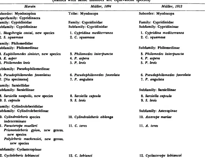
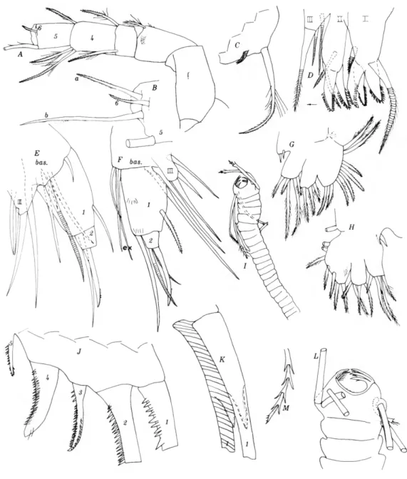
FIGURE 1.—Skogsbergia squamosa: a, anterior part of right valve, medial view; b, central muscle scars of left valve, lat- eral view; c, scalelike pattern on shell (from near posterior part of shell); d, anterior part of right valve, lateral view;
e, section of posteroventral margin of left valve, medial view; /, section of ventral margin of left valve, medial view;
g, posterior right valve, medial view; h, posterior left valve, medial view; i, left 1st antenna, lateral view; ;', right 2nd an- tenna, medial view; k, proximal part of endopodite of right 2nd antenna; /, right mandible, medial view; m, anterior margin of 2nd joint of endopodite of right mandible, medial view; n, left maxilla, lateral view; o, tip of right maxilla, medial view; p, ventral part of right 5th limb, posterior view.
(ah, n-p, ZMB 9151, lectotype; i-m, GMZ 24999.)
inferior tip of rostrum pointed; inner margin of incisur rounded (Figure \a,d); caudal process nar- row extending to middle of posterior margin (Fig- ure \g,h).
Ornamentation: Surface with scalelike reticula- tions (Figure Ic) posterior margin of scales con- vex; shell surface appearing smooth under low magnification; valves translucent with lateral eyes and muscle scars visible in external view.
Infold (Figure le,f): Infold broad and vestibule deep along anterior shell margin and in front of caudal process; infold narrow and vestibule shal- low along ventral margin; infold above incisur with about 16 bristles, below incisur and along ventral margin with about 25 bristles in row.
Hinge: Sclerotized inner ridge of caudal process of left value with knob at dorsal end which may interlock with socket on right valve.
Marginal pore canals: Numerous along free mar- gins. Similarly oriented canals also present along sclerotized inner bar of caudal process.
Central muscle scars (Figure 16): Consisting of about 15 individual scars; of these, 3 elongate an- terior scars slope downward posteriorly, whereas, posterior scars trend downward anteriorly produc- ing a roughly chevron-type muscle scar.
Size: $ (N-l instar), length 2.61 mm, height 1.69 mm. Muller (1894:207) gave the lengths of 2 fe- males as 3.3 mm, and of 2 males as 2.6 mm.
Sexual dimorphism: According to Muller (1894:
207), the shell of the male is similar to that of the female but smaller.
First antenna (Figure \i): 3rd joint with dorsal margin longer than ventral, dorsal bristle proximal to middle, and ventral bristle distal to middle; 4th joint with 2 bare terminal bristles, 1 ventral, 1 dor- sal; 5th joint separated from 6th by distinct suture;
sensory bristle of 5th joint with 8 long proximal and 3 or 4 short distal filaments; 6th joint with bare lateral bristle. 7th joint: a-bristle bare, b- bristle with about 3 filaments, c-bristle with short proximal filaments and 1 long and 1 short distal filament. 8th joint with 4 bristles: d- and e-bristles bare, f- and g-bristles with filaments.
Second antenna (Figure \j,k): Protopodite with medial bristle with short marginal spines. Endo- podite 2-jointed: 1st joint with 5 bristles consisting of a proximal group with 3 short bare bristles and 1 long bristle with marginal spines and a distal long ventral bristle with few marginal spines; 2nd
joint with long, faintly annulate, terminal bristle (2nd joint could be interpreted as being bulbous base of bristle). Exopodite: length of 2nd joint equal to combined lengths of joints 3-5; bristle of 2nd joint without curly hairs proximally and with about 14 ventral spines; joints 3-9 with basal spines, spines longer on distal joints; 9th joint with 4 bristles, 3 long and 1 short; bristles on joints 3-9 with natatory hairs.
Mandible (Figure \l,m): Coxale endite spinous.
Basale with 3 dorsal and 6 ventral bristles: 1 long spinous and 2 short bare a-bristles, 1 long c-bristle, and 1 long spinous and 1 short bare d-bristle. Exo- podite with acute tip and 2 ventral bristles, proxi- mal bristle longer of two. Endopodite: 1st joint with 2 long and 2 short distoventral bristles; 2nd joint with 3-4 ventral spinelike bristles, 1-2 distal to middle and 2 proximal to terminal tooth, and about 19 dorsal bristles; end-joint with 3 claws and 4 bristles. Ventral margin of basale on N-l $ with 8 bristles: 1 long and 2 short a-bristles, 1 short b-bristle close to a-bristles, 1 short and 1 long c- bristle, and 1 short bare and 1 long spinous d- bristle; ventral margin of 2nd joint of endopodite with 2 spinelike bristles distal to middle and 2 proximal to the reduced terminal tooth.
Maxilla (Figure \n,o): Precoxale with fringed marginal epipodial appendage; coxale with plu- mose bristle on anterior margin; lst-3rd endites hirsute, 1st with about 11 bristles, 2nd with about 7, 3rd with 7 distal and 1 proximal bristle; basale with bare ventral bristle (missing on some appen- dages); 1st joint of endopodite with 2 bare a-bris- tles and 3 /?-bristles; cutting edge on distal posterior corner of 1st joint consisting of ridge with 3 arcs;
end joint of endopodite short, with about 11 bris- tles; medial surface with clusters of fine hairs; exo- podite hirsute along posterior margin, with 1 plumose posterior bristle and 2 terminal bristles,
1 bare, 1 plumose.
Fifth limb (Figure \p): Epipodial appendage with 53-55 plumose bristles; endites I—III each with about 5 bristles. Exopodite: 1st joint with 6 curved teeth in row followed by conical tooth; 2nd joint with 4 curved clawlike teeth, 8 stout curved bare bristles, and 3 plumose bristles; 3rd joint hirsute, inner lobe with 2 distal and 1 proximal bristle, outer lobe with 2 distal bristles; 4th and 5th joints fused, hirsute with total of 4 bristles distally.
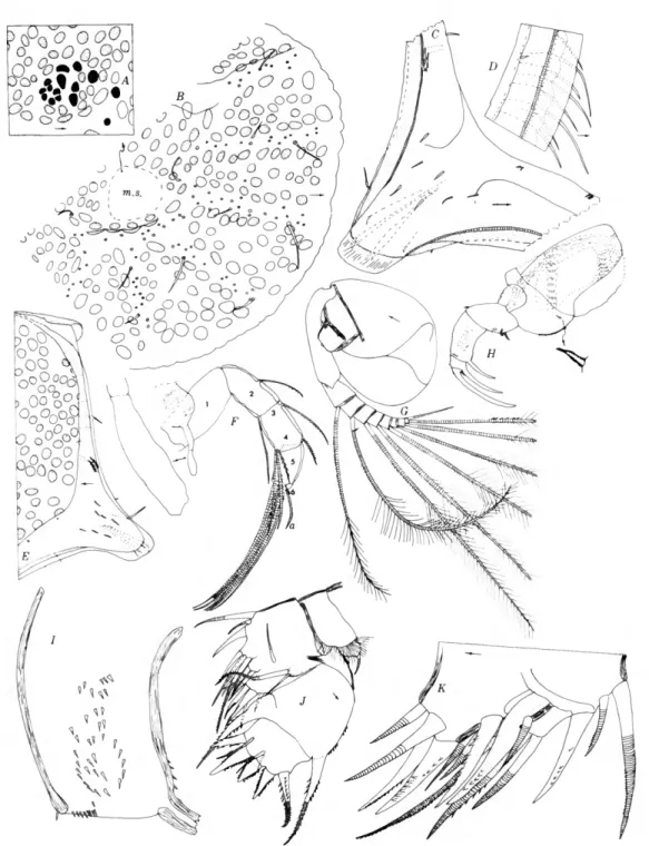
Sixth limb (Figure 2a): Epipodial appendage
FIGURE 2.—Skogsbergia squamosa: a, left 6th limb, lateral view; b, 7th limb; c, furca with brush- shaped organ and genitalia; d, detail of proximal part of furca; e, brush-shaped organ; /, an- terior part of animal with upper lip and rod-shaped organ; g, lateral eye; h, egg duster and detail of an egg showing nucleolus, germinal vesicle and cytoplasm containing yoke granules.
(a, d, e, f, h, ZMB 9151, lectotype; b, c, g, GMZ 24999.)
with 4 bare bristles. End joint with 15 bristles, the 2 posterior bristles longer than others. First endite with 1 long and 2 short bristles; 2nd endite with 2 long and 2 short bristles; 3rd endite with 3 long and 1 short bristle; 4th endite with 3 long and 2 short bristles. End joint with clusters of spines on lateral surface and longer spines along ventral mar- gin; medial surface with clusters of long spines.
Seventh limb (Figure 2b): Adult $ with 13 proximal and 15 distal bristles; comb with about 20 square-tipped teeth opposite short blunt cylin- drical tooth. N-l $ with 14 proximal and 14 distal bristles and only about 14 teeth in comb. All bris- tles with 2-6 distal bells and without marginal spines.
Furca (Figure 2c,d): Each lamella with 7 claws in adult $ , 6 in N-l $ ; 2nd and 3rd claws united with lamella; 4th claw more slender than 5th; ven- tral margin and lateral surface of each lamella proximal to claws bearing clusters of short spines;
proximal claws on right lamella slightly longer than on left; claws 1-5 with lateral and medial spines in row along concave margin, claws 6-7 with single row; claws 1-2 with minute spines distally along convex margin; claw 1 with large medial spines distally; several small spines present along anterior margin of each lamella.
Brush-shaped organ (Figure 2e): Short lobe bear- ing 8 bare annulate bristles situated above genital organs and behind each 7 th limb (observed on both adult $ and N-l $).
Genitalia (Figure 2c): Bilobed (attached to one of the lobes of the N-l $ is what appears to be a rhizopod similar to the genus Centropyxis.)
Eyes (Figure 2g): Lateral eyes with 29-34 omma- tidia, all but few divided; medial eye about half diameter of lateral eye.
Rod-shaped organ (Figure 2/): Short, pear- shaped with concentric ridges distally and terminal node. Conical projection present between organ and upper lip.
Upper lip (Figure 2/): Anterior lobe undivided and with glandular openings; posterior lobe di- vided, hirsute; lateral lobes small, hirsute. Lower lip helmet-shaped, hirsute.
Eggs (Figure 2h): Cluster of 13 round eggs pres- ent in each ovary of N-l $ instar.
Skogsbergia costai, new species
FIGURE 3
Cypridina mediterranea.—Mutter, 1894:206, pi. 1: fig. 16, pi.
2: figs. 1-2, 4-5, 8-20, 22-26, 33, pi. 34: figs. 21-24, 48, pi.
40: fig. 19 [not Costa, 1845].
Cypridina mediterranea [forma typica].—Brady and Norman, 1896:650, pi. 54: figs. 1, 2, pi. 55: figs. 1-11.
HOLOTYPE.—One female with eggs in ovaries in Zoological Museum, Berlin (ZMB 9150:1) ; valves, upper lip and some appendages preserved in alco- hol, remaining appendages on slides; collected by G. W. Miiller.
TYPE-LOCALITY.—Gulf of Naples. Miiller col- lected one specimen from the bottom and numer- ous specimens from the stomach of a fish "Mustelus laevis." The vial containing the holotype did not contain information concerning its exact source;
it is most likely from the fish stomach.
MATERIAL.—One complete female with eggs in the ovaries and 3 separated valves (ZMB 9150). I dissected the female and have designated it on la- bels as 9150 sp. 1 (holotype); the valves, some appendages and the upper lip are preserved in alco- hol, the remainng appendages are on slides. In ad- dition to the above, I briefly examined, while at the Zoological Station of Naples, about 21 specimens, including separated valves (GMZ 24997); most of these specimens were instars. The description below is based principally on the complete female with eggs, but the furca of a specimen from the Zoologi- cal Institute of Greifswald is also illustrated. All specimens collected by G. W. Miiller.
DISTRIBUTION.—Gulf of Naples.
DISCUSSION.—Miiller (1894:206) considered it probable that ostracods known at that time as Cy- pridina mediterranea belonged to two or more closely related species. He assigned the specimens collected by him in the Bay of Naples to C. medi- terranea because the outline of the shell conforms rather well with the sketch by Costa (1845, pi. 1:
fig. 2), and because it was collected in the same area. I do not believe that Miiller's specimens are conspecific with C. mediterranea Costa because the latter species has 59 bristles on the 7th appendage (Costa, 1845, fig. 12), whereas, the 7th appendages on Miiller's specimens have only 25-28 (Miiller, 1894, pi. 2: figs. 10-11; see Figure 3/E) . Also, C.
mediterranea Costa has 8 furcal claws (Costa, 1845, fig. 14) compared to 10 or 11 for the specimens col-
lected by Miiller (1894, pi. 2: fig. 25; see Figure Si).
Dr. G. Bonaduce kindly inquired at the University in Naples, which has in its collections ostra- codes collected by Costa, but specimens of C. medi- terranea could not be located. Because of this it is necessary to rely on the description of the species by Costa. I consider the above differences in the 7th appendage and the furca to be too great to be the result of poor observation or variability within a species. It is possible that the furca illustrated by Costa is from a juvenile specimen; however, if it is assumed that the well-developed 7th limb is from the same specimen, the possibility of the specimen being a juvenile diminishes.
Claus (1865:153, pi. 10: figs. 1,2-7) described the new species Cypridina messinensis from the Medi- terranean. In 1873 (p. 221, pi. 11: figs. 16-20) he referred C. messinensis to Cypridina mediterranea Costa. Brady and Norman (1896:650) and Miiller (1894:00 [with a "?"]; 1912:11) agreed that C. mes- sinensis was a synonym of C. mediterranea. The 6th limb of C. messinensis and S. costai differ in that C. messinensis has 10 bristles on the end joint and 3 bristles in place of the epipodial appendage (Claus, 1965, pi. 10: fig. 5) compared to 18 and 4, respectively, on S. costai (Miiller, 1894, pi. 2: fig.
13). In addition, the posterior process of the right valve of C. messinensis illustrated by Claus (1865, pi. 10: fig. 1) is considerably more prolonged than that of either S. costai or C. mediterranea. The same might be said of the shell of the specimen from Triest illustrated by Claus (1876, pi. 18: fig.
5) and identified by him as C. mediterranea Costa.
Sars (1888:208) referred specimens collected at Spezia in the Mediterranean to Cypridina mediter- ranea Costa. The appendages of Sars' specimens are quite similar to those of the specimens collected by Miiller in the Gulf of Naples, but as previously stressed by Miiller (1894:207), Sars' specimens are smaller. Sars (1888:208) gave the length of a fe- male with eggs in the brood chamber as 2.62 mm and of a male as 2.80 mm. Miiller (1894:206) gave the length of a female as 3.9 mm and of 2 males as 2.7 mm; Miiller (1894:206) stated that the size of the female varies but that, unfortunately, he could not determine whether the animals of different sizes bore eggs or were mature. The female that I de- scribe herein has eggs in the ovaries and is 3.14 mm long. It is known that differences in environment can cause a difference in size of specimens of the
same species, but it seems unlikely that the size could differ as much as found in the females col- lected in the Gulf of Naples and at Spezia. There- fore, I have not included Sars' species in the synonymy of S. costai.
Brady and Norman (1896:650) referred speci- mens from the Bay of Naples to Cypridina mediter- ranea. They recognized two forms of this species:
"typica" and "Var. b." Form "typica" was consid- ered by them to be close to C. mediterranea Costa;
"Var b" was considered close to C. messinensis Claus, which they designated (1896:652) as "C.
mediterranea var. messinensis (Claus)." Form "ty- pica" was referred to C. mediterranea Costa by Miiller (1912:11). I believe that the specimens designated "typica" by Brady and Norman are con- specific with those collected by Miiller and refer them herein to S. costai. This belief is based pri- marily on the number of bristles (29) on the 7 th limb (Brady and Norman, 1896, pi. 55: fig. 10), the number of furcal claws (11) (Brady and Norman, 1896, pi. 55: fig. 11), and on the shape of the cara- pace (Brady and Norman, 1896, pi. 54: figs. 1,2).
Brady and Norman (1896:650) and Muller (1912:
11) included in the synonymy of C. mediterranea Costa specimens referred to Bradycinetus brenda (Baird, 1850) by Brady (1871:292, pi. 26: fig. 6), who described the secondary branch of the male 2nd antenna as "largely developed and triarticu- late." Although the specimens are largely unknown, the morphology of the endopodite of the 2nd an- tenna of the male excludes it from the genus Skogs- bergia.
Skogsberg (1920:247) included "Cypridina medi- terranea, O. Costa, 1845, (G. W. Muller, 1894, p.
206, pi. 2, figs. 1, 2, 4, 5, 8-20, 22-27, 33)" in a list of species in his subgenus Vargula. Rome (1942:
7, pi. 1: figs. 1, 2) referred juveniles collected in the vicinity of Monaco to Cypridina mediterranea Costa. The 4th furcal claw illustrated by Rome (1942, pi. I: fig. 2) is much shorter relative to the 3rd and 5th claws than the 4th claw on either S.
costai or C. mediterranea Costa. Therefore, I think it necessary to consider the identification to be un- certain. Granata and Caporiacco (1949:6) refer ostracodes from the Mediterranean to Cypridina (Vargula) mediterranea (Costa). The ostracods are not described in sufficient detail to verify the identi- fication, but as the furca bears 10 claws, the specimens probably are related more closely to the
11 specimens of Miiller than those of Costa. However,
it would be necessary to examine the specimens be- fore attempting to reassign them. Poulsen (1962:
162) lists "Skogsbergia mediterranea (O. Costa)"
along with other species not investigated by him in the genus Skogsbergia. Cypridina mediterranea Costa has been reported from other areas in the Mediterranean, but either without accompanying descriptions or with insufficient description (e.g., Ramsch, 190a, 1906b; Brian, 1909; Hartmann, 1959; Reys, 1965a, 1965b; Rome, 1965).
DESCRIPTION OF FEMALE (details of most appen- dages are obscured by debris and therefore descrip- tion below is incomplete).—Carapace oval in lateral view with greatest height near middle and greatest length below middle, posterior evenly rounded (Figure 3a-d); inferior tip of rostrum narrowly rounded, inner margin of rostrum broadly rounded.
Ornamentation: Surface smooth without orna- mentation; valves translucent, lateral eyes and mus- cle scars visible in lateral view.
Infold (Figure 5b-d): Broad along anterior and posterior parts of shell, narrow ventrally; infold above incisur obsecure, but with 6 or more long bristles; below incisur and along ventral margin with about 21 bristles in row.
Hinge: Sclerotized ridge with knob at dorsal end present near inner margin of inner lamella in pos- terior part of left valve; sclerotized ridge present on inner lamella in posterodorsal part of right valve fits in front of knob on left shell.
Marginal pore canals: Numerous canals present along free margins.
Central muscle scars: Obscured in specimen ex- amined. Muller (1894, pi. 2: figs. 1, 3) illustrates valves with 11 elongate scars ventral to 1 oval scar.
Selvage: Narrow striated ridge with lamellar pro- longation present along anterior and ventral valve margins; lamellar prolongation with smooth mar- gin, unstriate except along lower margin of incisur.
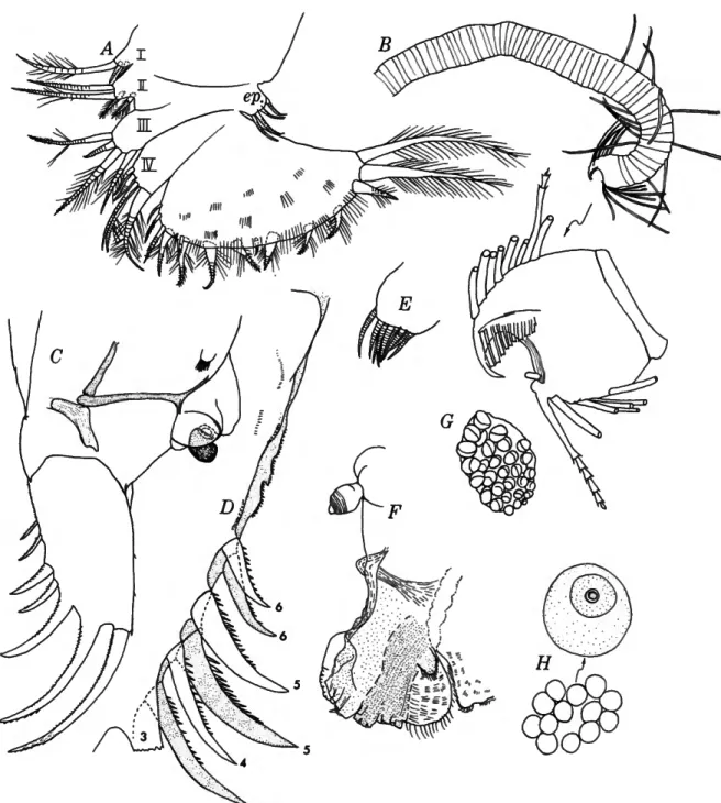
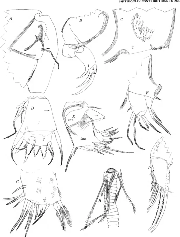
FIGURE 3.—Skogsbergia costai, new species, female: a, cara- pace showing position of central muscle scars and lateral eye;
b, rostral area left valve, medial view; c, posterior left valve, medial view; d, posterior margin right valve, medial view;
e, part of endopodite of 2nd antenna; /, basale of right man- dible, medial view; g, basale of left mandible, medial view;
h, lateral eye; i, furca; ;', claws 1-5 of left lamella of furca (teeth on posterior margins of claws not shown); k, 7th limb; /, upper lip; m, genitalia (spermatophores) n, cluster of eggs (oil globules shown in 1 egg), (i, GMZ 24997; a, h, j-n, ZMB 9150:1, holotype).
Size: Female holotype with eggs in ovaries, length 3.14 mm, height 1.95 mm. Muller (1894:206) gave the length of a female as 3.9 mm, and of 2 males as 2.7 mm.
First antenna: 3rd joint short with 1 dorsal and 1 ventral bristle; 4th joint with 1 dorsal and 1 ven- tral bristle distally; sensory bristle of 5th joint with about 12 marginal filaments, of these, distal 3 shorter than others; lateral bristle of 6th joint about 3 times length of 7th joint; end joints with bristles typical for genus.
Second antenna: Medial bristle of protopodite not observed. Short endopodite with weak suture separating 2 joints (Figure 3e): 1st joint with 1 long and several shorter bristles; 2nd joint with 1 long terminal bristle (2nd joint could be interpre- ted as bulbous base of bristle). Exopodite: bristle of 2nd joint with about 25 ventral spines, and about the same number of weaker spines dorsally (proximal hairs not observed); bristles of joints 3-9 with natory hairs; 9th joint with 3 long and 1 medium bristle; joints 5-8 with large basal spines;
joint 9 with large lateral spine; dorsodistal corner of joints 2-8 with minute spine, or comb of minute spines. Dorsal margin of protopodite with numer- ous protozoans attached.
Debris obscured appendages, especially the endo- podite. The 2nd antennae I examined is essentially similar to that illustrated by Muller (1894, pi. 2:
fig. 22). A few differences are noted below. The en- dopodite in Miiller's illustration has only 1 joint, whereas the specimen I examined has 2; however, additional specimens should be examined before definitely establishing which interpretation is cor- rect. Another difference is that the bristle on the 2nd joint of the exopodite on the specimen I exam- ined has both ventral and dorsal spines, whereas, this bristle in Miiller's illustration has only ventral spines. The ventral and dorsal spines could be seen quite clearly on the specimen I examined. I think it quite likely that the 4th joint of the exopodite has a basal spine as shown by Muller; I could not observe it on the appendages I examined, but this could be because of the debris.
Left mandible: Basale (Figure 3g): Ventral mar- gin with 3 a-bristles, 2 c-bristles, and 1 long d- bristle; dorsal margin with 1 bristle near middle and 2 subterminally. Ventral margin of 2nd endo- podite joint with 2 single spines distally and a group of 2 spines terminally.
Right mandible: Ventral margin of basale differs from left in having 1 short a-bristle, 2 c-bristles, and 1 short d-bristle (Figure 3/). The ventral margin of the basale of the right mandible illustrated by Miiller (1894, pi. 2: fig. 17) has 2 or 3 a-bristles, 1 c-bristle, and a long and short d-bristle. It is neces- sary to examine more specimens to determine the variability of bristle distribution on the basale.
Seventh limb (Figure Sk): Each limb with about 14 lateral and 13 distal bristles; terminal comb with about 12 or 14 blunt teeth.
Lateral eyes (Figure 3//): Large with 34-36 di- vided ommatidia.
Upper lip (Figure 3/): Consisting of unpaired anterior part and paired posterior part without tusks. An unpaired protuberence present above up- per lip and below anterior margins of 1st antennae.
Furca (Figure 3i,/): Each lamella with 10 or 11 claws, all separated from lamella; 4th claw slightly more slender than 5th; all claws with spines in row laterally along concave margin; claw 1 with broad medial spines distally and few short spines along distal convex margin.
Genitalia (Figure 3m): Consisting of 2 parts, each having 1 lobe (A spermatophore is connected to each lobe in Figure 3m).
PHILOMEDIDAE Muller, 1906
The Philomedidae contains two subfamilies, Philomedinae Muller, 1906, and Pseudophilome- dinae Kornicker, 1967a; both subfamilies are repre- sented in the Gulf of Naples.
Key to the Subfamilies of Philomedidae
Maxilla with 3 large endites; tooth on 2nd joint of 5th limb not prolonged PHILOMEDINAE Maxilla with 2 large and 1 reduced endite; tooth on 2nd joint of 5th limb prolonged, fanglike
PSEUDOPHILOMEDINAE
PHILOMEDINAE Muller, 1906
The Philomedinae is represented in the collec- tion by two genera Philomedes Liljeborg, 1853, and Euphilomedes Poulsen, 1962.
Philomedes Liljeborg, 1853
TYPE-SPECIES.—Philomedes longicornis Lilje- borg, 1853. Gender: Masculine.
Philomedes levis Muller, 1894
FIGURE 4
Philomedes levis Miiller, 1894:211, pi. 3: figs. 4, 15, 18, 29-31, 36; 1912;26, 30 [diagnosis].—Poulsen, 1962:345 [listed].
LECTOTYPE.—Female without eggs in the Zoologi- cal Museum of Berlin (ZMB 9153). I have desig- nated this specimen: 9153:1. The valves of the lectotype and some appendages are preserved in alcohol; the remaining appendages are on slides.
An instar that was in the same vial is herein desig- nated 9153:2. The instar which is a paralectotype, is preserved whole in alcohol.
TYPE-LOCALITY.—Gulf of Naples, Italy.
MATERIAL.—Two specimens (ZMB 9153) in vial containing label "9153 Philomedes levis n. sp.
Muller." The female in this vial has been selected as the lectotype and designated ZMB 9153:1. It is presently preserved in alcohol except for some ap- pendages on slides. The 2nd specimen, an instar preserved in alcohol, is designated ZMB 9153:2. In addition to the above I had the opportunity to ex- amine briefly about 14 specimens preserved in alco- hol GMZ 25010) at the Zoological Museum at Na- ples. The specimens are in poor condition.
DISTRIBUTION.—Gulf of Naples.
DESCRIPTION OF FEMALE (details of appendages and carapace are obscured by debris and radial crystals; therefore, the description is incomplete).—
Carapace oval in lateral view with greatest height
FIGURE 4.—Philomedes levis: a, carapace, length 1.21 mm; b, carapace, length 1.37 mm; c, part of anterior of left valve, lateral view; d, posterior left valve, medial view; e, muscle scars right valve, lateral view; /, anterior left valve, medial view (lamellar prolongation not shown); g, part of exopodite of 2nd antenna, medial view; h, left mandible, medial view (all bristles not shown); i, coxale endite and part of basale of left mandible, medial view; /, anterior of animal; k, ter- minal teeth of 5th limb; /, tip of 7th limb; m, exopodite of maxilla; n, furca. (a, c, e, ZMB 9153:2; b, d, f-n, ZMB 9153:1.)
13
14
and length near middle (Figure 4 a-c); linear an- terior margin of rostrum forming acute angle with ventral margin; inferior tip of rostrum with small protuberance; posterior margin of valve with caudal process at border with ventral margin.
Ornamentation: surface smooth without orna- mentation; valves translucent with muscle scars visible in lateral view.
Infold (Figure 4d,f): Broad along anterior and posterior parts of valve, narrow ventrally; anter- oventral part striate. (Bristles on infold are ob- scured by debris, but Miiller (1894, pi. 3: fig. 36) illustrates 19 on rostrum and 10 below rostrum on anteroventral part of infold.)
Central muscle scars (Figure 4e): 5 large ovoid scars forming slight arc over 6 smaller scars and 2 rows of elongate scars: anterior row with 3 scars, posterior row with 4. (The U-shaped scar shown by Miiller (1894, pi. 3: fig. 4) is incorrect; it consists of 2 ovoid scars with 2 smaller scars near their ven- tral margins.)
Selvage: Wide lamellar prolongation present along rostrum and below incisur; prolongation be- ing subdivided into broad platelets with blunt tips.
(The broad platelets are useful for recognizing this species.)
Size: $ lectotype, length 1.37 mm, height 0.91 mm; instar paralectotype, length 1.21 mm, height 0.81 mm.
First antenna: 2nd joint with 1 dorsal, 1 lateral, and 1 or 2 ventral bristles, about 5 clusters of spines along ventral margin, and numerous clusters on ventral surface; 2nd joint with 1 ventral and 2 dorsal bristles.
Second antenna: Endopodite 2-jointed: 1st joint with 6 bristles, 5 proximal, 1 distal; 2nd joint with 2 bristles, 1 proximal and 1 terminal, latter bristle recurved. Exopodite (Figure 4g): joints 2-8 with basal spines increasing in length on distal joints;
bristle of 2nd joint reaching well past joint 9; bris- tles of joints 2-4 without natatory hairs; 9th joint with 4 long and 2 or 3 short bristles.
Mandible (Figure 4h,i): Coxale endite bifurcate with several teeth near tips; basale with 3 or 4 stout medial spines with secondary teeth; exopodite reaching three-fourths length of 1st joint of endo- podite; end-joint of endopodite with 3 claws, 2 curved, 1 almost straight; straight dorsal claw not quite as long as curved ventral claw.
Maxilla: With 3 endites, 1st with about 10 bris-
tles, 2nd about 6; exopodite with 3 bristles, proxi- mal bristle about one-half length of 2 distal bristles (Figure 4m).
Fifth limb (Figure 4k): 1st exopodite joint with 4 or 5 stout teeth; 2nd exopodite joint with usual large triangular tooth.
Seventh limb (Figure 4/): About 5 bristles pres- ent in distal group and 12 in proximal group, each with 3-6 bells; 1 main peg present opposite comb (a few smaller pegs also may be present, but are difficult to see because of condition of appendage).
Furca (Figure 4n): Each lamella with 10 claws separated from lamella by suture; claws decreasing in length proximally on lamella except for last 4 claws, which are of about same length; claws 1 to 6 or 7 with teeth along concave margin; claw 1 with large tooth and several smaller proximal teeth on medial side; 1 or more rather long thick hairs pres- ent laterally near base of most claws; tuft of hair present laterally at base of claw 1; tips of claws 1-5 with blunt rounded tips.
Eyes (Figure 4;): Medial eye large; lateral eye minute.
Rod-shaped organ (Figure 4/): 2-jointed, elon- gate, tapering distally.
Upper lip (Figure 4;): Rounded with anterior process.
Euphilomedes Poulsen, 1962
TYPE-SPECIES.—Euphilomedes nodosus Poulsen, 1962, by subsequent designation (Kornicker, 1967a:
1). Gender: Masculine.
Euphilomedes asper (Miiller, 1894)
FIGURE 5
Philomedes aspera Muller, 1894:210-211, pi. 3: figs. 3, 17, 21, pi. 8: fig. 1; 1912:26, 27 [key, diagnosis].
Philomedes foveolata.—Brady and Norman, 1896:659-661, pi.
56: figs. 4, 5 [not Pseudophilomedes foveolata Muller, 1894].
Ph. (Ph.) aspera.—Skogsberg, 1920:351 [discussion].
Euphilomedes aspera.—Poulsen, 1962:363 [key].
Euphilomedes asper.—Kornicker, 1967a: 18, figs. 9, 10, lla-d, 12 [supplementary description].
LECTOTYPE.—One adult male in the Zoological Museum of Berlin (ZMB 9152). I have designated this specimen as 9152:1 because two separated valves also have the number 9152. The valves of the lectotype and its appendages are preserved in
alcohol, except for a 2nd antenna and one of the 7th limbs, which are on slides.
TYPE-LOCALITY.—Gulf of Naples, Italy.
MATERIAL.— (1) A small vial containing 1 whole ostracod and 1 left and 1 right valve and a label
"9152 Philomedes aspera n. sp. Miiller." This is the same material from the Zoological Museum of Ber- lin described by Kornicker (1967a: 19) but then a jar containing the specimens had the label "Type, Philomedes aspera G. W. Muller, Kat. Nr. 9152, Fundort Napoli." The whole specimen, an adult male designated the lectotype herein, was partly dissected and is presently in alcohol except for one 2nd antenna and one 7th limb, which are on slides.
I have designated the lectotype as 9152:1, the right valve as ZMB 9152:2, and the left valve as ZMB 9152:3. The right valve is probably from an adult male, the left from an instar. Both valves have been replaced in the same vial from which they were obtained. Most appendages of the lectotype and in- ternal features of its valves are partly obscured by debris. (2) Two instars from station B30, Gulf of Naples; collected by Harbans S. Puri and Gioac- chino Bonaduce in 1962 or 1963. (3) One instar, USNM 112803, from vicinity of Benta Palumma, Gulf of Naples; collected for L. S. Kornicker by technician at Naples Zoological Station, 21 April 1966. Sample reported to be from depth of 55 meters. (4) I also had the opportunity to examine briefly while at the Zoological Station at Naples in 1966, four of Miiller's Gulf of Naples specimens in the collection of the Zoological Institute of Greifs- wald (GMZ 25003). One of these specimens, an adult male, was dissected and some of its appen- dages are illustrated herein (Figure 5g,i,j,k).
DISTRIBUTION.—Gulf of Naples, Monaco (Rome, 1942, 1965). Marseille (Reys, 1964, 1965a, 1965b).
DISCUSSION.—A supplementary description of this species based mostly on specimens collected in the Gulf of Naples by A. M. Norman in 1887 and re- ported upon by Brady and Norman (1896:659), but also in part on specimens collected by G. W.
Muller, was published by Kornicker (1967a: 18).
The brief description below is based solely on Miiller's specimens. The adult male designated in the 1967 paper as ZMB 9152:1 is herein selected as the lectotype. The specimen is assigned the same number in the present paper. Kornicker (1967a: 18) was incorrect in listing Euphilomedes asper as a
"new combination" because it had previously been
assigned to Euphilomedes in a key to the genus by Poulsen (1962:363).
DESCRIPTION OF ADULT MALE.—Lateral outline oval, elongate with greatest height near middle, prominant rostrum, broad rostral incisur, and an- gular caudal process (Figure ba-c).
Ornamentation: Shell with numerous large oval pits (Figure 5b). Hairs with taper distributed on lateral surface of rostrum, some forming row near ventral margins of valves; additional hairs sparsely distributed on valve surface.
Infold: Broad at caudal process and behind rostrum; 12-13 bristles forming row on infold be- hind rostrum; infold below rostrum with small bristle followed by space and additional small bris- tles in row; infold anterior to caudal process with short bristles; infold along posteroventral and pos- terior margins with small bristles forming row on list.
Hinge: Linear along posterodorsal margin.
Marginal pore canals: Numerous along free mar- gins but faint.
Central muscle scars: Obscure, consisting of about 17 individual scars.
Selvage: With wide striate lamellar prolongation with slender spines along outer edge.
Size: Shell dimensions (in mm) are as follows:
Collector Remarks
Great- Great- est est length height
1.30 0.80 Muller adult male (lectotype) 1.28 0.78 Muller adult male
0.94 0.60 Muller instar 0.90 0.60 Puri and Bonaduce instar 0.76 0.49 P u" a nd Bonaduce instar 0.70 0.46 Kornicker instar
First antenna: 2nd joint with a dorsal, ventral, and lateral bristle; 3rd joint with 1 ventral and 2 dorsal bristles; 4th joint with 2 dorsal and 4 ventral bristles distally; 5th joint inserted ventrally between 4th and 6th joints and bearing sensory bristle with broad base and numerous filaments; 6th joint with medial bristle distally; end joints with 2 long stout c- and f-bristles, 1 short spinous dorsal a-bristle, 2 long b- and g-bristles with filaments, and 2 bare d- and e-bristles.
Second antenna (Figure 5d-f): Endopodite 3- jointed: 1st joint with 5 proximal and 1 distal bristle; 2nd joint elongate with 2 bristles near mid- dle; 3rd joint prehensile with 1 proximal and 2
16
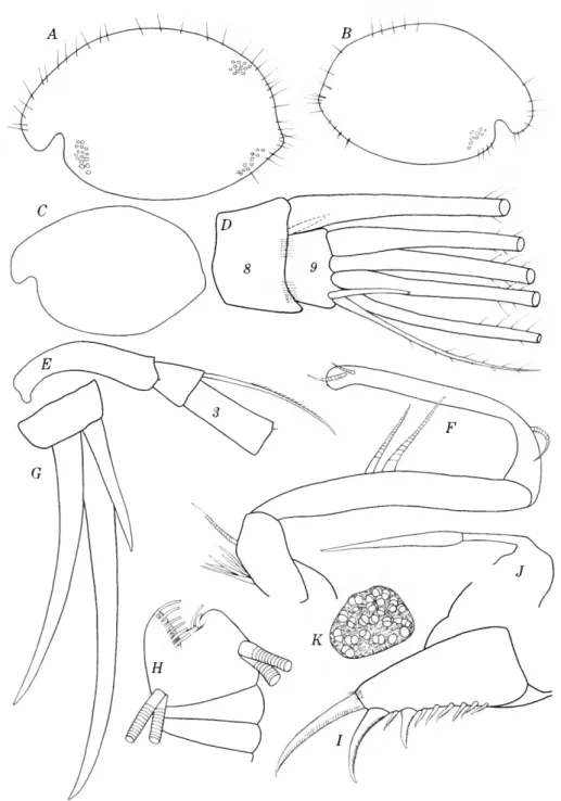
FIGURE 5.—Euphilomedes asper: a, carapace showing some punctae, length 0.90 mm; b, carapace showing some punctae, length 0.76 mm; c, carapace, length 0.70 mm; d, tip of 2nd antenna, medial view; e, part of exopodite of 2nd antenna, medial view; /, endopodite of left 2nd an- tenna; g, end claws on mandible (bristles not shown); h, tip of 7th limb; i, furca; /, outline of rod-shaped organ and medial eye; k, lateral eye. (/, ZMB 9152:1, from station B30; b, ZMB 9152:
2, from station B30; c, juvenile, USNM 112803; g, i, j , k, adult $, GMZ 250O3, a, d-f, h, ZMB 9152:1, lectotype).
distal bristles and ridges terminally. Exopodite: 1st joint with small medial spine; joints 3-8 with slender basal spines; bristle of 3rd joint with mar- ginal spines ventrally; joints 2-8 with short hairs in row distally on lateral margin; 9th joint with 4 long and 2 short bristles.
Mandible (Figure 5g): Basale: Ventral margin with 7 bristles; dorsal margin with 1 bristle near middle and 2 terminally; medial surface with about 5 short bristles proximally near ventral margin.
Exopodite reaching about middle of 1st endopo- dite joint, with 2 terminal bristles. Endopodite:lst joint with 2 short and 3 long distoventral bristles;
2nd joint with 2 dorsal bristles proximally, 5 or 6 dorsal bristles near middle, 2 ventral bristles subdis- tally and about 4 ventral bristles terminally; end joint with 2 long subequal claws, 1 short dorsal claw and about 4 bristles.
Seventh limb (Figure bh): Four long bristles in distal group, each with about 5 bells; 4 shorter bristles in proximal group, each with 3 or 4 bells;
terminal comb opposed by 2 blunt pegs.
Furca (Figure 5i): Each lamella with 10 or 11 claws, all separated from lamellae; claws 1, 2, 4 pri- mary, remaining claws secondary; 3rd claw about same length as 5th; primary claws with lateral and medial rows of spines; spines on each claw about same length; secondary claws with marginal spines;
clusters of long hairs present medially at base of some claws.
Eyes: Lateral eye (Figure 5k) large with about 26 divided ommatidia. Medial eye (Figure bj) about same size as lateral eye.
Rod-shaped organ (Figure 5;): Elongate with 2 joints; 2nd joint broad proximally and tapering distally.
Euphilomedes sinister, new species
FIGURES 6-7
Philomedes interpuncta.—Brzdy, 1868b:463, pi. 33: figs. 10- 13, pi. 41: fig. 3; 1871:293, pi. 26: figs. 1-5; 1872:60, pi. 1:
figs. 5.—Muller, 1894:210, pi. 3: figs. 1-2, 5-16, 19-20, 24- 28.—Brady and Norman, 1896:656, pi. 56: figs. 6-10, pi. 57.
—Fage, 1934:249, figs. 1-2.—Caraion, 1959:266, fig. 1 [not Baird, 1850].
ETYMOLOGY.—The specific name from the Latin sinister (left) refers to the posterior spines of the carapace being restricted to the left valve.
HOLOTYPE.—One female without eggs in the
Zoological Station of Naples. The valves and some appendages in alcohol; remaining appendages on slides; length 1.24 mm.
TYPE-LOCALITY.—Benta Palumma, station B27, 90 M, Gulf of Naples, Italy.
MATERIAL.—One female from station B27, Gulf of Naples, collected by Harbans S. Puri and Gioac- chino Bonaduce. I also had the opportunity to examine briefly while at the Zoological Station at Naples in 1966, three of Miiller's specimens in the collection of the Zoological Institute of Greifswald (GMZ 25008).
DISTRIBUTION.—Gulf of Naples; Loch Long, Fosse de Cap Breton (Bay of Biscay), Cumbrae (Brady and Norman, 1896:657); Bosporus, 41°25'S, 28°
59'E (Caraion, 1959:266).
DISCUSSION.—Muller (1894:205) assigned speci- mens of E. sinister collected in the Gulf of Naples to Philomedes interpuncta (Baird, 1850). The incom- plete description of the latter species (Baird, 1850:
257, pi. 17: figs. 8-10) makes it necessary to place it in the category "species dubia." However, the absence of secondary claws between the primary claws of the furca (Baird, 1850, pi. 17: fig. 8) indi- cates that Baird's species does not belong in the genus Euphilomedes. Unfortunately, the type- specimen of P. interpuncta has been lost (Loft- house, 1966).
According to Muller (1894:205), the left valves of females have an upper and lower spine posteri- orly, whereas, the male has only the lower spine.
Previously, Brady (1868b:464) had stated concern- ing specimens of E. sinister from the English coast, which he identified as Philomedes interpuncta (Baird), "There is much variety in the spinous armature of the posterior margin; in most cases one short spine exists near its lower extremity, and rarely one likewise at the upper angle; not infre- quently they are altogether wanting." Un- fortunately, I do not have a sufficient number of specimens to determine the degree of constancy of spines. However, I am inclined to believe that their absence would have been mentioned by Muller. In view of the diverse localities from which the species was reported by Brady (1868a, 1868b). I think it possible that specimens lacking spines may not be conspecific with E. sinister.
Specimens from Plymouth sound identified by Norman (1861:280, pi. 14: fig. 11) as Philomedes longicornis (Lilljeborg, 1853) differ from E. sinis-
ter in being without spines or in having a single spine on the upper posterior margin. Another dif- ference is that the surfaces of Norman's specimens are described as having large circular or subcircular pits, whereas, E. sinister has polygonal reticula- tions. Brady (1868b:464) stated that Norman's fig- ure and description were based on worn specimens;
however, Norman (1861:280) stated that he found specimens "in some numbers among dredged stuff sent to me by W. Webster, Esq., from Plymouth Sound . . . ." It would seem that if a number of specimens were collected, a lower spine, if originally present, would have remained on some specimens even if worn, because the lower spine is present on both males and females, whereas the upper spine is only on females. For the above reasons, I have not included Norman's specimens in E. sinister.
Lilljeborg (1875:3) placed specimens identified by Brady (1968b:463) as Philomedes interpuncta (Baird) in synonomy with Philomedes globosus (Lilljeborg). The absence of secondary claws alter- nating with primary claws on the furca of the lat- ter species makes the referral untenable.
Skogsberg (1920:398) considered the specimens identified by Norman (1861:280) as P. longicornis to belong to P. interpuncta (Baird). I consider the following statement in Norman's description of the species as an indication that Skogsberg's conclusion is incorrect, "I am indebted to Dr. Baird for point- ing out to me the identity of the Plymouth Ento- mostracan with Lilljeborg's species." This shows that Baird did not consider Norman's specimens to be conspecific with his species.
DESCRIPTION OF FEMALE.—Carapace oval in lat- eral view with greatest height near middle and truncate posterior (Figure 6a); inferior tip of broad rostrum with stout protuberance.
Ornamentation (Figure 6e): Posterior margin of
FIGURE 6.—Euphilomedes sinister, new species, female, holo- type, length 1.24 mm: a, carapace showing positions of mus- cle scars and lateral eye; b, adductor muscle scars right valve, lateral view; c, tip of rostrum, right valve, medial view;
d, anterior left valve, medial view; e, posterior left valve showing some surface reticulations, medial view; f, right 1st antenna, medial view (only a-bristle shown on end joints);
g, part of expodite of right 2nd antenna, medial view; h, tip of right 2nd antenna, medial view; i, endopodite of right 2nd antenna; ;, proximal part of 2nd antenna (not under cover glass); k, furca without claws, and genital area; /, anterior of animal; m, posteroventral margin of right valve, medial valve.
left valve with dorsal and ventral spine and toothed margin between spines; posterior margin of right valve without spines or teeth; surface of valves with reticulate pattern. Bristles present along margins and scattered over carapace.
Infold (Figure 6c-e,m): Broad along anterior and posteroventral parts of valve, narrow ventrally;
anteroventral part striate. Infold behind rostrum with 16-18 bristles, most with marginal spines; in- fold along anteroventral margin with 13 or 14 spinous bristles; infold along posteroventral mar- gin with 3 short bristles near middle and 3 at outer margin; list along posterior and posteroventral in- fold with about 33 bristles, some arranged singly and some in pairs.
Central muscle scars (Figure 6b): Six ovoid mus- cle scars forming arc behind and over 2 to 5 indis- tinct scars.
Selvage: Wide lamellar prolongation along an- terior, ventral, and posterior margins; prolongation divided on rostrum in area of protuberance at in- ferior corner; prolongation striate, with marginal fringe of long and short spines.
Size: $ holotype, length 1.24 mm, height 0.82 mm. (Miiller, 1894:210, gave length of both sexes as 1.27 mm.)
First antenna (Figure 6/): Joints 1-5 with clus- ters of hairs medially; 2nd joint with about 5 spines along dorsal margin and 3 bristles, 1 dorsal, 1 ven- tral, and 1 lateral, all with spines; 3rd joint with 3 bristles, 2 dorsal, 1 ventral, all with spines; 4th joint with 6 bristles, 2 dorsal, 4 ventral; 5th joint with filamentous sensory bristle ventrally; 6th joint with slender bristle with few long spines; a-bristle of 7th joint longer than bristle of 6th joint; bristles of joints 7 and 8 typical for family.
Second antenna (Figure 6g-/): Prodopodite with clusters of short spines on lateral surface. Endo- podite 2-jointed: 1st joint with 5 proximal and 1 distal bristle; 2nd joint with long spinous lateral bristle and 1 slender spine subterminally. Exopo- dite: 1st joint with small medial terminal spine;
terminal margin of joints 2-8 with comb of short spines; bristles of joints 2-5 with short ventral spines near middle and without natatory hairs;
bristles of joints 6-8 with natatory hairs and with- out ventral spines; joint 9 with 7 bristles, 4 ter- minally with natatory hairs, 3 distoventrally with short marginal spines (spinous bristles shorter than those with natatory hairs.)
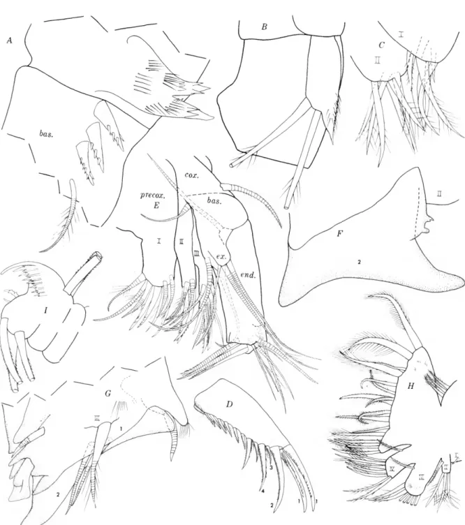
20
FIGURE 7.—Euphilomedes sinister, new species, female, length 1.24 mm: a, coxale endite and part of basale of right mandible, medial view; b, exopodite right mandible, lateral view; c, endites of 5th limb; d, furca; e, right maxilla, lateral view; /, outline of 2nd joint of right 5th limb, anterior view; g, distal end left 5th limb, anterior view; h, 6th limb; i, tip of 7th limb.