lnvertebr. Taxon., 1995,9,1231-63
Revision of the Genus Ogygioses (Palaeosetidae)
Donald Ray DavisA,Ole Karsholl', Niels P. KristensenB and Ebbe S. Nielsenc ADepartment of Entomology, National Museum of Natural History,
Smithsonian Institution, Washington DC 20560, USA.
BZoological Museum, Universitetsparken IS, DK-2100, Copenhagen 0, Denmark.
cDivision of Entomology, CSIRO, PO Box 1700, Canberra, ACT 2601, Australia.
I
Abstract The systematics and external morphology of the oriental genus Ogygioses are reviewed. Monophyly of the genus is demonstrated by the two segmented labial pal pi, loss of the jugal lobe in the forewing, stalking of the forewing R4 with the base of R2 + 3 and a specialised scale arrangement on the forewing base. Sculpturing of the egg chorion differs from that of other Exoporia in exhibiting elongate tubercles regularly dispersed over the surface. The genus is retained tentatively in the Paleosetidae on the basis of the loss of all tibial spurs, including the epiphysis, and the extreme reduction of the maxillae. Four species are recognised: O. caliginosa Issiki & Stringer, O. eurata Issiki & Stringer, O. issikii Davis, sp. nov., from Taiwan, and 0 luangensis Kristensen, sp. nov., from Thailand. Adults are diurnal with the males forming hovering groups, or leks, similar to some species of Hepialidae.I
Introduction
In 1932, Syuti Issiki and Herbert Stringer published (in two parts) a small but important paper describing two primitive hepialoid genera from the Oriental Region. Cenustes lutata [immediately synonymised in the second part under C. minutus (Hampson») was described from Assam, India, together with a related genus, Ogygioses, consisting of two species from Taiwan. The only New World genus associated with these genera is Osrhoes from Colombia (Robinson and Nielsen 1984; Kristensen and Nielsen 1994). The relationship of all three taxa to the Australian Palaeoses is tentative at present.
This study is intended as an update on the systematics and integumental morphology of one of these problematic genera, Ogygioses , in anticipation of a general review of all members of the questionably monophyletic family Paleosetidae. We here report the occurrence of the genus in continental Asia, based on the finding of a new species in north
eastern Thailand. Bouin-fixed material of the latter taxon has been available, and histological sections have enhanced our understanding of some details of the integumental structure. Further findings on Ogygioses soft anatomy will be published elsewhere.
Materials and Methods
It should be noted that the Japanese place names on the labels of the Taiwanese specimens collected by Professor Syuti Issiki have been transliterated in this paper to currently used Chinese names as listed in Chu and Yamanaka (1973, 1975).
0818-0164/95/061231 $10.00
1232 D. R. Davis el al.
The acronyms of the institutions listed in this study are as follows:
AN[C Australian National Insect Collection, CSIRO, Canberra, Australia BMNH The Natural History Museum, London, UK
FSCA Florida State Collection of Arthropods, Gainesville. PL, USA KMNH Kitakyushu Museum of Natural History, Kitakyushu, Japan
TPM Taiwan Provincial Museum. Taipei, Taiwan, Republic of China
USNM National Museum of Natural History (formerly United States National Museum), Smithsonian Institution, Wa hington, DC. USA
ZMUC Zoological Museum. University of Copenhagen, Denmark
Biology Distributioll
Taiwan can be roughly partitioned into three major altitudinal zones with the plains below 100 m occupying only 3 1·3% of tOLal land area, the are's between 100 and 1000 m totalling 37 ·2%, and the high mountains above 1000 m occupying 31·5% (Li et at. 1975).
The chief feature of the island i a high mountain system extcnding nearly the entire lcngth of the country from norlh to soulh. In all, 48 peaks exceed 3000 m in height with the highest, Yu han, rcaching 3997 m. In addition to the central mountain system, which i ' divided into approximately rour distinct ranges. there exists along the eastern coast the lower, nearly parallel Haian, or Coastal Range. Ogygioses is currently known on ly from the central mountain system of Taiwan.
Li et af. (1975) subdivided the Taiwanese nora into seven major zones: I, coastal-tropical:
2, subtropical forest (below 500 m); 3. warm temperate mi)(ed forest 700-1800 m); 4. cool temperate coniferous forest ( 1800-2500 m); 5, cold temperate coniferous forest; 6, subalpine coniferous forest (3000-3500 m); and 7, alpine (above 3800 m). Ogygioses spp. have been collected from an elevation of 300 m (near Kwanlzuling). and possibly as low as 100 m (Wulai), to as high as 2400 m on Mt AJishan. Their habitats thus occur in at I ast three major botanical zones. including subtropical. wann temperate nd cool temperate foresLs.
Posi.tioned as it is, with the Tropic of Cancer dividing the island. Taiwan possesses a generally tropical-subtropical climate. Consequently, the summers are long and the winters mild, with the mean monthly temperalllre of the coldest month (February) ranging from 14·8°C In the north (Taipei) LO 20·5°C in the south (Hengchun). Adult capture rec rds
r
Ogyginses ref! t this favourable clim, te in ranging from March to Octob r.
Outside Taiwan, Ogygioscs is known only from the Phu Luang Wildlife Sanctuary, south
west of Loei in north-eastern Thailand_ Thc sanctuary is shunted 011 an elevated plate u, with fairly steep sides above lowlands, which are now largely deforested (Round 1988). The lOp of th plateau, 1400- 1500 m above sea level, is covered by low evergreen forest with numerous glades.
Life History
No species has been reared and, consequently, little has been published on the life history of Ogygioses. The eggs are white when laid, but become black within a few hours, as is typical of Hepialidae (Stokoe and Stovin 1948; Madge 1954; d'Aguilar 1966) and Mnesarchaeidae (Gibbs 1979)_Chauvin and Barbier (1979) report that the colour change is the result of the melanisation and sclerotisaLion of the vitelline envelope. The eggs may not be scattered in flight, as generally reported for Hepialidae, but may be deposited on the host or some nearby substrate. The few eggs collected of Ogygioses caLiginosa were attached randomly to the inside wa lls of a vial by the enclosed fema.le (Heppner, personal communication). Aspects of the adult behaviour of O. caLiginosa have been summarised by Kuroko (1990) and Heppner (1987). These reporls, for the most part, agree with the unpublished observations received from K. Ueda and the late Professor S. ,lssiki.
Adults hav e bee n encountered over an 8-month period from spring to fall (March-October). Flight activity is strictly diurnal and most often noted from midday to early afternoon. No specimens have been collected at light. Adults have been observed flying in semi-shaded areas on overcast days and in periods of full sun (Heppner 1987). Ueda
Revisior
Fig. I.
Photogr;
Fig. 2.
this area
(in lilt.) (person forested
D. R. Davis el al. Revision of Ogygioses 1233
al Museum),
; wi th the plai ns 100 an d 1000 IT!
(Li I!f ai, 1975).
the entire length with the highest, :h is divided into Ihe lower, nearly from the central
•was tal-tropic,lI;
·1 00 111) ; 4. cool IfeSt; 6, subalpine
'S pp. havc been as low as 100 m
lea,t three major JfesLS.
iWJn possesses a gand the winl rs 'Y) ranging from
~plUrc records or )anctuary, sou tll
lled plak au. wi th d 1988). The lO p Teen forest with
,n the life history few hours, as is lilar 1966) and colour change is he eggs may not
~ited on the has t
II were attached 'pncr, person aI
I summarised by agree with the 'iki.
sprin g to fall from midday to
! been observed ,ncr 1987). Ueda
l'ig. 1. Habitat of Ogygioses caligillrl.\'(I, Mt Alishan. 2400 m. Aduhs were collected along path in ravine.
Photograph courtesy of K. Ueda.
Fig. 2. Lekking site of Ogygioses caliginosa, Mt Alishan, 2400 m. Adults were observed swarming in this area as well as resting on broad-leaf, unidentified shrubs. Photograph courtesy of K. Ueda.
(in litt.) has noticed males hovering under bushes during heavy rains. Both Ueda and Issiki (personal communication) have noticed that males tend to swarm in groups, or leks, often in forested ravines (Figs 1-2). Kuroko (1990) observed swarming males of O. caliginosa along
1234 D. R. Davis el al.
steep, moist banks bordering an asphalt road above 2000 m on Mt Alishan. The swarms usually contained 10-20 males which occupied a flying space 10-30 cm in width and 10-15 cm in height. Kuroko noted that each individual while swarming tended to fly in a pendulum-like sideways swinging motion on an arc about 10-20 cm long. The duration of swarming varied according to climatic conditions, from as brief as 1·5 min to as long as 20 min. On Mt Alishan, swarming was observed by Kuroko to occur between 1400 and 1800 hours, most often under low light conditions with optimum illumination between 3000 and 4500 lux. Ueda (in litt.) has compared the swinging, pendulum-like motion of hovering males to the similar flight of hovering male Hepialus hecta (L.) in Hokkaido, Japan. Issiki observed (personal communication) Ogygioses most commonly along mountain streams between 600 and 2000 m. He usually found them flying in shade c. I m above the ground in small swarms of 10-20 individuals, but sometimes solitary. The presence of prominent hair penci.ls, both on the hindwing and hindtibia of the males, suggests that females are attracted to swarming males by olfactory substances as well as by visual stimuli. Both lekking behaviour and pheromone production by males are means of long-distance female attraction developed in some Hepialidae (Mallet 1984). Wagner and Rosovsky (1991) have shown that the reversed calling system is a derived condition in Hepialidae and is present only in the most advanced genera.
The Thai species, O. luangensis, has been observed only within a quite small (about lOx 50 m) rocky area with trees growing to 5-10 m height. This area was more humid than the surrounding forest, with trunks and branches overgrown by bryophytes and epiphytic ferns.
While some males were observed flying (singly) on a sunny afternoon, most specimens (including the four females) were found on a morning in moist weather; they were then sitting on the fern- and bryophyte-covered branches. The species may be somehow associated with this periphyton. Specimens were found partly by direct searching, and partly by sweep-netting and beating.
Heppner (1987) described the normal flight of O. caliginosa as relatively s'low, straight, and very much resembling that of Trichoptera. He also observed that when disturbed in flight, as by a missed net swing, a moth would drop straight to the forest floor, possibly feigning death. This behaviour is similar to that observed by Mallet (1984) in a few 'bumping' incidents involving female Hepialis humuli striking a lekking male. After contact in flight, both male and female fell to the ground where they mated. Kuroko (1990) found mating pairs of O. caliginosa on grass leaves near the swarming site.
The lekking behaviour of the males may partially explain the overwhe1lming predominance of male Ogygioses to females in collections. Swarming, diurnal males obviously are more likely to attract the attention of collectors, who tend, as a result, to collect mostly males.
Family Relationship
As discussed by Kristensen (1978a), the genera currently assigned to Paleosetidae differ from all other Hepialoidea in lacking the inter-M crossvein. Evidence supporting the possibility that this crossvein has been secondarily lost in these genera is equivocal (Kristensen and Nielsen 1994). Issiki and Stringer (1932b) originally proposed Gellustes and Ogygioses in the Palaeosetidae largely on the basis of venational similarities and the extreme reduction or loss of tibial spurs and maxillae. Kristensen (1978a) suggests maxil'lary reduction as an autapomorphy for the family, noting that the maxilla can also be extremely reduced in most Hepialidae. Except for the foregoing regressive characters, no other apomorphies have been found to demonstrate the monophyly of the family.
As in other genera assigned to the Palaeosetidae, the maxillary vestiges in Ogygioses are smaller than the mandibles (Figs II, 26). The dense microtrichiation of the latter is known from other hepialoid clades as well; it is illustrated for Afrotheora and Fraus by Nielsen and Scobie (1986) and Nielsen and Kristensen (1989), respectively. As in Palaeoses, the labial pal pus has a long terminal segment, evidently formed by fusion of segments 2 and 3; in Osrhoes (Kristensen and Nielsen 1994) and Gentlstes (lssiki and Stringer 1932b), a three
segmented pal pus is retained. Shared by Ogygioses, Genustes and Palaeoses is the presence
Revision
Figs 3-:
(17·6 m {I/C/ngen
of two (1978b Superfi
proces~
1235 D. R. Davis el al.
\lishan. The swarms
11 in width and 10-15 g tended to fly in a long. The duration of
min to as long as 20 ween 1400 and 1800 on between 3000 and : motion of hovering
~kkaido, Japan. Issiki ng mountain streams n above the ground in nee of prominent hair :females are attracted timuli. Both lekking ince female attraction 991) have shown that is present on ly in the uite small (about lOx more humid than the :s and epiphytic ferns.
oon, most specimens
~thcr; they were then es may be somehow t searching, and partly atively slow, straight, lat when di sturbed in
forest floor, possibly dIet (1984) in a few 19 male. After contact
Kuroko (1990) found the overwhelming ming, diurnal males , tend, as a result, to
to Paleosetidae differ lenee supporting the genera is equivocal roposed Genusles and ifitics and the extreme ) suggests maxillary
;an also be extremely characters, no other lily.
igcs in Ogygioses are of the latter is known Fralls by Nielsen and
Palaeoses, the labial segments 2 and 3; in nger 1932b), a three
leases is the presence
Revision of Ogygioses
3
5 6
7
9 10
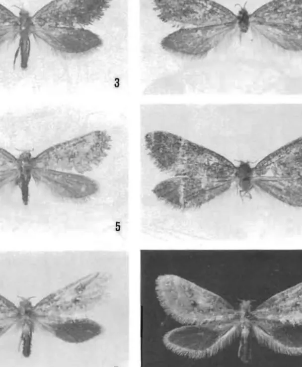
Figs 3-10. Ogygioses adults (wingspan in parentheses): 3, O. ca/iginosa (15-4 mm); 4, O. caliginosa (17·6 mm); 5. O. eurala (16 mm); 6, O. eurala (17·6 mm); 7-8, O. issikii, holotype (16 mm); 9, O.
iuangensis, holotype (13·8 mm); 10, O./uangensis, paratype (15·7 mm).
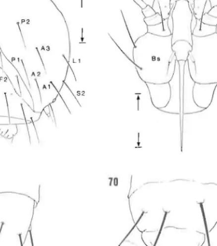
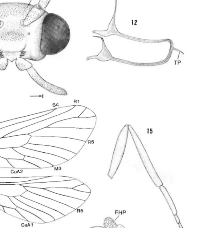
of two pairs of cranial nipples (Figs 11,21) immediately caudad to the antenna. Kristensen (1978b) reports these structures in other Hepialoidea, with three pairs present in Anomoses.
Superficially, they appear to be sensory in function (Kristensen 1978b) and resemble paired processes present on the pronotum (Figs 16, 22) of several Hepialoidea.
4
l
1236 D. R. Davis et a/. Revision of Og.
Among the previously described Paleosetidae, Ogygioses shows closest affinities to Genusles with regard to the development of a tibial hair pencil and similar forewing pattern. In the latter at least the 'bands' of small bluish white spots may count as a genuine synapomorphy; such distinct spots are absent from the mottled forewings of Osrhoes, and
19
14
Figs 11-15. Ogygioses euraw, head and thoracic structure: 11, head, anterior view; 12, tentorium; 13, wing venation, male; 14, fore, mid and hindlegs, male; 15, hindleg, female. eN, cranial nipple; FHP, follicles of tibial hair pencil; IS. intercalary sclerite; TP. posteromedian tentorial process. All scales
=
0·5 mm.
Figs 16-20.
16, prothora:
view (0·5 m ventral view primary fllre arm; TB. ter!
1237 D. R. Davis el al.
llVS closest affinities to similar forewing pattern.
~ay count as a genuine rcwings of OS/-hoes, and
. /
"J;;
. ~ TPrview; 12, tentorium; 13, eN, cranial nipple; FHP, rial process. All scales
=
Revision of Ogygioses
AP
20 19
T
1
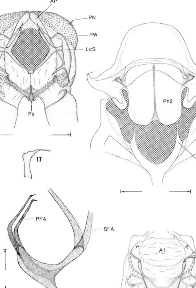
Figs 16-20. Ogygioses caliginosa, thoracic and abdominal structure (scale lengths in parentheses):
16, prothorax, anterior view (0·5 mm); 17, prothoracic furca, lateral view; 18, mesothorax, posterior view (0·5 mm); 19, metathoracic furasternum, lateral view (0·25 mm); 20, abdominal segments 1-2, ventral view (0·5 mm). A, abdominal segment; FS, furcal stem; LcS, laterocervical sclerite; PFA, primary furcal arm; PN, pronotal nipple; Ps, prosternum; PW, pronotal wart; SFA, secondary furcal arm; TB, tergal process.
.
1238 D. R. Davis el at.
Figs 2] -28. Ogygioses ca/iginosa, head structure (scale lengths in parentheses): 21, cranial nipple (25
flm) ; 22 , pronotal nipple (20 flm); 23, antenna, sixth segment (27 flm); 24, sensilla coeloconica, paired, in 23 (4·3 fl 01); 25, intercalary sclerite (6 )1m); 26, mandible (left), maxilla (30 )1m); 27, apical (second) labial palpus (20 flm); 28, sensilla trichodea at apex of labial palpus; note lack of sensory pit (17·6 )1m).
S, scape; P, pedicel. Bar scale for all photographs as shown in 21.
Revision of Ogyg.
the forewings hind wing of the
1978h), hut tl
relation sh ip he sim ilar hair pc V iette, 1951 in Puermytralls [' strongly rem il in vag inated hel even speak of • T he male geo their mo nophy (tergum X), la rel ativ ely simr
Osrhoes. The p and Ogygioses middle, and th e (or almost so) Osrhaes arise L
slende r, and fr Palaeases in he Ogygioses • monophyly of . constituent spc:
on the forewini a minute dis rlI represent a vc;
imilady notie groundplans 01 genital similari course, anotilci for similar fo r signi ficance ca sclerotiscd hin from a few Icp probahly he c modification a can test the vai By far the!
antennal intert 1978b) tentati' 'primiti ve lepi apart from a ( footnote 5) str among palaeo:
intercalary sci ' primitive typl reversal.
The 'ptero!
Ogygioses sp spacious laeur upper and low in the lacuna; eonccn tration but also aroun
1239 D. R. Davis el at.
): 21. cranial nipple (25 'lia cocloconica, paired,
m); 27. apical (second) I' sensory pit ([7·6 11m).
Revision of Ogygioses
the forewings of Palaeoses are without any distinct marking . The hair pencil of the hindwing of the Ogygioses male is superficially similar to that of Anomosetidae (Kristensen 1978b), but the hair ultrastructure is not; there are no other indications of a close relationship between these two taxa. Another hepialoid taxon from which a superficially similar hair pencil on the male has been reported is the neotropical genus Puermytrans Victle, 1951 in the Hepialidae s. str. (Nielsen and Robinson 1983). It is intriguing, that the Puermylrans male also has, in the posterior part of the forewing base, a scale pocket strong,ly reminiscent of that in Ogygioses laungensis. However. since this pocket is invaginated behind, not in, vein I A, strict homology seems ruled out; one cannot, therefore, even speak of 'underlying' synapomorphy here.
The male genitalia or all four genera differ strikingly, revealing little evidence to support their monophyly. Both Palaeoses (Davis, in press) and Osrhoe possess tergal processes (tergum X ), lacking in Ogygioses and Genustes. The valvae of thc lalter two genera are relatively simple (unlobed) in contrast to the prominently lobed valvae of Palaeoses and Osrhoes. The pseudoteguminal arms differ in all four genera, with the condition in Genusles and Ogygioses most similar. Here the pseudoteguminal plates are not synscleritous in the middle, and the arms arise widely separated; they taper gradually and are joined to the apex (or almost so) by the membranous body wall. The pseudoteguminal arms in Palaeoses and Osrhoes arise close together with those of Osrhoes more elongate with long apical portions, slender, and rree, as well as spined. The trulleum of Ogygioses is very simil r to that of Palaeoses in being well sclerotised and finely tapered.
Ogygioses currently consists or four species restricted to Taiwan and Thailand. The monophyly of OgYKioses is clearly demonstrated by a suite or notable synapomorphies of its conslitu nt species, including the forewing Rs configuration, the unique scale arrangement on the forewing base, and the loss of the jugal lobe [as noted by lssiki and Stringer (1932b), a minute disruption of the rorewing anal margin is evident in some specimens that may represent a vestigial jugum]. The close resemblance in genital structure of both sexes is similarly noticeable, but since few inferences can so far be made about details in the groundplans of hepialoid and palaeosetid genitalia, the phylogenetic significance of these genital similarities remains largely uncertain. The presence of spiny hindgut sclerites is, of course, another remarkable similarity shared by the Ogygioses species, but a careful search for similar formations in other hepialoids is obviously needed, before their phylogenetic significance can be assessed. One cannot easily attribute any physiological significance to sclerotised hindgut plates in non-feeding moths; however, such plates have been recorded from a few lepidopteran larvae (Dauberschmidt 1933), and their presence in adults can most probably be explained in terms of a takeover or the larval proctodael intima with little modification at metamorphosis. Obviously, only a future examination of Ogygioses larvae can test the validity of this conjecture.
By far the greatest surprise that emerged from the present study is the difference in the antennal intercalary sclerite of the Thailand and Taiwanese Ogygioses. Kristensen (I 978a, 1978 b) tentatively considered the retention in 'the P laeosetidae' of what we here call the 'primitive lepidopteran type' intercalary sclerite to be a plesiomorphy, that set this family apart from a clade comprising the other hepialoid ramilies. However, Kristensen (1978a:
footnote 5) stressed that the character in question had then been checked only in Ogygioses among palaeosetids. Our subsequent observations have revealed, that a 'typical hepialoid' intercalary sclerite is also prcscnt in Osrhoes and Palaeoses. It is possible, then, that the 'primitive type' intercalary sclerite in Taiwanese Ogygioses is an autapomorphic character reversal.
T he 'pterostigma' reported by Issiki and Stringer in both fore and hindwings of fixed Ogygioses specimens is not a noticeable sclerotisation of the wing membrane, It is a spacious lacuna (Fig. 78) in which epidermal cell bridges here and there extend between the upper and lower wing walls, and after fixation coagulated haemolymph forms a spongy mass in the lacuna; the wing surface here also has dense sca.le-sockets. Staining emphasises the concentration of these sockets not only in the subcostal regions shown by Issiki and Stringer but a'iso around most of the perimeter of both wings.
1240 D. R. Davis el al.
Species Inter-relationships
It is straightforward to assume that a sister-group relationship exists between Ogygioses luangensis and the Taiwanese Ogygioses species. However, although the latter assemblage is phenetically homogeneous, concrete evidence for its monophyly is not obvious. If, as suggested above, the 'primitive type' antennal intercalary sclerite in Taiwanese Ogygioses is indeed a character reversal, then this trait is an autapomorphy supporting the monophyly of the three species.
Ogygioses Issiki & Stringer
Ogygioses Issiki & Stringer, 1932a: 72. - Issiki and Stringer, 19320: 73; Kristensen, 1978a: 287;
Kristensen, 19780: 14; Davis, 1992: 62.
Type-species: Ogygioses caliginosa lssiki & Stringer, original designation.
Redescription Adults (Figs 3-10)
Relatively small moths with broad wings, often with 2-3 indistinct bands of small white spots obliquely traversing yellowish to dark brown forewings; wingspan 12-4-19·2 mm;
jugum absent; anal margin of hindwing with an elongate dorsal fold and hairpencil.
Head (Fig. II). Generally with rough vestiture; scales over frons erect and piliform with simple apices; vertex with divergent piliform scales concentrated immediately posterior to antenna, where they project over eyes, and caudad, tending to obscure (in unrubbed specimens) dense, appressed layer of broad, minutely serrated scales that cover most of vertex. Dorsal antennaI condyle of cranium distinctly developed, strongly melanised.
Antenna (Fig. 23) simple, filiform, extremely short, c. 0·15-0·2 the length of forewing, 17
segmented; scape cylindrical, enlarged, about twice the diameter of pedicel and 3 x the diameter of f1agellomere, without pecten, with scattered erect, piliform to slender scales.
Intercalary sclerite in lateral scapo-pedicellar membrane either (0. luangensis) of which might be termed the 'normal hepialoid type', viz. elongate, smooth and partly in vaginated into a pocket in ·the membrane (Fig. 77), or (Taiwanese species) that might be termed the 'primitive lepidopteran type', viz, a small, microtrichiated and superficially located plate (Fig. 25) fused to opposing rims of scape and pedicel. Flagellum mostly covered with moderately broad scales that gradually become more slender and sparse toward apex; each flagellomere (Fig. 23) densely covered with numerous microtrichia c. 0·2 the diameter of the segment; sensilla trichodea less numerous, evenly scattered, c. equal to segment diameter, arising from relatively large, shallow follicles; sensilla coeloconica (Fig. 24) reduced, indistinct; sensory pit shallow, without encirclement of specialised spines, randomly scattered over f1agellomere. Chaetosemata absent. Eyes moderately large but widely spaced (Fig. II); interocular index c. 0·64; eye index c. 1·0; cornea without interfacetal microsetae.
Vertex with 1-2 pairs of small cranial nipples (Figs 11,21) directly caudad to antenna.
Anterior tentorial pits arising high on frons (Fig. II), closer to antenna than to mandible;
dorsal arms of tentorium well developed, arising well anterior (Fig. 12); corporotentorium with prominent posteromedian tentorial process. Labrum undifferentiated; pilifer absent.
Mandible (Figs II, 26) reduced to a minute lobe covered with microtrichia. Maxilla (Fig.
26) reduced even smaller to a slender, minute lobe immediately below (caudad) and mesad of mandible; lobe probably rudiment of haustellum. Labial palpus 2-segmented; terminal segment elongate, nearly 3 x the length of short, triangular basal segment; terminal sensory pit (organ of vom Rath) not developed, with c. 12-14 short sensilla trichodea loosely concentrated subapically and arising from flush surface (Figs 27, 28); dorsum and distal half of terminal palpal segment also with numerous, large shallow sockets (Fig. 28), the smaller ones being the sockets of piliform scales, the larger sockets possibly functioning as other sensilla. Palpus arising from a prominent median lobe (prelabium).
Revision of Ogyg
Figs 29-36.
scalcs at lowe longitudinal ri scak structure of hindwing d (250 [.1111); 36 photographs a
i
.
D. R. Davis el al. Revision of Ogygioses 124 1
iSIS between Ogygioses Ihe lalter assemblage is is not obvious. I f, as faiwanese Ogygioses is fling the monophyly of
I; Kristensen, 1978a: 287;
ct bands of small white 19span 12-4-19·2 mm;
nd hairpellcil.
rons ereci and piliform
~ immediately posterior obscure (in unrubbed .Ies Ihal cover most of tl, slrongly melanised . lenglll of forewing, 17
of pedicel and 3 x Ihe :'orm to slender scales.
luan gel/sis) of which and paflly in vagi nated
H miglll be termed the erficially located plate mostly covered with Irsc toward apex; each 0·2 Ihe diameter of the to segment diameter, ca (Fig. 24) reduced,
;ed spines, ran domly .rge bUI widely spaced nlcrfaeetal microsetae . y caudad to antenna.
nna Ihan 10 mandible;
12); corporotentorium tiated; pilifer absent.
Ilrichia. Maxilla (Fig.
v (caudad) and mesad -segmented; terminal neOl; terminal sensory lIa Irichodea loosely lorsum and dislal half (Fig. 28), Ihe smaller funclioning as other
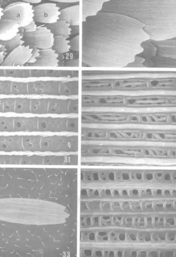
Figs 29-36. Ogygious caliginosa, wing structure (scale lengths in parentheses): 29, dorsal forewing sca les at lower distal corner of discal cell ( 100 J.lm); 30, detail of white scale (a) of 29, note absence of longitudinal ridge dimorphism (38 J.lm); 31, scale struclure of white scale; see 'a' of 29 (3 ~m); 32, sca le structure of brown scale; see 'b' of 29 (3 ~m). Ogygioses eurala, wing structure: 33, dorsal scale of hindwing discal cell (43 J.lm); 34. scale structure of 33 (3·8 ~ m); 35, hindwing, anal fold of the male (250 pm); 36, detail of anal fold of the male, open to show hair pencil (250 ~m). Bar scale for all photographs as shown in 29.
l
1242 D. R. Davis el aL.
Thorax. Proth rax (Fig. 16) with a thin, mostly int rnally inflected, laminate prosternum (discrimen): length of profureal arms (Fig. 17) c. 3·5 x the width of conical base;
laterocervical sclerite with relatively slender elongate median-lateral extension; pronotal wart extending to below dorsal apex of pleural wart, with acute. pronotal nipple (Figs 16.
22); anterior plate a sclcrotised dorsal arch distinctly divided at midline by a low internal sulcus but not extended caudad between pronotal warts. Mesothorax (Fig. 18) with moderately long, acute mesofurcal arms; phragma with shallow ven tromedian notch.
Metafurcasternum (Fig. 19) moderately laterally compresscd. with a relatively broad venlral keel that tapers to elongate. caudally projecting primary furcal arms; anterior process anterior to primary arms absent; common base f primary fueal arms arising midway on furcal stem; secondary arms separating weI! above common base of primary arms. Wingspan 12-4-19·2 mm. Forewing moderately br ad, length c. 2·8 x the width; retinaculum absen t;
jugum abs nt; microtrichia distributed over all wing surfaces; for wing spinarea (sens /L Minet I 89) small, but distinctive (Fig. 80). Venation hOnlon urous (Fig. 13) with humeral crossvein present; Sc unbranched (but site of vestigial Sc fork indicat d by n rve branching, Fig. 79). R4 connate with base of R2 + 3; R5 connatc with base of R2 + 3 + 4; discal cell relatively short, c. 0·5 the length of forewing; accessory 'ell absent, base of M nOl forked within cell; inler-M cross-vein (between M2 and 3) ahsent; base of CuP present to cross-vein or slightly beyond; I A straight, without basal loop. not reaching margin. Basal part of I A in
O. luangellsis with scale-bearing pocket (Figs 83-89). S(al covering of forewing upperside
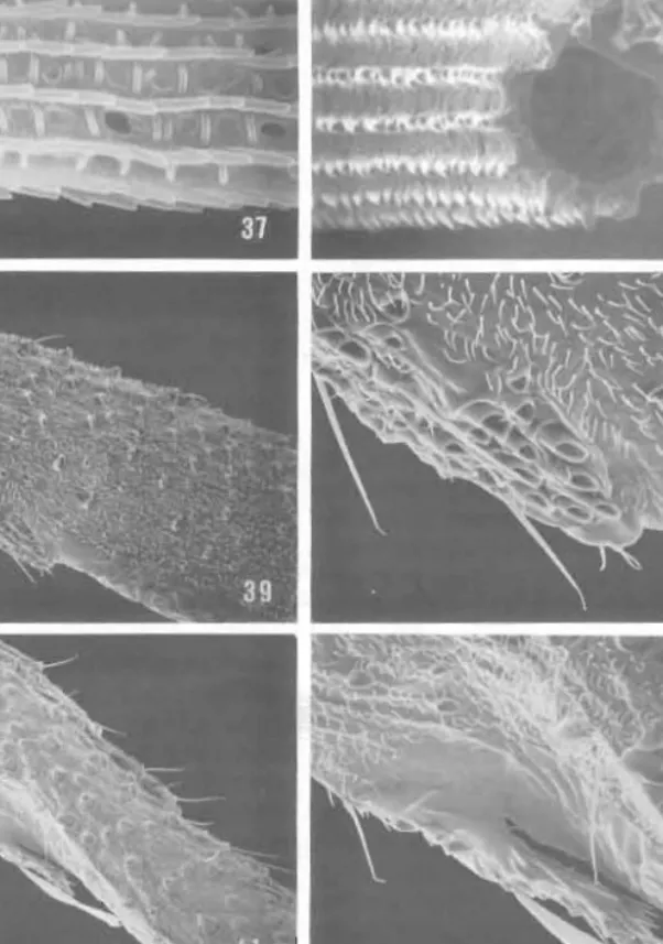
strikingly specialised (generic autapomorphy): in basal third, scale in front of M + eu largely arranged in 'whorl' of concenlric arcs (Fig. 82); toward midwidth of wing, scale axes becoming about normal to that of wing xis, or scale apices bing even directed more or less distinctly toward wing basco Eventually . cale rows 'straighten ut', so scales come to be oriented more or less parallel to long axis of wing, as is usual in Lepidoptera. Forewing pattern r'mging from pale goldcn yellowish brown to dark brownish fuscous. with numerous small bluish white spots bordering perimeter of wing and traversing wing in 2-3 indistinct blique bands; also irrorated by darker brown scales; white scales (iridcscing light blue) slightly broader than all other wing scales, with usual ly 6-dcntate apices, indistinct ridge dimorphism. and reduced windows (Figs 29-31); yellowish to brown scales 4-5 dentate, with moderate longitudinal ridge dimorphism (2-6 secondary ridges between each primary riJge), and elongate, large, rectangular windows traversed by irre ular cross ribs (Figs 29, 32). Hindwing with length C. 2·6 x maximum width; frenulum absent; Sc simple; radius as in rorewing. with R2 and 3 dividing near apex, R4 stalked at ba 'c with R2+3; R5 arising from base or R2+3+4; discal cell short, C. 0·5 lhe total wing length; base of M not forked within cell; inter-M crossvein absent; anal lobe or male with an elongate dorsal rold (Figs 35, 36) containing a hair pencil; pencil composed of extremely long, tubular (holl w) . cales (Figs 37, 38) capable of being raised vertically from the pocket; general surface of hindwing n oSlly covered by a relatively sparse, single layer of moderately slender, 3-4 dentate scales Wilh rather pronounced ridge dimorphism (Figs 33, 34); darker scales over distal two-thirds or O. issikii hindwing as br ad as forewing scales, usually with 5-dentute apices. Legs as shown (Fig. 14), with tarsi relatively short, C. equal to I ngth of tibia; first (basal) tarsomcre the 10lJgesl. C. 2 x the length of second on fore and midJeg ,nd 3 x on hind leg; all tibial spurs (including epiphysis) abs nt; foreleg with median Libial notch or short spine as vestigial epiphysis (Figs 39-42); hindtibia of male enlarged. particularly in Taiwanese species, with pr minent hair pencil (c. equal to length of tibia) arising near base and lying along a men branous, longitudinal, dorsal groove (Figs 14,47); scales of peneil tubular with sinuale Ivngitudinal ridges (Figs 48, 49); membranous groove apparently capable of inflation and covered with spindle-shaped androc nia with low relief, reticulate surface pallern (Figs 50-52) !ackino distinct longitudinal ribs and scutes; hindtibia of femal nol swollen, without hair pencil (Fig. 15). Pretarsus of all legs similar, with well-developed, simple claws, a broad arolium on a relatively short base, a symmetrical pair of moderately developed pulvilli (Figs 43. 44); unguitract r plate relatively slender with C. 6--7 somewhat staggered vertical row. of microscutes (Fig. 43); pseudempodial seta variable, reduced (Fig. 45) to moderately well developcd.
Revision of O!
Figs 37-44 hair pencil fo reti bia, v 11m); 42. d scale for nl
1243 D. R. Davis et at.
lIy inflected, laminate he width of conical base;
.eral cxte ns io n; pronotul lronotal nipple (Figs 16,
~idline by a low internal Ilthorax (Fig. 18) wi th
~v vcntromedian notch.
I relatively broad ventral arms: anterior proc ess lrms arising midway an primary arms. Wingspan dth; retinaculum absent;
)rewing spinarea (sen su s (Fig. 13) with hum ral ated by nerve branch i ng, if' R2 + 3 + 4; discal cell It, base of M not forked :IIP present to cross-vein rgin. Basal part of I A in Ig of t'orewing uppcrside lies in front of M + Cu Vidth of win g. scale axes len directed more or less
" so scales come to be Lepidoptera. Forewing fuscolls, with numerous g wing in 2-3 indistinct
; (ifldcscing light blue) apices, indistinct ridge ,wn scales 4-5 dentale.
; between each primary liar cross ribs (Figs 29,
;Sc simple; radius as in R2+ ; R5 arising from of Mnot forked within o['lal fold (Figs 35, 36) ,r (hollow) scales (Figs 11 surface of hindwing Ider, 3-4 dentate scales s over distal two-thirds lentate apices. Legs as , first (basal) Larsomere ( on hin<lleg; all tibial llch or short spi ne as icularly in Ta iwa nese 19 near base and lying : of penCil III ular with tly (,:;lpable of inflation c sur ace pattern (Figs Ie not swo llen, withuu t oped, simple claws, a tely developed pulvilli
"hat ·taggered vertical Fig. 45) [0 moderately
Revision of Ogygioses
Figs 37-44. Ogysioses caiigillosa. scale and leg structure (scale lenglhs in parentheses): 37. detail of hair pencil scale in 36 (2·5 flIn); 38, cross-sectional view of hair pencil sc~le in 3 7 (2·5 flOl ); 39, foretibia, vestigial epiphysis (86 flm): 40, detail of 39 ('27 flm); 41. foretibia, ve tigial epiphysis (86 flOl); 42. detail of 41 (30 !l0l); 43, foreleg pretarsus, ventral view (20 flIn); 44, lateral view of 43. Bar scale for all photographs as shown in 37.
I
1244 D. R. Davis el ai. Revision of G
Ahdomer marg ins; v area. and 111 membranoll area of trOll surface (Fi§ 55. 56); tre hindtibial I
defined. na
In re sclera swo\l n. c1~ caudad of f
Ma it' ge.
sterno. in h;
somewhat · removable sternum) st rim \1f poc:
. pecic '. ne species (Pi~
whi h cxte.
slraighllor...
{(opographi body wal l inlcrsegmc:
commonpJ·
slightly di ( ielsen an d()fsal s lri Pseudotcal without dl f
a
P,\lror
I pseudoteguarms
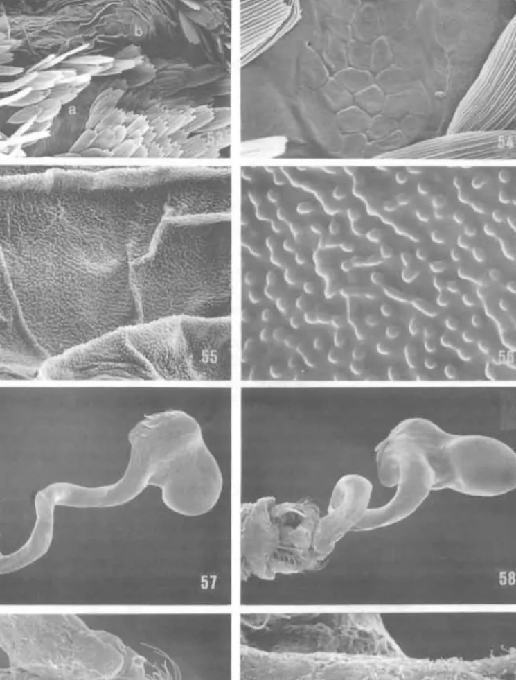
loose (C. 0·5 the minut<!ly bl broadened heneath an with morpl purall I m rou nded a~FemaLe lat ral or r almost to eopulatrix asymmetri IX plate, Figs 45-52. Ogygioses caiiginosa. leg structure (scale lengths in parentheses): 45, dorsal view of 43
Ductus bu showing reduced pseudempodial seta (arrow) ( 10 J.lm); 46, foreleg showing rough scaling along tarsal
spicules: ( segments (0·5 mm); 47, hindtibia, hair pencil of the male (0·3 mm); 48, hair pencil scales of 47 (13·6
membrana 11m); 49, detail of single hair pencil scale in 48 (3 11m); 50, hindtibia of the male showing dense array of
underlying androconia. hair pencil removed (0-43 mm); 51 . androconia of 50 (120 11 m); 52, surface small. seJ detail of androconial scale, 51 (6 11m). FNP, fo.ll icles of hair pencil. Bar scale for all photographs as spermath shown in 45.
1245 D. R. Davis el al.
,es): 45. dorsal view of 43 rough scajing along tarsal r pencil scales of 47 (13·6 ale showing dense array of 50 (120 pm); 52. surface ale for all photographs as
Revision of Ogygioses
Abdomen (Fig. 20). First tergum mostly membranous with strongly sclerotised lateral margins; vestiture sparse, largely naked with scatt red small. slender scales over medial area. and more densely clothed with longer, piliform scales laterally. First sternum entirely membranous. Pleural region of A2 with a naked, slightly hollow trough (Fig. 53); ventral area of trough with a small (c. 75 11 111 long) ovoid, tuberculate plate po sessing a reticulate surface (Fig. 54); surface of trough mostly covered with anastomosing microtubercles (Figs 55, 56); trough may receive hindwing hair pencil fold when wing is folded, as well as hindtibial hair pencil. Second sternum partially membranous with a more or less well
defined. narrow 'H' -shaped ternite that posses es a largely membranous anterior half and more sClerotised posterior half; a pair of rod-like anterior arms terminating in slightly swollen. clavate apices that articulate with latero-ventral pro csses from tergum I (Fig. 20) caudad of first abdominal spiracle. Spiracles present on first 8 segments.
Male geni/(Jlia (Figs 91,93-99). Thin-walled eighLh sternum differing from preceding sterna in having the anterior rim well melanised, strcngthened by transvcr e suture (wiLh somewhat variable course); median group of strong setae (unlike adjacent scnles not easily removable "fter KOH maceration) present on posterior sternal area. Vinculum (ninth sternum) strongly reflexed caudally, forming a vincular pocket (Figs 91. 97); posteroventral rim of pockct (i.e. topographically caudal margin of vinculum) bilobed in Taiwanese species, nearly traight in O. II/angensis. The incular pocket is deeper in the Taiwanese species (Fig. 97) than in O. luallgensis (Fig. 91); on the other hand the vinculum apodeme, which extend forward from the baLlom of the pocket, is markedly longer in the laller. It is straightforward to assume. that the 'vinculum' portion that is morphologically anterior to (topographically posterior to/below) the apodemal invagination line, is a scJerotisation of (he body wall area which at some ancestral stage formed the bulk of the ventral VIII-IX intersegmental membrane; simi lar modifications of the ventral VIII-IX transition arc commonplace in hepialoids: internal lateral ridges of vinculum Fig. 94) eiLher parallel or slighlly divergent towanl dorsal caudal margin. Tegumen (ninth tergum) membranous (Nielsen and Kristensen 1989). Tergum X almost entirely membranous, except for J slender dorsal strip; small scale groups may demarcate the site of vestigial tergal lobes. Pseudoteguminal plates with strong internal crests along lateral margins (Pig. 93). but wiLhout differentiation of 'intermediate plates' laterally; plates well separated. consisting of a pair of lateral, triangular sclerite ' tapering caudally as a pair f extremely slender pseudoteguminal arms thal converge but do not join posteriorly (Figs 94-96); plates and arm; loosely connected dorsally by a sheet of membrane. Trulleum reduced to an elongate (c. 0·5 the lenglh of valva), well sclerotised spine-like process tapering Lo an acute or minutely bulbous apex; apical area more or less distinctly corrugated. JuxLa articulaling with broadened anterior base of trul leum. an elongate plate with median strengthening ridge.
beneath and mesad of pseudoleguminal plates, then widening toward ventral articulation with morphologicaUy caudal margin of vinculum. Valva simple, densely selose. Wilh almost parallel margins or (0 . /uangensis ) somewhat widening before tapering abruptly to a rounded apex. Phallu entirely membranous; gonopore immediately dorsad to trulleum.
Female genitalia. Ninth sternum mooerat Iy well developed, evenly rounded, without lateral or medial lob s, setose. Subanal plates (Fig. 101) slender, paired, extending caudally almost to midline where they are narrowly separatcd by shallOW intergenital cleft. Bursa copulatrix elongate, c. 0·8 the length of expanded abdomen. Antrum conical and strongly asymmetrical: the sclerotised antrum floor, which posteriorly is continuous with the sternum IX plate, has the tapering anterior apex strongly directed toward the anImal's right side.
Ductus bursae slender, about same diameter from ostium to corpus, wal.ls with scattered spicules; corpus bursae relatively small, usually gradually, sometimes abruptly enlarged, membranous, occasionally with wrinkled walls. Spermatheca (Figs 100, 103) arising from small, sclerotised, cupuliform papilla on an elongate moderately broad duct; entire spermatheca elongate, often exceeding length of bursa copulatrix, with efferent
I
1246 D. R. Davis el at. Revision of ()
(fertilisati with vesicle c pu la tri..
d velopmer
Viscera.
the exami n plates on til as 0, luanG low sclerO •
DiSCUlision Largely OUl1d to dj differences;
population:
yellOW f ro rccogni 'cd straight cO {"(II( illosa outline. T Subscquen ductus spe indicate tr.<
The proble of rno t fc these, all ' rubbed (lh O. caligi/l~
same spec female wi~
large scrie
r
O. I!11/', TaipingsheA gene colOllr eve browOIsh t Future em
As more f . permalhc ductus Wll
an identl tOO, 102).
represent '
Figs 53-60 pleura, 53·
(area 'u' o' caligilwsa , (O·3~ mm) spermatopt
1247 D. R. Davis el at. Revision of Ogygioses
(fertilisation) canal variable, tightly coiled in c. 7-8 whorls at anterior end near junction with vesicle to merely sinuate; utriculus long and slender, more than half the length of bursa copulaLrix. Dorsal plate (tergum JX + X, ielsen and Kristensen 1989) similar in development to sternum IX, setose, shallowly bilobed.
Viscera. An unexpected detail in the visceral anatomy of Ogygioses was noted during the examination of KOH-treatcd abdomen preparations: the presence of spiny, sclerotised plates on the cuticular intima of the anteriorm 5t part of the hindgut in O. caliginosa as well as O. luangensis. The plates are arranged in a ingle circle around the gut. Each plate is a low sclerotised dome with a few (occasionally a single) blunt pines on top (Figs 90, 92).
DisclIssion
Largely due to unusual uniformity of genital morphology, no reliable character ha' been found to distinguish males of the three recognised Taiwanese species or Ogygioses. Minor differences in the orientation of internal ridges in the vinculum (Figs 94, 95) vary within populations. Ogygioses issikii, known only from males, is easily distinguished by its pale yellow forewings and distinctly marked hindwings (Figs 7, 8). Issiki and Stringer (I 32) recognised two species on the basis of two features: forewings ochraceous orange with a straight costa (0 . eurala), and forewings bronzy fus us with slightly sinu Ie costa (0. caliginosa). No significant difference was observed in this study with regard to costal outline. T he type series of O. caligillos(l and O. eurata also consist only of males.
Subsequent collections of females include at least two species based on differences in the ductus spermathecae (Figs 100. (03). The uniform browl1lsh hindwings of these females indicate thaL they most probably represent O. caLiginosa and O. eural£l and not O. issikii.
The problem is, then, one of association, which is complicated further by the poor condition of most females examined. Only J I females from Taiwan were available for study. Five of these, all with mostly sinuate ductus spermalhecae, were collected on Alishan. Although rubbed (three are preserved in alcohol), these females most resemble the darker holotype of O. caligillosa. lIeppner collected seven males swarming with one female near Liukuei of the same species as from Alishan (with sinuaLe ductus spermathecae). Similarly, the single female with a coiled ductus from Hassenzan (= Pahsienshan), and particularly the relatively large series of associated males from the same population. most resemble the paler holotype of O. ellra/a. The latter association also is true f the single female examined from Taipingshan and the three pale males collected at Taiheisan (= Tuipingshan).
A general attempt was made to group all Taiwanese males of Ogyg ioses according t colour even though there exists an almost continuous range from pale yellowish brown to brownish fuscous. Without associated females, identification of males remains que tion, ble.
Future emphasis should be directed toward collecting more pairs ill copula at lekking sites.
As more females become avai lable for study, the morphological variation of the ductus spermathecae should be evaluated. No intermediacy b tween the muILi-coileu and sinuute uuctus was noted in the II females dissected. Within females possessing multi-coiled duClS and identified as O. ellrala. variation in the form of the corpus bursae was observed (Figs 100, 102). This difference could indicate another species, or, as treated in this report, merely represent within-species variation.
Figs 53-60. Ogygioses, male structure (scale lengths in parentheses). O. eurata, second abdominal pleura, 53-56: 53. naked 'trough' (100 11m); 54. reticulate surface and pores of A2 tuberculate plate (area 'a' of 53) (20 11m); 55. surface of trough (urea 'b' of 53) (10 11m); 56. detail of 55 (2 rim). O.
caiigill()sa, spermatophore extruded from abdomen, 57-60: 57, lateral view (0·43 111 m); 58, ventral view (0·33 mm); 59, ventral iew of genitalia and segment VIll (120 11 m); 60. exposed fibrous tissue of spermatophore stalk (38 11m). Bar scale for al.1 photographs as shown in 53.
:
1248 D. R. Davis el al.
Key of Species of Ogygioses
I. Forewing (Figs 3-8) In both sexes without prominent light central spot and postdiscal band;
hindtibia of the male (Fig. 14) strongly swollen; male genitalia ( igs 94, 95, 98) with valvae almost parallel-sided, pseudotcguminal arms slender. with simple pointed apices; female genitalia (Fig. 10 104) without prominent broadening of ductus bursae; Taiwan ... . 2 Male forewing (Fig. 9) with prominenl. light yellowish central spot and postdiscal band; hindlibia of the male (Fig. 81) not markedly swo llen; male gemtalia (Fig. 99) with valvllc distinctly broadened subapically. p~eudo-teguminal arm stout, strongly melanised. With concavity in apical region; female genitalia (Figs 105, 106) with prominent broadening of ductus bursae; continental South-east Asia .. .. . . . . . ... O. Irwngel1sis, sp. nov.
2. Ilindwing, including fringe. uniformly grey to brown (Figs 3,5) ... 3 Hindwing with apical two-thirds dark brown; basal third and fringe conlrastingly white to cream (Figs 7-8) ... ... .. . . . .. . .... O. issikii, sp. nov.
3. Forewing usually paler (Figs 5, 6). often with Iij!hl brown to yellowish suffUSIOn; femllle with multi- w iled ductus spermathecae (Figs 100. 102) .... . . .. . . ... .. O. ellrala Issiki & Stringer Forewing generally darker nnd less contrnslingly marked (Figs 3. 4); tluctu: sperm the ae mo'lly sinunte, with no more than a single eoil (Fig. (03) ... O. caligillosa Issiki & Stringer
Ogygioses caliginosa Issiki and Stringer (Figs 3, 4 .16-32, 37-52,57-76. 78,94, 103, 104)
Ogysioses caligil1l1sa Issiki & Stringer. 1932a: 72. -Issiki and Stringer, 1932b: 74; Heppner. 1987:
13; Dnvis, 1992: 62.
Material Examined
Holotype. Taiwan, Nantou District: Ranlaisan [=? LantashanJ. 3-5.vi.1927, S. Issiki, sltde 16602 (USNM).
Other lila/erial /!Xl/milled. Taiwan: specific locality unknown. 25, Mar. 1927, S. Issiki (USNM).
Daamn [present name = '!]: I 't, 25.vi.1927, S. Issiki, slides 16603, 30617 (USNM). Chlpyi District:
Alishan, 2400 m, 10, I '? IO.vii.1981, K. eda, ,Iides ORO 3834. 3835 (KMNH); 20 , 19. slide 30596 (USNM); 90, 2<;>, 28.vi.1987, H. Kuroko. slides 31178. 31179 (USNM1; 2274 m, I'i' . 17-23.vi.l988, H. Kuroko, slilJe NPK 914 (ZMUC). Fennchlhwu: 1450 m. 60. 2-4.vii.1985, 1.
Heppner and Wang, slide ORO 3684 (FSCA). Taltaka [TungpuJ: 60, 8. vi.l943. S. rssiki, slide 30595 (USNM). llualien DIstrict: Higasinok [Tungnunkao], 40, 2-3.vi.1943, S. l~~iki. ~Iides 16154,30976 (USNM). Tzu en: 20, 4-6.vi.I982, T. Tunube (KMNH). Koashiung District: Shanpin Forestry SIn. nr Liukud, 750 m, 80. 12, 29.IV.-3.v_1989, J. Heppner and H. Wang, slides ORO 3845-3847. 31119 (FSCA. USNM). Nantou District: Lushnn. 10'. J9.iii.1982, S. lin himOlo, slide DRD 3833 (FSCA).
Musya [Jenni = Wushel: 10 . 2J.v. 1947 S. Issiki. slide NPK 916 (ZMUC). I
a,
slides 30568, 3108 .~ 11 17 (USNM). Rantalsan ['1 Randaizan
=
LantashanJ: 20 p:uatypes). Mar. 1927. S. Issiki. (BMNH);10 (hololype). 12 (p:uatype). CI-5.vi.1927. 10. IS.v.1933, S. Issiki. slides 16602,30593 (USNM).
Taichung District: Baibaru IMeiyuan], 10, 23.iii. 1943, S. Issiki, slide NPK 917 (ZMUC), 1($. I '? , slides 30967. 31172 (USNM ). Tolnan Distl'"iet: Kansirei [Kunnt7.uling, 300 m ], I'?, 16.'(.1934. S.
Is iki, slide 30594 (USNM).
Redescription Male (Fig. 3)
Wingspan 13·5-15 rom. Head with scattered, erect, piliform, cream to dark brown scales over frons and rear of head; vertex covered by broad, appressed, equally mixed cream and brown scales. Antennal scape with broad to slender, cream scales irrorated with dark brown to fuscous; flagellum completely covered with moderately broad, mostly brown scales dorsally and cream ventrally. Labial pal pus very rough, with long, divergent, slender cream scales heavily intermixed with brown to fuscous. Thorax with pro- and mesonota gener;.tlly covered with smooth, broad, cream scales irrorated with brown to fuscous laterally;
occasionally mesonota almost entirely brown; long, erect pili form brown to cream scales prominent at sides and posterior; venter sparsely covered with long, slender, mostly dull white scales. Forewing generally bronzy brown to fuscous, irrorated with small spots of
Revision of G
white scale but more or 14 spots bo of hind (do costa and a predominan venter of fo in colour. I dorsal and scales beco similar but smoother, u pencil crea Abdomen d
fHale gel converge di minutely 1'0 caudally; VI apex; abruf.
elliptical, s\
slender, slif generally sr
F efllale I Wingspz Hindwing \ hair pencil
Female without mc internal spi bursae. Du(
coil before
Egg (Fig Length ( when laid, with a regu pm in dian diameter ra with 2 pore vitelline en'
First illS Maximu epicranial r cleft and 5 lateral rim segments; , and 2 stout III minute, length to I sensillum a palpi 3-se£
1249 D. R. Davis el at.
ntral spot and postctiscal band;
In (Figs 94, 95, 98) wilh valvae simple poi nted apices; fema le , bursae; Taiwan ... ... 2 and postdiscal band; hincttibia of
°ig. 99 ) with valvae di tinctly IJalliscd. with concavity in apical ing of ductu bursae; continental .. . O. h/{Illgell.\'i.~ . sp. nov. . .. . . .. .. 3
~e contrastingly white to eream .. ... .. . O. issikii. sp. nov.
Ish ~uflusion: female with multi
. O. eurata Issiki & Stringer 4); ductus spermathccae mostly . 0 ca/igil/Qsa Issiki & Stringer
get )3. IO·l)
ngcr 1932[,: 74; Heppner. 1987:
·5vil927, S. Issiki. slide 16602
Mar 1927. S. Issikl (USNM).
D617 (U5NM). Chillyi District:
,,815 (K.1vl H); 20'. I '? slide 1\ 179 (LlSNM); 2274 m, I"' , 1450 m. 66 , 2-4.Vli.1985, J. 8 vi. 1943. S. Issiki. slide 10595
~. S Issiki. slides 16154, 30976 ISlrlCl Shanpin Forestry Stn. nr
sliue DRD 3845-3847, 3 11 19 noto, slide DRD 3833 (FSCA).
flle). 10. slides 30568. 31083 • . Mar. 1927, S. Issiki. (BMNH):
sl id~s 16602.30593 (USNM).
eNPK~17(ZMUC). lo , 19, Ilg, ~oo mi. 1'2 , 16.x.1934. $.
cream to dark brown scales d, equally mixed cream and :s irrorated with dark brown ,road, mostly brown scales 19, divergent, slender cream Jro· and mesonota generally rown to fuscous laterally;
orm brown to cream scales
1 long, slender, mostly dull rorated with small spots of
Revision of Ogygioses
white scales with blue iridescence and dark brown scales; white spots scattered over wing but more or less arranged in 3 oblique rows (subterminal, medial and basal) also as many as 14 spots bordering entire wing margin from basal third of costa around termen to basal third of hind (dorsal) margin; dark brown to fuscous scaling often concentrated along base of costa and along a subterminal arch from below apex to basal half of hindmargin; fringe predominantly brownish fuscous to light brown interrupted by white at each marginal spot;
venter of forewing uniformly light brown. hindwing uniformly grayish brown ; fringe similar in colour. Foreleg with tibia and tarsus extremely rough scaled (Fig. 46) primarily over dorsal and lateral surfaces; scales mostly long, slender and brown irrorated with cream;
scales becoming shorter and cream to white ventrally and toward apex of tarsus. Midleg similar but generally paler, with more white scaling toward tarsus . hindleg generally smoother, uniformly light brown, becoming paler over tarsus and venter of tibia; tibial hair pencil cream; clusters of long piliform scales at apex of tibia and some tarsomeres. Abdomen dark brown to fuscous dorsally and laterally; white to buff ventrally .
Male genitalia (Fig. 94). Pseudoteguminal arms very slender, tapering to acute apices that converge distally but are well separated by membrane. Trulleum likewise tapering to a slender, minutcly rounded to acute apex. Vinculum with internal ridges either parallel or divergent caudally; ventrocaudal margin of vinculum bilobed. Valvae about as broad at base as near apex; abruptly narrowing to subacute apex. Spermatophore (Figs 57-60) terminating in an elliptical, swollen sac about 0·50 mm long and 0·23 mm in diameter; extending from the sac a slcnder, slightly coiled tube more than twice the length of the sac. Surface of spermatophore generally smooth except for fibrous texture where interior wall exposed (Fig. 60).
Female (Fig. 4)
Wingspan 13·5-19 mm. Similar in general colour to male but with less distinct pattern.
Hindwing with anal pocket and pencil absent. Hindtibia normal, not swollen and without hair pencil (Fig. 15).
Female genitalia (Figs 103, 104). Caudal margin of sternum IX evenly rounded, without median depression evident in 0. luangensis. Ductus bursae evenly tubular, with internal spicules evenly distributed throughout length, to elongate, smooth-walled corpus bursae. Ductus spermathecae mostly sinuate, with efferent canal forming only one complete coil before vesicle; caudal end of efferent canal terminating at a sclerotised ring.
Egg (Figs 61-66)
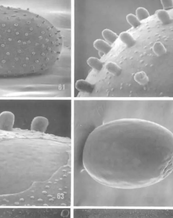
Length 0·37-0·40 mm; width 0·25-0·27 mm (n = 3, from a single female). Eggs white when laid, turning black within a few hours. Chorion extremely thin, transparent, studded with a regular array of relatively prominent, rounded tubercles up to 13 I-lm in Icngth and 9 I-lm in diameter (Figs 61-62), numerous, minute, low. rounded swellings c. 2-2·5 I-lm in diameter randomly scattered over chorion between tuberculcs (Fig. 62). Micropyle elliptical with 2 pores at opposite ends (Fig. 66). surrounded by 9 obscurely defincd plates. Underlying vitelline envelope rigid and smooth except for wrinklcd arca at micropylar (?) end (Fig. 64).
First instal' larva (Figs 67-76)
Maximum length 0·85 mm. Head with frontoclypeus extending nearly half the distance to epicranial notch. A3 located high on cranium dorsolaterally to PI. Labrum with deep medial elcft and 5 pairs of setae. Three pairs of rounded stemmata (Nos 3-5) closely bordering latcral rim of antennal socket. Antenna (Figs 71-72) with 3 short, strongly retracted segments; apex of II bearing I long, tactile, trichoid sensillum, 2 small sensilla trichoidca, and 2 stout sensilla basiconica about twice the length of the small trichoid sensilla. Segment III minute, bearing two sensilla: an elongate, digitate sensillum basiconicum about equal in length to largest basiconic sensillum of II but less than half its diameter, and a second sensillum about equal in length but with its basal half stout and distal half slender. Maxillary pal pi 3-segmented (Figs 68, 73); apical segment elongate, exceeding combined length of
l
1250 D. R. Davis el ai. Revis
basal two segments, with 3 sensilla basiconica clustered dorsally near mid length (Fig. 74). Dislr Labial palpus 2-segmented, with slender apical segment equalling length of stouter base. Ttl Spinneret extremely long and slender as in Hepialidae (Wagner et al. 1989), extend ing to 2400 prosternum (Fig. 68). Thorax with relatively long sle nder legs (Fig. 69); tarsus slender,
elongate, c. 2·3 x length of tibia; claw elongate, with secondary dorsal spines (Fig. 75).
Abdomen with long, slender prolegs (Fig. 70) on A3-6 and 10 bearing 6-7 crochets in a uniordinal circle (Fig. 76).
Figs 61-66. Ogygioses caiiginosa, egg structure (scale lengths in parentheses): 61, egg with chorion mostly intact (136 flm); 62, detail of chorion surface (27 flm); 63, thin chorion partially torn exposing vitelline envelope (20 flm); 64, egg with chorion completely removed (136 flm); 65, micropyle area (38 fl m); 66, micropyle with 9 peripheral plates (15 flm). Bar scale for all photographs as shown in 61 .
Figs view
Fligl;
A capll
DisCl
S,
Tai\l
of lh insta plan eggs dcnu therE
.'
1251 D. R. Davis el al.
ncar midlength (Fig. 74).
Ig length of ~touter base.
'I al. 1989), extending to (Fig. 69); tarsus slenJer,
dorsall spines (Fig. 75).
learing 6-7 crochets in a
~ses): 61 , egg with chorion ion partiaJiy torn exposing
m); 65, micropyle area (38 raphs as shown in 61.
Rev;sion of Ogygioses
Distribution
This species has been found in the Central Mountain Range from 300 m (Kwantzuling) to 2400 m on Alishan.
1
Figs 67-70. Ogygioses caligil10sa IIrst instar larva (scale lengths in parentheses): 67. head. dorsal view (75 ~I11); 68. labium and maxilla, ventral view (43 ~m)
as.
basisternite; 69, leg. T2; 70, pro1cg, A4.Flight Period
Adults have been collected from March to as late as October. The greatest number of captures has been in March (n = 7), April-May (/I = II), June (n = 30) and July (n = 8).
Discltssion
Seven mal.es and one female were collected by sweeping by J. Heppner near Liukuei, Taiwan. After mating with a male inside a vial, the female attached several eggs to the sides of the container. Within 6-7 days, several of these hatched and a few eggs along with first instar larvae were preserved in 70% EtOH. Attempts to rear the larvae on several local plants, including moss, were unsuccessful (Heppner, personal communication). Only six eggs and two larvae were available for this study. Both larvae were shrivelled and partially denuded as a result of being slide-mounted, Fungal hyphae also covered the specimens, thereby obscuring the chaetotaxy.
I
1252 D. R. Davis et af. Revision of
aS
Figs 71-76. Ogygioses caliginosa, larval structure, first instar (scale lengths in parentheses): 71, antenna (12 ~m); 72, antenna (8·6 ~m); 73, maxilla, ventral view (20 ~m), subapical sensilla (arrow);
74, maxillary palpus, subapical sensilla (3 ~m); 75, pretarsal claw, T2 (5 ~m); 76, crochets, AS (5 ~m).
Bar scale for all photographs as shown in 71 .
The egg chorion of Ogygioses is extre mely thin and easily loosened from the underlying vitelline envelope by immersion in alcohol. Most of the six eggs examined had lost much and in some cases all of their chorion (Figs 63, 64). The tuberculate sculpturing of
Ogygioses ( Hepialidae ( Wagner el a, tubercles in
Stemmat:
represent au instars shoul
Material Ex.
HO!lItype.
Other ma 30559 (USN Kirisato [Tu 6-10.v.1942.
DRD 3802 C Sankakuho 30616 (USt-
7.vi.1942. S Issiki, slides:
S. Issiki (U~
(USNM). R;
Wulai): 10
Redescript!
Male (F
Wings~
in pattern brown sca, band. Hinc dorsally aT' Male g.
Fema/~
W,ingsl1
Female with smOQ canal fore
Dislributi.
This sl (KwantZLl
Flight Pe Adult
May (n
![Figs 2] - 28. Ogyg ioses ca/iginosa, head structure (scale lengths in parentheses): 21, cranial nipple (25](https://thumb-ap.123doks.com/thumbv2/123dok/11185617.0/8.918.184.732.187.999/figs-ogyg-iginosa-structure-lengths-parentheses-cranial-nipple.webp)