This brings the virus membrane and cell membrane into close proximity, which triggers fusion between the two membranes (Gallo et al., 2001). The final product of reverse transcription is a double-stranded linear DNA that forms part of the PIC, called a provirus (Miller et al., 1997). Multiple Tats lead to strong up-regulation of viral genome transcription (Wei et al., 1998).
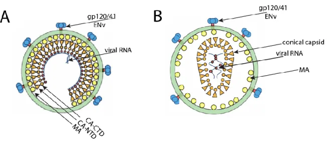
The carboxyl terminus protrudes toward the center of the viral particle (Fuller et al., 1997; Wilk et al., 2001). Imaging studies of HIV-1 CA mutants in target cells further provided evidence for a role for reverse transcription in facilitating uncoating, suggesting that variations in capsid stability can affect reverse transcription (Hulme et al., 2011). Recent studies have shown that the uncoating process can occur simultaneously with reverse transcription: that is, reverse transcription and uncoating could be coupled (Hulme et al., 2011).
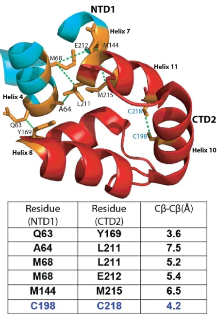
TRIM5 binds to the capsid of HIV-1 and apparently inhibits infectivity by accelerating uncoating (Stremlau et al., 2004; Stremlau et al., 2006). Two endogenous cysteines form an intramolecular disulfide bond between residues C198 and C218 (Mateu, 2002; Pornillos et al., 2009). This structure is based on data from the X-ray crystal structure of the hexamer (PBD 3H4E), Pornillos et al., 2009.
Recent studies using X-ray crystallography have provided a model for capsid assembly with a more detailed structure of the HIV-1 fullerene core (Ganser-Pornillos et al., 2007; Pornillos et al., 2009).
CTD-CTD dimerization interface: The CTD-CTD dimer interface is formed between hexamers of the mature HIV-1 capsid when helix 9 dimerizes with a neighboring helix 9
CTD-CTD dimerization interface: The CTD-CTD dimer interface is formed between hexamers of the mature HIV-1 capsid when helix 9 dimerizes with a neighboring helix 9.
CTD-CTD threefold axis: Although not evident by the X-ray crystal structure, cryoEM modeling predicts the presence of a CTD-CTD interface located at a three-fold axis of
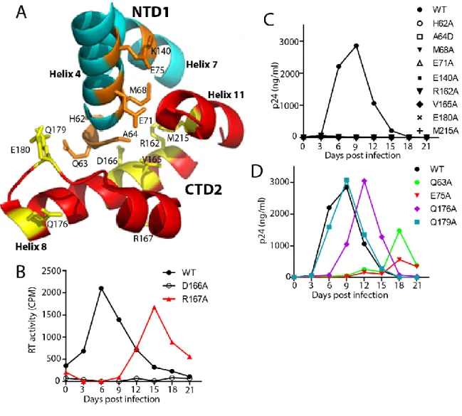
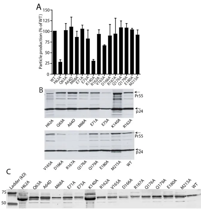
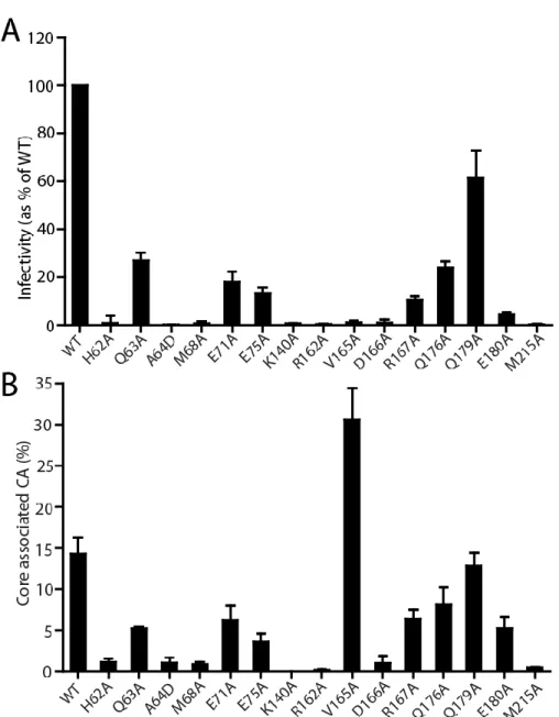
I also showed how the HIV-1 capsid is assembled and the role of the trimer interface in assembly. To study the role of the NTD-CTD interface in HIV-1 capsid structure and function, I generated a panel of HIV-1 proviruses containing single substitutions at the NTD-CTD interface. In contrast, most mutants of the NTD-CTD interface showed levels of core-associated CA lower than wild type (Figure 2-5B).
My results show that the reduced infectivity of the NTD-CTD interface mutants is associated with impaired reverse transcription. Overall, the effects of the mutations on capsid morphology suggest that residues at the NTD-CTD interface are important for proper capsid structure as well as stability. In this chapter I focused on the role of the NTD-CTD intersubunit interface in HIV-1 capsid structure and function.
However, the role of the NTD-CTD interface in the virion and its contribution to capsid function had not been. My engineered disulfide cross-linking data confirm that the NTD-CTD interface is a component of the native HIV-1 capsid network. H62 occupies the hydrophobic core of the NTD-CTD interface forming stacking interactions with F32 and Y145 (Ganser-Pornillos et al., 2007; Pornillos et al., 2009).
Most of the substitutions at the NTD-CTD interface produced virions with unstable capsids, all of which were poorly infectious. Point mutations of the hydrophobic residues at the trimer interface alter the level of CA associated with purified HIV-1 cores. We conclude that the hydrophobic interactions at the CTD-trimer interface are critical for formation and stability of the viral capsid.
Previous studies of the structure of the tubular HIV-1 capsid assembly observed strong density at the trimer interface at 16 Å resolution. A detailed comparison of the density maps of CA-SP1-NC and CA is shown in Figure 4-1D-I. Superposition of the CA monomer with CA-SP1-NC protein revealed that the NTDs of both structures aligned well, except for the CTD which did not (Figure 4-2C).
Overall, a comparison of intersubunit interfaces in CA and CA-SP1-NC tubes indicates that the trimer interface is altered or absent in CA-SP1-NC. –C, upper panel) Superposition of CA-NTD models at the NTD interfaces (A&B) and CA-CTD models at the trimer interface (C), with the assembled CA structure (gold) and that of the assembled CA-SP1-NC structure (blue). Recent mutational analysis of the hinge region has shown that the NTD-CTD linker sequence is critical for proper capsid assembly (Jiang et al., 2011a).
Much is known about the structure of the mature HIV-1 capsid and the various interfaces present.
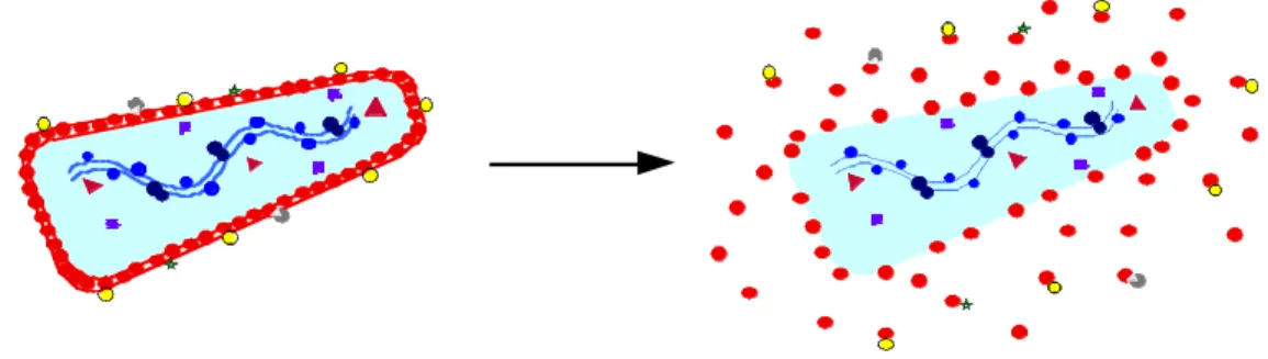
Disassembly and reassembly model of maturation
Protons that are protected by participating in subunit interactions are not available for amide exchange. Monroe et al., (2010) showed with immature virus-like particles, rapid deuterium exchange occurred at peptide residues participating in a mature virus-like NTD-CTD interface, indicating that NTD interactions -CTDs are not yet formed in the immature virus. Furthermore, a recent study suggested that the position of the CA NTD in the immature HIV-1 virus is quite different from its position in the mature virus, arguing that the interactions that exist in the immature should be different from those in the mature virus. Briggs et al., 2009).
The implication arising from this model is that contacts occurring in the immature capsid must be different from those in the mature capsid, but it can also be argued that major conformational changes occur during maturation. Taken together, this suggests that the immature capsid can break apart and then reassemble de novo to form the mature capsid, and that the capsid interfaces in the immature capsid may be different from those in the mature capsid.
Subunit rearrangement model of maturation
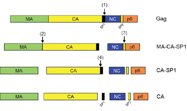
It has long been hypothesized that cleavage at the MA-CA junction leads to β-focle formation. Engineering these mutations into viruses containing engineered disulfide cross-links can be used to analyze whether the formation of any of the interfaces is perturbed. For each virus, the level of core-bound CA was determined as a percentage of the total CA in the gradient (based on ELISA).
Characterization of the invariant residue 51 mutations of human immunodeficiency virus type 1 capsid protein on in vitro CA assembly and infectivity. Residues in the HIV-1 capsid assembly inhibitor binding site are essential for maintaining the assembly-competent quaternary structure of the capsid protein. A retroviral chimeric capsid protein reveals the role of the N-terminal beta-hairpin in mature nuclear assembly.
Evidence for a functional link between core disassembly of human immunodeficiency virus type 1 and nuclear import of the virus. Structure of the full-length capsid protein of HIV-1 in a conformationally trapped, unassembled state induced by small molecule binding. Structural requirements for human immunodeficiency virus type 1 core recognition during host restriction in owl monkey cells.
Domains of the human immunodeficiency virus type 1 matrix and gp41 cytoplasmic tail required for envelope incorporation into virions. Inhibition of early stage events in HIV-1 replication by small molecule targeting of the HIV-1 capsid. Kinetic analysis of the role of intersubunit interactions in human immunodeficiency virus type 1 capsid protein assembly in vitro.
Conformational stability of dimeric and monomeric forms of the C-terminal domain of human immunodeficiency virus-1 capsid protein. Electron cryotomography of immature HIV-1 virions reveals the structure of the CA and SP1 Gag shells. Coupling of human immunodeficiency virus type 1 fusion with virion maturation: a novel role of the gp41 cytoplasmic tail.