The Role of Peroxidasin in Basement Membrane Physiology and Human Disease
By
Abraham Scott McCall Dissertation
Submitted to the Faculty of the Graduate School of Vanderbilt University
in partial fulfillment of the requirements for the degree of
DOCTOR OF PHILOSOPHY in
Pharmacology August, 2015 Nashville, Tennessee
Approved:
David G. Harrison, M.D. Sean S. Davies, Ph.D.
Ambra Pozzi, Ph.D.
Dan M. Roden, M.D.
Billy G. Hudson, Ph.D.
.
ii
PHARMACOLOGY The Role of Peroxidasin in Basement Membrane Physiology and Human Disease
Abraham Scott McCall
Dissertation under the direction of Professor Billy G. Hudson
Basement membranes are a distinct form of extracellular matrix responsible for signal transduction and mechanical integrity throughout development, in mature tissues, and during wound healing. The collagen IV scaffold of basement membranes relies on a sulfilimine crosslink (S=N) between methionine and lysine for its essential function of maintaining basement membrane architecture. The sulfilimine crosslink is formed by the heme peroxidase, peroxidasin. The precise mechanism by which peroxidasin forms sulfilimine crosslinks, or if this crosslinking process is involved in disease, remains largely unknown. Biochemical investigation of peroxidasin-catalyzed formation of the crosslink found that bromide (Br-) appeared to be the preferred enzymatic cofactor through its conversion to hypobromous acid (HOBr). Through my development of Br-free salts, purified proteins, and in vitro cell culture models of basement membranes, Br- was shown to be essential to the formation physiologically observed levels of sulfilimine crosslink. I further investigated the underlying mechanism of sulfilimine formation with chemical crosslinking, mass spectrometry analysis, and modeling.
These approaches cumulatively supported the presence of a S-Br+ (bromosulfonium-ion) intermediate by the crosslinked methionine of the NC1 domain of collagen IV as the key reaction intermediate and energetic basis for bromines role in sulfilimine crosslinking. The essentiality of Br was therefore tested in vivo in Drosophila by developing novel Br-free culture techniques. I found that dietary Br-deficiency is lethal in Drosophila while Br- replenishment restores viability, demonstrating a physiologic Br- requirement. Importantly, through electron and fluorescence microscopy, I was also able to show that Br-deficient flies phenocopy the developmental and basement membrane defects observed in peroxidasin mutants which indicates a functional connection between Br-, collagen IV, and peroxidasin. These data collectively established that Br- is required for sulfilimine crosslinking of collagen IV, an event critical for basement membrane assembly and tissue development. Thus, bromine is an essential trace element for animals through the enzymatic activity of peroxidasin, and Br- deficiency may be relevant to basement membrane alterations observed in patients undergoing dialysis, receiving total parenteral nutrition, and in some smoking related disease. I also sought to test the hypothesis that anti-peroxidasin autoantibodies occur in a specific rapidly progressive glomerulonephritis known as Goodpasture’s disease (GP). Goodpasture’s Disease is characterized by anti-collagen IV NC1 antibodies and the sulfilimine crosslink is thought to modulate immunogenicity of the collagen IV epitopes. Many GP patients have concurrent autoantibodies which recognize myeloperoxidase (MPO), a structurally related heme peroxidase to peroxidasin. Through testing multiple independent patient cohorts by immunoassay, I found anti-peroxidasin autoantibodies in GP patient sera, both before and at the time of clinical presentation.
Unexpectedly, the anti-peroxidasin specific antibodies cross-react with coated, but not native MPO, accounting for a subset of the historically characterized dual-positive (anti- collagen IV and anti-MPO) patients. I also found that anti-peroxidasin antibodies inhibited HOBr production, suggesting a possible contribution of these antibodies in GP pathogenesis within this subset of anti-peroxidasin positive patients. These studies demonstrate chemical, biochemical, and tissue level evidence for the role of peroxidasin and Br- in the assembly of sulfilimine-crosslinked collagen IV scaffolds in basement membranes and peroxidasin’s potential role in disease, including as a novel autoantigen in a subset Goodpasture’s disease patients.
iii
Acknowledgements
I would first like to thank my mentor Billy G. Hudson. During my time in the lab, he pushed me to excel both experimentally and intellectually in a way no one had before. I’m incredibly grateful for the opportunities he provided me while I was under his guidance; they made my time in graduate school everything I could have hoped. The constant pursuit of novelty from truly fundamental biology, especially with chemical detail, is the biggest lesson I will carry forward from our time working together.
Next, I would like to sincerely thank my committee, Drs. David Harrison, Ambra Pozzi, Sean Davies, and Dan Roden.
My path through graduate school would charitably be described as unorthodox, and their willingness to work with me through this process has meant a great deal. The flexibility, time given, and generosity shown was genuinely appreciated.
I am excited for these mentoring relationships to extend beyond graduate school.
I would like to explicitly acknowledge and thank ‘Team Bromine’ for their contributions to the final manuscript and to my training in the process. Dr. Gautam Bhave and I worked closely on many aspects of the project, including the design of Br-free Chloride purification apparatus and subsequent experimental designs of all Br-free purification and production methods for proteins, media, and Drosophila culture conditions. Dr. Bhave’s previous fundamental work on peroxidasin, both in terms of its biochemistry and the establishing the laboratory techniques required for its study, enabled this work to proceed at the astounding speed it did. The technical advances required with Br-free materials in this work were substantial, and while I conducted the experiments, it was our ability to work together that really brought the Br-free experimental concept to fruition. Dr. Chris Cummings performed initial experiments on peroxidasin cofactor preference which initiated our group’s collective interest in Br as a cofactor. He kindly allowed me to present our collaborative work in Figure 6A and D, and an experiment he performed in Figure6B to facilitate my explanation of the progression of the experiments. The drafting of the manuscript was a long process, but working with Dr. Cummings on many of the drafts was a great experience. Dr. Roberto Vanacore and I worked together on the mass spectrometry analysis, which would not have been possible without his expertise. The time he took to teach me how to conduct the analysis which ultimately appeared in the paper was a foundational piece of my training in the lab. Our work proved to be the backbone of my ability to quantify the mechanistic aspects of the sulfilimine crosslinking. Dr. Andrea Page-McCaw was instrumental in guiding the in vivo fly work, working closely with Dr. Bhave and me to develop the Br-free culture techniques, genetics, and assays which we ultimately used. Since I performed all of Drosophila work in her lab, I genuinely appreciated her hospitality, concern for my success, and mentorship in a field of science to which I was completely new. It was a privilege to be able to work this group of people.
Dr. Vadim Pedchenko, Dr. Stephen Olson, Dr. Agnes Fogo, and Ms. Ellen Donnert also deserve recognition for their resources, assistance, and insight, with the anti-peroxidasin antibody project. I am also grateful to K.L. Rose at Vanderbilt Mass Spectrometry Resource Center; J. Clanton, X. Wang, and E. Shannon for assistance with Drosophila in the Page- McCaw lab; J. Williams for assistance with Electron Microscopy and sample preparation, performed with use of the VUMC Cell Imaging Shared Resource; and the Bloomington Drosophila Stock Center, Yale FlyTrap, and the VDRC for fly stocks;
and S. Vaughn in the VUMC Hormone Assay and Analytical core for her assistance with cytokine assays.
My other colleagues in the Hudson lab, C. Jones-Paris, A. Fidler, K. Brown, C. Darris, S. Chetykrin, I. Ero-Tolliver, S.
Colon, N. Danylevych were exceptional resources, friends, and support throughout my time in the lab. The technical assistance and advice of P. Todd and M. Rafi was invaluable.
Dr. Terry Dermody, Dr. Larry Swift, and Ms. Melissa Krasnove in the Medical Scientist Training Program have provided important guidance, advice, and kindness throughout my training. It is deeply appreciated. I also am grateful for the support of Dr. Joey Barnett, Dr. Vsevolod Gurevich, Dr. Christine Konradi, and Ms. Karen Geig in the Department of Pharmacology.
Finally, I would like to thank my family, my fiancée Natalie, and my past mentors (S. Kraft and G. Conrad), without whom none of this would have been possible nor would it mean so much.
Sources of Funding: This work was supported by NIH R01 DK18381, NIH F30 DK100094, NIGMS T32 GM07347 to the Vanderbilt Medical-Scientist Training Program and the Canby Robinson Society of the Vanderbilt University Medical Center.
iv
Table of Contents
Acknowledgements ... iii
List of Figures ... vi
List of Tables ... viii
Chapter I. Introduction ... 1
Basement membrane architecture ... 1
Collagen IV composition and function in the basement membrane ... 6
Sulfilimine crosslink within the NC1 hexamer of collagen IV ... 8
Peroxidasin forms the sulfilimine crosslink ... 9
Goodpasture’s disease and collagen IV ... 12
Major questions addressed by this work ... 15
II. Materials and Methods ... 17
Materials ... 17
Biochemical Methods ... 22
Drosophila Methods ... 28
Human Antibody Methods ... 40
III. Bromide is a required cofactor for peroxidasin mediated collagen IV sulfilimine bond formation ... 49
Abstract ... 49
Introduction ... 50
Results ... 51
Structural basis for sulfilimine crosslink heterogeneity in the collagen IV scaffold ... 51
Bromide is required for sulfilimine formation ... 52
Peroxidasin catalyzes sulfilimine crosslink formation via Br- ... 57
Modelling thiocyanate effects on peroxidasin mediated sulfilimine crosslinking in smokers ... 61
Discussion... 66
IV. The chemical basis for selection of Br- over Cl- as the cofactor in peroxidasin mediated NC1 sulfilimine crosslink formation ... 70
Abstract ... 70
Introduction ... 71
Results ... 72
Chemical oxidations and sulfoxide formation ... 72
v
Model for S=N Bond Formation within the NC1 Hexamer and
Thermodynamic Calculations ... 73
Initial Sulfilimine formation potentiates a second crosslink through changing the apparent amine concentration ... 82
Discussion... 87
V. Bromide is essential for Drosophila development and normal basement membrane ultrastructure ... 89
Abstract ... 89
Introduction ... 90
Results ... 91
Bromide is essential for Drosophila development ... 91
Br-deficiency decreases sulfilimine content ... 92
Tissue Defects with Br Deficiency ... 92
Br-Peroxidasin-Collagen IV interaction in the ‘molecular corset’ ... 96
Discussion... 97
VI. Novel Anti-Peroxidasin antibodies in preclinical and clinical Goodpasture’s disease ... 106
Abstract ... 106
Introduction ... 107
Results ... 109
Anti-Peroxidasin Antibodies in Pre-Clinical and Clinical Goodpasture’s Disease ... 109
Anti-Peroxidasin Antibody Specificity ... 115
Cytokines in Pre-Clinical Goodpasture’s Disease Patients ... 116
Peroxidasin Inhibition by Anti-Peroxidasin Antibodies ... 122
Discussion... 124
VII. Conclusions and Future Directions ... 127
Essential function of Bromide-Peroxidasin-collagen IV axis in the basement membranes of Animals... 127
Mechanistic and Chemical Role of Bromide in Sulfilimine Formation... 128
Bromine Essentiality and Physiology ... 128
Recent developments since the establishment of Br as an essential trace element ... 133
Clinical Implications of Bromide Deficiency ... 134
Peroxidasin as a novel autoantigen ... 138
Peroxidasin and Disease ... 143
References ... 145
vi List of Figures
Figure 1. Tissues architecture and basement membrane composition and crosslinking ... 5 Figure 2. Peroxidasin forms the sulfilimine crosslink in the collagen IV NC1 domain ...10 Figure 3. Measurement of sulfilimine crosslink content within NC1 domains of collagen IV scaffolds...53 Figure 4. Quantitation of sulfilimine (S=N) crosslinked peptides (associated with Figure 3) ...56 Figure 5. HOBr reproduces physiologic levels of S=N crosslinking within the NC1 Hexamer (Associated with Figure 3) ...56 Figure 6 Bromide is the required cofactor for sulfilimine crosslink formation ...58 Figure 7. Purification apparatus and setup for the purification of Br-Free Chloride Salts and (associated with Figure 6) ...59 Figure 8. Peroxidasin uses physiologic Br- levels to form sulfilimine crosslinks ...62 Figure 9. Model of the interaction between serum Br and SCN- on sulfilimine crosslinking in normal individuals and smokers ...69 Figure 10. Chemical mechanism of sulfilimine (S=N) formation within the NC1 hexamer ...75 Figure 11. Met93-sulfoxide is formed as a result of HOCl oxidation and is a ‘dead-end’ for sulfilimine crosslink formation in the NC1 (Assocoated with Figure 10) ...78 Figure 12. Analysis of S=N formation via a halo-sulfonium intermediate. (Associated with Figure 10) ...83 Figure 13. Bromide is essential for development and basement membrane architecture in Drosophila. (Previous page) ...95 Figure 14. Br- and peroxidasin interact in vivo to strengthen collagen IV scaffolds. ...98 Figure 15: Phenotypic and genetic analysis of the role of Br- and Pxn in Drosophila (Associated with Figure 13 and Figure 14) ... 103 Figure 16: Antibodies recognizing peroxidasin are present in Goodpasture’s disease patients before and at the time of diagnosis. ... 111 Figure 17: Time resolved Anti-peroxidasin and Anti-MPO antibodies in pre-diagnosis GP
patients by ELISA from the DoDSR. ... 113
vii
Figure 18: Anti-α3 IgG and IgM levels in pre-diagnosis GP patients from the DoDSR by ELISA.
... 114 Figure 19: The subset of patients recognizing peroxidasin has anti-peroxidasin specific
antibodies. ... 118 Figure 20: GP patients have TH17 cytokines before diagnosis and elevated focal peroxidasin levels in crescentic glomeruli. ... 119 Figure 21: Peroxidasin is inhibitable by patient anti-peroxidasin antibodies in vitro. ... 123 Figure 22. Model of the essentiality of bromine in forming collagen IV sulfilimine crosslinks. .. 130 Figure 23. Schematic of Br function at a range of concentrations demonstrating deficient, optimal, and toxic concentration ranges ... 131 Figure 24 Model of anti-peroxidasin antibodies within the context of Goodpasture’s disease pathogenesis ... 140
viii List of Tables
Table 1. ICP-MS analysis of reagent grade and purified salts used experimentally. ...45
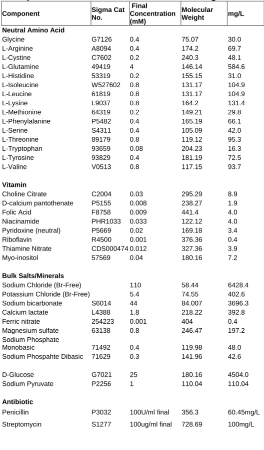
Table 2. Components of Br-Free Dulbecco’s Modified Eagle Media ...46
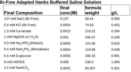
Table 3. Br-Free Adapted Hanks Buffered Saline Solution ...47
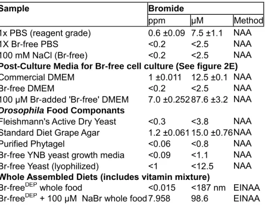
Table 4. Neutron Activation Analysis of Media and Fly Diet Components ...48
Table 5. Analysis of a two-equal and independent crosslinking event model ...79
Table 6. Fly Vitamin and Mineral Additive Mixture ... 104
Table 7. Neutron Activation Analysis of Fly Diet Components and Drosophila tissues ... 105
Table 8: Pre-diagnosis Cytokine differences in pre-diagnosis GP patients vs matched controls. ... 121
1 Chapter I
Introduction
Basement membrane architecture
Basement membranes are a conserved, unique, and specialized form of extracellular matrix responsible for signal transduction and mechanical integrity in epithelial and endothelial tissues (Bissell, 1997; Daley and Yamada, 2013; Morrissey and Sherwood, 2015). Through the ability to bind and control growth factor gradients such as bone morphogenic proteins (Wang et al., 2008) as well as constrain the physical growth of cells as part of tissue development (Haigo and Bilder, 2011), and even guide the differentiation of stem cells into functional organs such as kidneys and lungs (Song et al., 2013), the basement membrane possess a unique functional niche across all animal tissue types (Figure 1A). Another essential feature is the critical role re-establishment of the basement has in appropriate wound healing after injury, which seems to represent the bifurcation between scarring and regeneration of a wound (Thorning and Vracko, 1977; Vracko, 1974). These features collectively support the idea that the basement membrane serves as a key nexus in tissue development and homeostasis.
The main constituents (by mass) of the basement membrane are collagen IV, laminin, perlecan, nidogen, and several other glycosaminoglycans. (Hynes and Naba, 2012; Yurchenco, 2011)(Figure 1B). Based on the particular demands of the tissue,
2
additional components can be doped into the matrix to aid in organ specific function. For example, the heparan sulfates in the glomerular basement membrane(GBM) of the kidney assist in providing an inherent negative charge to the GBM and endowing some filtration specificity which helps the kidney exclude albumin from the urine (Groffen et al., 1999) (Figure 1C).
Basement membrane properties are also fine-tuned through the step-wise secretion and subsequent selection of subunits for the main integral basement membrane proteins. For example, during development, laminin is deposited as the initial and indispensable component of the basement membrane and then followed by collagen IV when additional integrity is required during organogenesis (Poschl et al., 2004). In addition to the change in protein content, different gene products yielding different isoforms of integral membrane proteins can be expressed through development before forming the mature basement membrane network. In the glomerular basement membrane for example (Figure 1C), the ubiquitous α112 predominant network of collagen IV (composed of two α1 and one α2 chain of collagen IV as a α112 protomer) is replaced by the α345-predominant network in the mature basement membrane (Abrahamson et al., 2013). While the exact reasons these different collagen chains are incorporated is unclear, the tissue specific expression patterns of basement membrane proteins have profound end-organ functional effects and contribute to multiple conditions. (Boudreau et al., 1995; Hudson et al., 2003)
Covalent crosslinking is another consistent and foundational feature of basement membranes.(Figure 1B) Collagen IV, crosslinked at multiple places along the length of the molecule, requires at least two enzymes, peroxidasin (PXDN) and lysyl oxidase
3
family members (LOX) to be fully integrated in the matrix (Bhave et al., 2012; Bignon et al., 2011). The recent discovery of the sulfilimine bond (S=N) as the crosslink within the carboxy terminus non-collagenous 1 domain (NC1) of collagen IV, and the ensuing discovery of peroxidasin as the enzyme responsible, has thrown this post-translational modification into sharp relief as fundamental to collagen IV function within the basement membrane. Laminin shares the need for crosslinking to achieve its optimal function within the basement membrane, with another unique enzyme (QSOX- quiescin sulphydryl oxidase) required for the formation of different disulfide mediated crosslinks (Ilani et al., 2013). The overall degree of crosslinking strongly influences the mechanical properties of the basement membrane, its biologic activity, as well as its longevity in vivo (Chaudhuri et al., 2014; Levental et al., 2009).
The extracellular, insoluble, and highly crosslinked nature of the basement membrane necessitates that the turnover of these networks is at a different pace from intracellular proteins. While the typical intracellular protein has a half-life of hours to days (Eden et al., 2011), tissue to tissue variability of basement membrane half-life can vary from weeks to decades, with high tissue specificity.(Walker, 1972b, 1973) Through designed experiments in rats and inadvertent experiments (silver toxicity) in humans, the colonic basement membrane appears to have a half-life measured in weeks (likely around 1-2 weeks)(Walker, 1972a; Walker, 1972b), while the glomerular basement membrane appears to have half-life on the order of several months
4
5
Figure 1. Tissues architecture and basement membrane composition and crosslinking
Figure on Previous Page
(A)An overview of tissue organization in animals, focusing on the interaction of cell layers with the underlying basement membrane
(B) The major protein components, their crosslinking, and the enzymes responsible for that crosslinking. The integral membrane components, defined as those protein with an intrinsic structural role in the basement and the crosslinks found within those proteins.
The enzymes responsible for crosslinking are noted below the respective location of the crosslink in the protein. The collagen protomer showing the 7S N-terminal region,
locations within the triple helical portion of the collagen protomer responsible for disulfide formation with other collagen IV protomers once in the matrix, and the c-
terminal NC1 domain (Non-Collagenous 1 domain) which binds another NC1 domain to form a collagen IV NC1 hexamer in the basement membrane. The sulfilimine crosslink in the NC1 hexamer occurs between a juxtaposing lysine and methionine. QSOX (Quiescin-sulfhydryl oxidase), LOX (Lysyl-oxidase family members including the LOX- like enzymes), PXDN (peroxidasin). The enzyme responsible for collagen IV disulfide crosslinking is unknown.
(C) An overview of the glomerular basement membrane (GBM) within the glomerulus and its surrounding cell types. A simplified model of the mature collagen IV network within basement membrane and peroxidasin localization to facilitate sulfilimine crosslinking. The 7S domain
6
(approximately 100 days (Price and Spiro, 1977)). These widely variable rates of basement membrane turnover suggest an additional level of regulation for the basement membrane and the corresponding epithelial cell layer responsible for its deposition.
The interplay of composition, crosslink status, and half-life of the basement membrane couples with the ability of the cell to sense its mechanical environment to appropriately drive differentiation during development and tissue homeostasis. In an important example of all the aforementioned features, the environment facilitated by the basement membrane (both in terms of mechanical strength and composition) is essential to maintaining an appropriate intestinal stem cell niche (Amcheslavsky et al., 2009; Watt and Huck, 2013). As part of this overall picture of basement membrane synthesis and architecture, collagen IV is an essential structural role with important signaling contributions.
Collagen IV composition and function in the basement membrane
Collagen IV, in its final form in the basement membrane, is composed of two protomers joining head to head via the carboxy terminus NC1 domains, and four protomers associating as part of the amino terminus 7S domain (Figure 1B-C). These two separate points of attachment facilitate the “network forming” behavior of collagen IV. Each promoter is the combination of three individual α chains. The most ubiquitous protomeric form, and therefore network, found within all basement membranes in the human body is α112.(Khoshnoodi et al., 2008) As mentioned earlier, other collagen IV networks are expressed in a tissue specific manner. In total, collagen IV genes (COL4A1-6) encode six distinct chains for which the products are referred to as α-
7
chains (e.g. α1-6), expressed differentially, with specific stoichiometries for final protomers. The typical composition of a protomer is two α chains derived from α1, 3, or 5 and one chain from α2, 4, or 6 to produce the common forms of protomeric collagen IV α112 and α345 . The specific chain compositions are thought based on recognition of α2 NC1 domain for the conjugate NC1 domain in α1 (and likely α4 chain selecting α3 and α5 by homology) to limit the number of permutations observed within the matrix.
(Khoshnoodi et al., 2006). While α112 is ubiquitous and the main collagen IV expressed early in development, the other networks identified, such as α345 (expressed in the glomerular basement membrane, lung, testes, eye and ear), and the recently identified α112556 (found in the aorta) appear in the final, functionally mature basement membranes. (Hudson, 2004; Robertson et al., 2014). The specific role of the collagen IV chains in the basement membrane has been dissected through the use of genetic knockout of α1 and α2, which somewhat surprisingly did not prevent basement membrane deposition, however did result in early embryonic lethality in mice through an apparent disruption of basement membrane integrity at later developmental stages (Poschl et al., 2004). Gene mutations within α1 chain (COL4A1) in humans produce a range of phenotypes, with the common themes of small vessel/microvessel fragility (resulting in hemorrhagic stroke) and porencephaly depending on the severity of the mutation. (Gould et al., 2005; Gould et al., 2006; Weng et al., 2012) Genetic defects mainly in COL4A5 result in Alports syndrome, characterized by hematuria, hearing defects, and ultrastructurally, a thickened GBM, corresponding exquisitely with the site specific expression of the α345 network. (Hudson, 2004; Hudson et al., 2003) These differences in collagen IV mutation phenotypes highlight the specificity of collagen IV
8
chains in the different basement membranes in the body and multiple functional roles demanded of a collagen IV network.
Sulfilimine crosslink within the NC1 hexamer of collagen IV
The Non-Collagenous 1 domain (NC1 domain) of collagen IV molecule is unique in both its biochemistry and role within the overall collagen IV molecule. Typically, basement membrane components are studied through their liberation from the insoluble matrix via detergents and bacterial collagenase digestion. The globular conformation of the NC1 domain makes it immune to proteolysis by the standard bacterial collagenases which target the triple-helical portion of the collagen IV molecule. This feature of the NC1 domain results in it being the most soluble and easily accessible portion of matric derived collagen IV to study. For these reasons, the early study of collagen IV recognized post-translational heterogeneity in the NC1 domains derived from many primary tissue sources, with multiple seemingly dimeric NC1 bands identified by SDS- PAGE which varied significantly based on the tissue source of the basement membrane preparation (Langeveld et al., 1988; Weber and Pullig, 1992). These studies led eventually to the publication of competing crystal structure publications of the NC1 hexamer by the Hudson and Than groups (Sundaramoorthy et al., 2002; Than et al., 2002). Analysis of these crystal structures indicated that the basis for the dimerization of the NC1 domain by SDS-PAGE was not a supposed disulfide linkage (Weber et al., 1988), but rather that a linkage between methionine and lysine was possible (Sundaramoorthy et al., 2002; Than et al., 2002). After purification of the putative crosslinked peptide by high-performance liquid chromatography, high resolution mass spectrometry identified the S=N crosslink through the absence of 2 proton equivalents
9
in the crosslinked peptide and the identification of an olefin elimination product and a methyl-sulfenamide fragment on CID (collision induced dissociation) MS3 analysis (Vanacore et al., 2009) (Figure 2A). These data represented the first known instance of a sulfilimine crosslink in biology. After the initial identification, both the salient residues (Met93 and Hyl211 (or Lys211 in α2-like collagen IV chains)) as well as the S=N bond itself were found to be almost completely conserved in all true animals, suggesting that the S=N crosslink is essential for collagen IV function (Fidler et al., 2014).
Peroxidasin forms the sulfilimine crosslink
Given the discovery of a novel biological bond, the next natural question was to identify which enzyme was responsible for sulfilimine formation. Through the use of azide click chemistry and mass spectrometry, peroxidasin was identified as a basement membrane peroxidase. With the additional evidence of peroxidase inhibitors (phloroglucinol, methimazole, and 3-aminotriazole) ability to block S=N crosslinking, peroxidasin appeared to be the most likely candidate. (Bhave et al., 2012) Recombinant expression of peroxidasin revealed that it was capable of forming S=N crosslinks in isolated NC1 hexamer and producing hypohalous acids (HOCl and HOBr) in a hydrogen peroxide dependent manner. Both hypohalous acids were also shown to chemically be sufficient to form varying degrees of S=N crosslink. Through in vitro and in vivo methods in drosophila melanogaster, peroxidasin was identified as the enzyme necessary and sufficient for the formation of the S=N bond (Bhave et al., 2012) (Figure 2A).
Originally discovered in drosophila (Nelson et al., 1994), peroxidasin is a 500KDa trimeric heme peroxidase with a complex multidomain structure including IgG-like, Leucine Rich Repeat (LRR), Von Willebrand factor type C-like domain. (Figure 2B) The
10
Figure 2. Peroxidasin forms the sulfilimine crosslink in the collagen IV NC1 domain
(A)Peroxidasin mediated crosslinking of the NC1 hexamer based on the data of Bhave et al. (Bhave et al., 2012) showing the unresolved identity of the halide cofactor and resulting hypohalous acid species involved in sulfilimine formation.
(B) The domain structure of peroxidasin
11
peroxidase domain, having the strongest similarity to LPO (Lactoperoxidase), and appearing to be one of the oldest evolutionary peroxidases, corresponds well over evolutionary time to collagen IV. (Fidler et al., 2014; Soudi et al., 2012) Recent interest has shown peroxidasin to be a heavily N-glycosylated protein with key disulfide links as part of each of the four domains. (Lázár et al., 2015; Soudi et al., 2015) Consistent with data generated by Bhave et al. (Bhave et al., 2012), the homotrimeric structure was confirmed by mass spectrometry. Additional mutagenesis work by Peterfi’s group showed that Cys1315 was essential for the homo-oligimerization observed in the protein. (Lázár et al., 2015)
The in vivo function of peroxidasin has this far been studied through the hypomorphic drosophila allele (Pxnf07229) as the genetic loss of function model for
peroxidasin; however there are also several consanguineous families who have homozygous truncation mutations at multiple points within the gene. These mutants manifest mainly with anterior eye segment dysgenesis phenotypes, including corneal opacity, cataracts, and in the worst cases, mircopthalmia (Choi et al., 2015; Khan et al., 2011; Yan et al., 2014). It is notable here that no further interrogation into the phenotypes of these individuals was carried out, leaving significant room for our understanding of peroxidasin-null humans and their physiology. However, several additional mouse knockout models (C1272X homozygous truncation mutants) have similarly demonstrated ophthalmologic phenotypes resembling the human homozygous mutants (Lázár et al., 2015; Yan et al., 2014). Beyond the eye manifestations, there has also been evidence in Caenorhabditis elegans suggesting a role for peroxidasin in functional axon guidance through development (Gotenstein et al., 2010; Lee et al.,
12
2015). In addition to the mounting developmental evidence of peroxidasin function, a unilateral ureteral obstruction model of kidney injury in mice demonstrated elevation in peroxidasin content in the injured kidney, suggesting potential involvement in the response to injury and its potential resolution (Peterfi et al., 2009). The knowledge of peroxidasin’s key role in the formation of the S=N bond coupled with the growing knowledge of what its physiologic absence can mean for an organism, underscores the need for deeper knowledge about the mechanism of action of peroxidasin both in vitro and in vivo to understand its contribution to basement membrane function.
Goodpasture’s disease and collagen IV
Goodpasture’s disease (GP) is a rapidly progressive autoimmune glomerulonephritis with concurrent alveolar hemorrhage which can result in death if left untreated. Within the kidney, the glomerulus is the functional filtration unit responsible for organ function.
Glomerular architecture relies on the vascular endothelium, glomerular basement membrane (GBM), and visceral epithelial cells known as podocytes to maintain optimal filtration efficacy and selectivity (Figure 1C). The kidney and lung injury which are pathognomonic for Goodpasture’s disease are driven through inflammatory responses against the specific type of collagen IV networks in those specific basement membranes. The autoimmune response in Goodpasture’s disease is directed against the NC1 domain of α3 and α5 collagen IV (Pedchenko et al., 2010). The α345 collagen IV network, as previously mentioned, has a very confined tissue expression window predominately limited to the glomerular basement membrane and alveolar basement membrane. The pathogenicity of α3 autoantibodies can then be very rationally
13
understood because of the localization of the α345 collagen IV network to these tissues.
The localization of IgG to the glomerular basement membrane in these patients creates hallmark linear staining of the GBM by indirect immunofluorescence of kidney biopsies.
(Pedchenko et al., 2010) The spatial proximity of the S=N crosslink to the autoimmune epitope in GP has led to the intuitive thought that the two features might be linked in disease pathogenesis. In fact, the overall recognition of the NC1 hexamer by patient autoantibodies is influenced by the relative degree of S=N crosslinking (measured as dimerization of the NC1 domain in this study) in the target α345 hexamer. (Borza et al., 2005; Pedchenko et al., 2010)
Independent of any perturbation of S=N crosslink status, GP autoantibodies isolated from patient’s serum and nephrectomies were some of the first autoantibodies shown to be uniquely pathogenic in a passive transfer to non-human primates. (Lerner et al., 1967) These data have formed the foundation or the understanding of GP autoantibody pathogenicity. Epidemiologically, GP occurs in two distinct temporal groups: young individuals in their early 20’s or older individuals in their mid-50’s, with the latter group having significant percentages with concurrent Anti-MPO (myeloperoxidase) autoantibodies at the time of diagnosis (Yang et al., 2007a). While the overall significance of the coincidence of Anti-MPO and Goodpasture’s disease is controversial, it is clear that there is a meaningful overlap in these two serologic findings amongst this patient cohort. (Levy et al., 2004a) Other epidemiologic clues come from a strong contribution of HLA-DRB1*15:01 to the development of Goodpasture’s disease, suggesting multiple potential contributing immunologic pathways to the final clinical picture. (Ooi et al., 2013) Despite the genetic predisposition, a dwindling CD4+ T-cell
14
population and Anti-GBM antibody load after the acute disease episode potentially accounts for the very infrequent disease recurrence, a rarity amongst autoimmune conditions which are normally life-long (Salama et al., 2001). The multi-factorial suppression of B-cells observed in bone marrow for the suppression of autoreactive α3NC1 B-cells bolsters the idea of complex regulatory mechanisms. (Clark et al., 2011) One potential explanation for the changes in cellular immunity toward α3NC1 is differential antigen processing by antigen presenting cell lysosomes. Typically, the autoreactive T cell epitopes in α3NC1 are quickly degraded in the early endosomal pathway by Cathepsin D, (Zou et al., 2007) however, a change in the classically oxidizing endosomes could change antigen processing significantly enough to enable a robust CD4+ T Cell response. Given our limited knowledge of how a sulfilimine crosslink would behave in an oxidizing endosomal compartment, the potential contribution of the sulfilimine crosslink to differential T-cell epitope display remains open-ended. As a whole, the epidemiologic and immunologic evidence about Goodpasture’s disease is complex and conspicuously multifactorial
The molecular determinants and epitopes of the α3 and α5 NC1 domains’
recognition by Goodpasture autoantibodies is well understood, however many lingering questions exist about Goodpasture’s disease pathogenesis. The major questions are threefold: 1) Is the S=N involved in altered antigen presentation? 2) What changes occur in the autoimmune milieu that facilitates the development and then regression of such a robust autoimmune process? 3) is the coincidence with Anti-MPO antibodies important in disease pathogenesis?
15 Major questions addressed by this work
The recently discovered S=N crosslink between a methionine and a (hydroxy)lysine within the NC1 domain has emerged as an essential component of collagen IV function across all phyla of animals. Peroxidasin, as the enzyme required to form the S=N crosslink, is similarly conserved (Fidler et al., 2014). However, the precise mechanism and terminal oxidant manufactured by the enzyme is unclear. While peroxidasin is known to be important for basement membrane function and sulfilimine crosslinking (Bhave et al., 2012), we have thus far not been able to demonstrate directly that S=N crosslink deficiency within collagen IV causes changes in the ultrastructure of the basement membrane.
Thus, peroxidasin has recently emerged as an important enzyme with key unresolved questions about the mechanism of crosslink formation within normal basement membrane physiology and any potential involvement in disease. Because of the potential role of the S=N crosslink in Goodpasture’s disease, this condition offers a potentially unique window into the peroxidasin-collagen IV functional complex.
Within this context, we sought to address the following questions:
What is peroxidasin’s cofactor for crosslinking collagen IV?
How is S=N formation occurring in the NC1 with that cofactor?
Based on cofactor identity, what is the underlying chemistry driving S=N bond formation chemistry within the NC1 domain?
16
Once the cofactor is identified, what are the biologic consequences of its perturbation and what direct effects on the basement membrane will this cause?
Based on the coincidence of Anti-MPO and the interaction of S=N crosslink with GP recognition, is peroxidasin involved in GP disease?
17 Chapter II
Materials and Methods
The following chapters of this work contain many experiments which rely on shared techniques, materials, and analytical methods. To avoid redundancy and provide an easily accessible resource for the comprehensive materials and methods used, they have been assembled here.
These materials and methods were published in part here: McCall, A.S., Cummings, C.F., Bhave, G., Vanacore, R., Page-McCaw, A., and Hudson, B.G. (2014). Bromine is an essential trace element for assembly of collagen IV scaffolds in tissue development and architecture. Cell 157, 1380-1392.
Materials
Chemicals: Reagent grade chloride salts were purchased from Thermo Fisher Scientific (Whaltham, MA), cell culture reagents from Mediatech (Manassas, VA), and all other chemicals from Sigma-Aldrich (St. Louis, MO) except where specified.
Br-Free Salt Purification: All reagents were used as supplied, without further purification. The glass reaction Chamber was designed by Gautam Bhave and built by
Adams and Chittenden Glass Company (Berkeley, CA
http://www.adamschittenden.com/). All dishware and Teflon was acid washed and then rinsed with Millipore purified H2O. This method was adapted from a similar method published previously (Joy et al., 1973).
18
Ultrapure Br-Free NaCl Preparation: 210 ml of 50% w/w NaOH solution (J.T. Baker, 4.014 mols, 1.0 eq) was measured using a polypropylene graduated cylinder and decanted into a 250 ml Teflon beaker (Chemglass Inc.) containing a large caliber Teflon coated stir-bar. Separately, 1710 ml of 37.25% w/w HCl solution (Macron Chemicals, 20.07 mols, 5.0 eq) was carefully poured into the outer moat of the reaction chamber.
Silicon high-vacuum grease (DuPont Chemicals) was then spread uniformly along the top rim of the reaction container. The Teflon beaker containing the NaOH solution was placed into the holder in the reaction chamber and the lid sealed (Figure 6). The assembled reaction chamber was then placed into an oven pre-equilibrated to 30oC with a magnetic stir-plate inside set to vigorously stir the NaOH solution. The reaction was allowed to proceed for 48 hrs. without removal of the sealed lid. At this point, the chamber was removed from the oven, lid removed, and pH of the solution in the Teflon beaker was tested using litmus paper. If the stir bar was trapped by precipitation of NaCl, a Teflon-coated magnetic rod was used to pulverize the accumulated salt and enable stirring. The pH was retested after mixing; if still above pH 7.0, reaction was placed back into the oven in 24hr increments until neutralization occurred. If the stir bar had stopped, only the pH measured after the breakup of accumulated salt was used to determine reaction progression because a pH gradient can develop without active stirring. Total reaction times were typically 96 hours. Extreme caution was used to ensure no liquid in the outer mote spilled into the Teflon beaker.
Once neutralization of the NaOH solution occurred via vapor diffusion of HCl, that solution was filtered through a Buchner funnel (crude yield: 202.8 g NaCl wet weight).
The crude precipitate was then re-dissolved in 18 MΩ Millipore purified H2O (100ml/40g
19
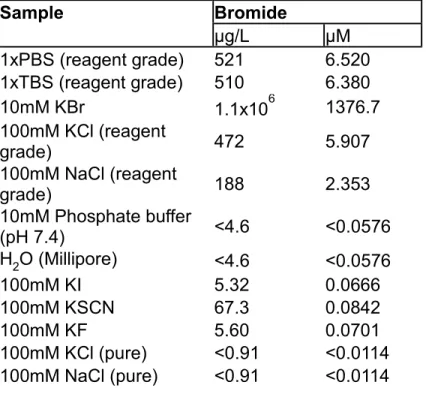
crude precipitate) and hot filtered again through a Buchner funnel after boiling. After filtering, the volume of the water was reduced by heating to an approximate volume of 85 ml with vigorous stirring, then removed from heat and allowed to cool slowly to room temperature. This recrystallized NaCl was filtered with a glass-frit, and placed under high vacuum with heating to 100oC to completely dry the sample. Overall Yield: 156.7 g NaCl (2.681 mols, 66.8% yield). Final NaCl purity was tested by ICP-MS showing a Br concentration of <11nM/100mM NaCl solution, below the lower detection limit for this method (see Table 1) .
Ultrapure Br-Free KCl Preparation: The reaction and purification were performed exactly as above with 210 ml of 45% w/w KOH solution. Yield: 87.2 g (1.124 mols, 47.0%). Purity of the KCl was tested by ICP-MS showing a Br concentration of
<11nM/100mM KCl solution, below the lower detection limit for this method of analysis (see Table 1).
Collagen IV Production, Isolation of Collagen IV Matrix, and Purification NC1 Hexamer:
Matrix Production: PFHR9 cells were plated at high density and maintained at confluency for 5-8 days in the presence of 50μg/ml ascorbic acid, with media changes every 24-36 hours. If desired, S=N bond formation was inhibited by the sustained presence of 1mM KI or 50 μM phloroglucinol (PHG), with inhibitor treatment being initiated upon confluency (typically 24hours after plating). Cultured cells and matrix were washed in 1x PBS before being scraped in 1% (w/v) sodium deoxycholate lysis buffer.
The lysate/matrix mixture was sonicated to sheer genomic DNA, the insoluble material
20
was pelleted by centrifugation (20,000xg), washed in 1M NaCl plus 10 mM Tris HCl , again pelleted via centrifugation, and ultimately washed (by addition of buffer and vortexing for 30 sec., followed by centrifugation) into the experimentally indicated buffer twice. NC1 Hexamer Purification: If purification of NC1 domain was desired, the matrix was washed into 50 mM Tris HCl pH 7.4. NC1 hexamers were excised from the collagenous matrix by digestion of purified matrix with bacterial collagenase (Worthington Biochemical Corporation) in a buffer containing 5 mM CaCl2, 5 mM Benzamidine, 25 mM 6-aminocaproic acid, and 0.4 mM phenylmethylsulfonyl fluoride (PMSF). The supernatant from the collagenase digest was dialyzed against 50 mM Tris HCl followed by separation by DEAE chromatography in 50mM Tris-HCl (pH 7.5). NC1 hexamers remain unbound in this condition, and were collected in the flow-through fractions. DEAE-purified NC1 was further refined by gel filtration (SuperdexTM 200, 10/300GL, GE Healthcare) to isolate purified NC1 hexamers. If Br-Free hexamers were needed, the S200 column was equilibrated in a 10 mM Phosphate buffer with 100 mM Br-Free NaCl. Table 1 contains the ICP-MS analysis of phosphate buffer demonstrating the absence of Br contamination.
Purification of Recombinant Human Peroxidasin (hPXDN): Peroxidasin was purified from stably transfected HEK293 as described previously (Bhave et al., 2012) and adapted to obtain purified peroxidasin free of bromide contamination with only the changes noted below to the protocol. Buffers used in the purification protocol were made with Br-Free NaCl. The mono-Q anion exchange column (GE Life Sciences) was washed for 10 column volumes in 0.3 M sucrose, 0.1 M NaCl(Br-Free), 20 mM Tris
21
Acetate (pH 8.5). The sucrose gradient for ultracentrifugation was poured from 5% and 20% sucrose solutions of 50 mM NaCl(Br-Free), 3 mM hexadecyltrimethylammonium chloride, and 10 mM phosphate buffer (pH 7.5).
Preparation of Hypohalous Acids: 10mM reagent hypochlorite (OCl-) (Sigma Aldrich) was used alone after dilution into required reaction buffer (concentration of HOCl confirmed spectrophotometrically ε292 = 350 M-1cm-1 ) or mixed with and equal volume of 12mM NaBr or KBr, and allowed to react for 60 seconds at high pH before being diluted into the reaction buffer at pH 7.4. HOBr was always prepared less than 15 minutes before use and concentration quantified spectrophotometrically (ε329 = 332 M-
1cm-1).
Phytagel Purification: In order to limit any bromide contamination present in commercial Phytagel (Gellan), we decided solubilize the phytagel to liberate any trapped bromide. Phytagel (7.5 g) (Sigma Aldrich) was added to 500 ml of 5mM EDTA (1.5% w/v final) solution at 60oC with stirring. Concentrated NaOH (268 μl, 5.5 mmol) was added drop wise resulting in a complete solubilization of the Phytagel. [Doner and Douds demonstrated that 1.1 mmol OH- was needed to solubilize 1.5g of Phytagel(Doner and Douds, 1995) in the absence of divalent cations]. Concentrated H3PO4 (300 μl) was then added (final pH 5.5) and the flask removed from heat. 2 volumes (1L) of isopropanol were added and the mixture was shaken vigorously. A stringy precipitate was visible immediately upon addition of the isopropanol. The
22
solution and precipitate were allowed to stand until cool, filtered over a glass frit, and washed with another volume of isopropanol, then left under high vacuum for 24 hours.
6.81g (90.8% yield) of a stringy white solid (Sodium Gellanate) was recovered, and stored at room temperature. Bromide content in the purified phytagel was tested by Neutron Activation Analysis and found to be less than the detection limit afforded by this method (<0.06 ppm, <0.8 μM).
Biochemical Methods
Determination of Sulfilimine Crosslink Number and Oxidation State of Methionine in NC1 Dimeric Subunits: To determine the number of S=N crosslinks in NC1 dimers, NC1 hexamers (native and chemically oxidized) from HR9 matrix were fractionated on 12% SDS-PAGE. After staining with BioSafe Coomassie (BioRad), gel images were acquired and analyzed in a ChemiDocTM XRS+ imaging system using Image Lab 2.0 software. Dimeric NC1 subunits D1 and D2 showing equal intensities were excised out of the gel and prepared for “in-gel” trypsin digestion protocol(Vanacore et al., 2009). To establish the oxidation state of methionine 89, 91, and 93 in NC1 domains after oxidation with HOBr or HOCl, gel bands corresponding to the monomeric subunits of each sample were excised out of the gel and processed by “in-gel” trypsin digestion as described above. Peptides were dried and stored at -20 ˚C until MS analyses were performed.
High-Resolution Mass Spectrometry: For mass spectrometric analysis of tryptic digests of NC1 subunits, dry samples were reconstituted in 0.1% formic acid and loaded
23
onto a capillary reverse phase analytical column (360 µm O.D. x 100um I.D.) using an Eksigent NanoLC Ultra HPLC and autosampler. The analytical column was packed with 20 cm of C18 reverse phase material (Jupiter, 3µm beads, 300 Å, Phenomenex), equipped with a laser-pulled emitter tip. Peptides were gradient-eluted at a flow rate of 500 nL/min, and the mobile phase solvents consisted of 0.1% formic acid, 99.9% water (solvent A) and 0.1% formic acid, 99.9% acetonitrile (solvent B). A 90-minute gradient was performed and consisted of the following: 0-10 min, 2% B; 10-50 min, 2 - 35% B;
50-60 min, 35-90% B; 60-65 min, 90% B; 65-70 min 90-2% B; 70-90 min, 2% B. Upon gradient-elution, peptides were mass analyzed on a LTQ Orbitrap Velos mass spectrometer (Thermo Fischer Scientific, San Jose, CA), equipped with a nanoelectrospray ionization source. The instrument was operated using a data- dependent method in which full scan (m/z 400-2000) spectra were acquired with the Orbitrap as the mass analyzer (resolution 60,000), and the three most abundant ions in each MS scan were selected for fragmentation in the LTQ. These were followed by data-dependent MS3 analysis of the three most abundant ions in each MS2 scan. An isolation width of 2 m/z, activation time of 30 ms, and 35% normalized collision energy were used to generate MS2 and MS3 spectra. The MSn AGC target value was set to 1e4 or 2e4, and the maximum injection time was either 150 ms or 250 ms, respectively.
For MS2 scans, a minimum threshold of 500 was used to trigger data-dependent spectra, while a threshold of 500 or 100 was used to trigger MS3 spectra. Dynamic exclusion was enabled, with an exclusion duration time of 60 ms.
24
Peptide identification and Bioinformatics: Two strategies were followed for the identification of sulfilimine-cross-linked peptides. First, raw data files were manually searched for sulfilimine-cross-linked peptides using the QualBrowser of Thermo Xcalibur 2.1 software. To calculate the monoisotopic mass of the sulfilimine-cross-linked peptides, the mass of two hydrogen atoms was subtracted from the sum of the masses for Met93-containing peptide and Lys211-containing peptide, each generated using GPMAW version 8.00sr1 (Lighthouse Data, Denmark), as previously shown by Vanacore et al(Vanacore et al., 2009). Second, sulfilimine-cross-linked peptides not found manually were identified by searching the mouse subset of the Uniref100 database (www.uniprot.org) using either SEQUESTTM (Thermo Electron, San Jose, CA) on a high speed, multiprocessor Linux cluster in the Advanced Computing Center for Research & Education at Vanderbilt University or using Myrimatch algorithm on a 2 six- core Intel Xeon processor HP Proliant DL160 G6 server running Windows server 2008 R2 enterprise using the Bumbershoot suite(Tabb et al., 2007). The search files were adapted for the identification of peptides that included the dynamic amino acid modifications of interest ( i.e. –48.0034 on Met, +45.9877 or +61.9826 on Lys as well as and oxidation of methionine to sulfoxide (+15.9949) and/or sulfone (+31.9898)) and the static modification of cysteine by carbamidomethylation (+57.021). The resulting files obtained from SequestTM and/or Myrimatch were assembled on IDPicker(Holman et al., 2012; Tabb et al., 2007) and/or Scaffold (Proteome Software, Portland, OR). MS3 spectra matching peptides containing modified Lys211 or Met93 were either confirmed by manual evaluation or by processing the raw data files through ScanRanker(Ma et al., 2011), and the spectra annotated with IonMatcher(Tabb et al., 2007).
25
The abundance of S=N cross-linked and Met oxidized peptides was quantified by generating extracted ion chromatograms (XICs) for precursors (all ionic forms) within each data set using Xcalibur software. The area of the peak containing modified peptide(s) was normalized to the total ion current over the entire chromatographic separation.
Cell Culture Screening of Halides: PFHR-9 cells were grown in the presence of halide salts for up to 7 days-post confluency. NC1 domains were biochemically isolated as above, and visualized by SDS-PAGE. Densitometry was conducted using ImageJ software (NIH), and dose-response curves fit in GraphOad Prism 5.1 using non-linear regression.
In Vitro Isolated Matrix Reactions: Collagen IV matrix was grown under 1mM KI for inhibition of crosslinking, and purified with 1% deoxycholate and 1M NaCl washes.
Insoluble pellets were then subjected to five washes in the appropriate reaction buffer to remove contaminating endogenous halides. Reactions were initiated upon addition of 1mM H2O2, stored at -20° post-reaction. The collagenous matrix was digested with collagenase prior to separation of the soluble NC1 domains by non-reducing SDS- PAGE.
Reactions of Purified Human Peroxidasin and other peroxidases with the NC1 Hexamer under Br-Free Conditions: Peroxidasin (27.25 nM) and NC1 hexamer(1.3 μM) [both prepared Br-Free vide supra] were reacted in the presence of either reagent grade phosphate buffered saline (PBS) or Br-Free PBS composed 136 mM NaCl(Br-
26
Free), 2.68 mM KCl(Br-Free), 10 mM Na2HPO4, 2 mM KH2PO4 [NAA analysis of these PBS mixtures appears in Table 4, which demonstrated a Br concentration of <2.5 μM, the detection limit for the analysis, and each component was individually demonstrated to have <11 nM Br contamination by ICP-MS(Table S1)]. The reaction was initiated when H2O2 (10 μM) was added and the mixture placed at 37OC for 10 minutes. The reaction was quenched by the simultaneous addition of 5 mM phloroglucinol, 0.2 mg/ml Bovine catalase, and 10 mM methionine in a premixed solution.
The dose response of purified peroxidasin to Br- was accomplished in an identical way with Br-Free PBS as the buffer. For the reactions involving Myeloperoxidase (MPO), Eosinophil peroxidase (EPO), (purchased from Calbiochem), each enzyme was obtained commercially, and dialyzed against 10000 volumes of Br- free PBS to ensure minimal Br contamination.
Chemical oxidation of NC1 domains with HOCl and HOBr:
Dose response: HOCl and HOBr, prepared as noted above, were added to uncross- linked purified NC1 hexamer (833 nM) and reacted for 5 minutes at 37OC and quenched by adding methionine to 1 mM. Preoxidation: For the HOCl preoxidation experiment, HOCl (in indicated concentrations) was added to 1.3 μM purified NC1 hexamer in 1x Br- Free PBS for 1 minute at 37OC, at which point 8.5 mol eq HOBr (66 μM final concentration) was added and the solution reacted for 1 additional minute at 37OC.
Methionine was added to 20 mM to quench the reaction. For the H2O2 preoxidation, NC1 Hexamer (1.3 μM) in Br-Free PBS was mixed with H2O2 in indicated concentrations and incubated at 37OC for 14 hours. After incubation, a 0.02 mg/ml final
27
concentration of catalase was added to consume residual H2O2 for 10 minutes at 37OC, followed by treatment with 8.5 mol eq HOBr (66 μM final concentration) for 1 minute at 37OC. Reactions were then quenched with 20 mM methionine.
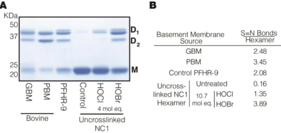
Stoichiometric Oxidations for S=O and S=N product comparisons: 1.3 μM purified NC1 hexamer in 1x Br-Free PBS was oxidized with 10.7 mol eq (83.3 μM) of HOBr or HOCl for 1 minute at 37OC then immediately quenched with 20 mM methionine. All NC1 domains were then subjected to non-reducing SDS-PAGE on a 12% gel and
Coomassie stained (50% Methanol, 10% Glacial Acetic acid, 0.01% Coomassie Brilliant Blue) for 30 minutes, rinsed with distilled water, and destained for 1 hour (15%
methanol, 10% Glacial Acetic acid) enabling consistent quantification between experiments.
Br-Free cell culture of PFHR-9 cells:
Serum Dialysis: 27.5 ml Fetal Bovine Serum (Atlanta Biologicals, Norcross, GA) was dialyzed (10,000 MW cutoff dialysis tubing) against Br-Free HBSS (
Table 3) twice (2.5L/dialysis) for 12 hours each at 4oC.
Media Preparation: The components listed in Table 2 were combined and the dialyzed serum was added. The final Br-Free media (DMEM +5% FBS) was sterile filtered (0.22 μm pore) and stored at 4oC until use.
Culture Method: Passage 9-14 PFHR-9 cells, grown to confluency in a 15cm culture dish were trypsinized, centrifuged at 1000xg to form a pellet, and the supernatant removed. Br-Free DMEM was used resuspend and wash the cell pellet twice(10
28
ml/rinse), and then the cells split 1:4 into 10 cm culture dishes containing 12 ml of either standard media (commercial DMEM + normal 5% serum) as the control, Br-free media + 5% Br-Free Dialyzed FBS , or 100 μM NaBr +Br-free media + 5% Br-free Dialyzed FBS (Br-added). 24 hours after plating, 50 μg/ml ascorbic acid was added to the cultures. Media was changed every 24 hours and the cells were cultured for 5 days at 37oC in 10% CO2. After 5 days, the plates were harvested for matrix. Cells were rinsed with ice cold PBS with 50μM PHG followed by the application of a 1% deoxycholate, 10 mM Tris HCl, 1 mM EDTA lysis buffer with 50μM PHG, 0.5 mM PMSF, and Aprotinin, Leupeptin and Pepstatin (Sigma) (10μg/ml) added. The insoluble matrix and this buffer were scraped and then sonicated until mostly homogenous, then centrifuged (20000xg for 20 minutes). The supernatant was removed and the pellet washed by vortexing in a 1M NaCl, 50 mM Tris (pH 7.4), 0.5 mM PMSF, 50μM PHG solution for 10 minutes. The sample was re-pelleted and a collagenase digestion was performed as explained above (collagen NC1 purification) with the addition of 0.5 mM PMSF and 50μM PHG to the collagenase buffer. The supernatant following the collagenase digestion was subjected to SDS-PAGE (12% gel) and Coomassie blue staining.
Each media used for culture, including 5% Serum, was analyzed by NAA for Br content (Table 4). Standard commercial media contained 1.00±0.011 ppm (12.5±0.1 μM), Br-free Media contained < 0.2 ppm (<2.5 μM, the detection limit of the analysis), and Br-added contained 7.0 ±0.252 ppm (87.6 ±3.2 μM).
Drosophila Methods
Yeast Culture in Br-Free Conditions: Yeast were cultured in an adapted Yeast Nitrogen Base (YNB) media. Once the solution was made, it was sterile filtered and
29
stored at 4oC until use. Yeast were started by placing active dry pellets (~ 30 pellets) of Saccharomyces cervesea (Type II)(Sigma Aldrich, St. Louis, MO) in 5 ml sterile distilled water and inoculating in 50 ml Br-free YNB starter cultures at 30oC and 230 rpm for 26 hours. From this starter culture, 500 μl was used to inoculate 500 ml of the same Br-free YNB. These 500 ml flasks were grown under the same conditions as above until an OD600=1.5 was observed (~27 hours). The yeast was then harvested in a series of centrifugations at 4000xg for 25 minutes at 4oC. Once pelleted, the yeast was re- suspended in 25 ml Br-Free PBS and re-centrifuged, and the supernatant was removed.
The yeast pellet was the re-suspended in 15 ml of Millipore distilled water and transferred to a lyophilization chamber where the yeast paste was snap frozen around the periphery of the chamber in a dry ice/ethanol bath, followed by a 48 hour lyophilization process. The yeast powder was then stored at room temperature until use.
Total dry yield was 6.47 g/ 6 L culture media (1.08g Dry Yeast/L).
Phytagel Semi-Solid Plate Preparation: Dishes were prepared in sizes appropriate for either larval rearing (35 mm dish, 2 ml phytagel mixture) or for egg deposition/adult feeding (55 mm dish, 10 ml phytagel mixture). To prepare the phytagel mixture, a solution was formulated to mimic the bulk salts present in grape juice (as the standard source of nutrients and salts) as well as to provide sufficient divalent cations to enable phytagel polymerization. This polymerization solution consisted of Br-free KCl (5 mM), Br-free NaCl (5 mM), KH2PO4 (1 mM), and MgSO4 (5 mM). To the polymerization solution, 18.1% w/v sucrose and 0.5% w/v purified sodium gellanate (prepared as described in Materials) were added to the desired final volume and the suspension vortexed and microwaved in 15 second intervals until boiling. Once the phytagel had