LOUIS S. KORNICKER
m
i
SMITHSONIAN CONTRIBUTIONS TO ZOOLOGY • NUMBER 505
SERIES PUBLICATIONS OF THE SMITHSONIAN INSTITUTION
Emphasis upon publication as a means of "diffusing knowledge" was expressed by the first Secretary of the Smithsonian. In his formal plan for the Institution, Joseph Henry outlined a program that included the following statement: "It is proposed to publish a series of reports, giving an account of the new discoveries in science, and of the changes made from year to year in all branches of knowledge." This theme of basic research has been adhered to through the years by thousands of titles issued in series publications under the Smithsonian imprint, commencing with Smithsonian Contributions to Knowledge in 1848 and continuing with the following active series:
Smithsonian Contributions to Anthropology Smithsonian Contributions to Astrophysics
Smithsonian Contributions to Botany Smithsonian Contributions to the Earth Sciences Smithsonian Contributions to the Marine Sciences
Smithsonian Contributions to Paleobiology Smithsonian Contributions to Zoology
Smithsonian Folklife Studies Smithsonian Studies in Air and Space Smithsonian Studies in History and Technology
In these series, the Institution publishes small papers and full-scale monographs that report the research and collections of its various museums and bureaux or of professional colleagues in the world of science and scholarship. The publications are distributed by mailing lists to libraries, universities, and similar institutions throughout the world.
Papers or monographs submitted for series publication are received by the Smithsonian Institution Press, subject to its own review for format and style, only through departments of the various Smithsonian museums or bureaux, where the manuscripts are given substantive review.
Press requirements for manuscript and art preparation are outlined on the inside back cover.
Robert McC. Adams Secretary
Smithsonian Institution
S M I T H S O N I A N C O N T R I B U T I O N S T O Z O O L O G Y • N U M B E R 5 0 5
Myodocopid Ostracoda of Enewetak and Bikini Atolls
Louis S. Kornicker
SMITHSONIAN INSTITUTION PRESS Washington, D.C.
1991
A B S T R A C T
Komicker, Louis S. Myodocopid Ostracoda of Enewetak and Bikini Atolls. Smithsonian Contributions to Zoology, number 505, 140 pages, 71 figures, 7 tables, 1991.—Samples collected at Bikini Atoll in 1946 and at Enewetak Atoll in 1969 and 1979, mainly in benthic traps and bottom trawl samples, contained a diverse assortment of mydocopid ostracodes: 17 species (3 described previously, 12 new, and 2 left in open nomenclature) in 15 genera (2 new). Keys are supplied for genera in the Sarsiellinae and to species in 3 genera. Three species from Enewetak Atoll were previously known from either Hawaii, Fiji, or the Philippine Islands. Of the 15 genera collected at Enewetak and Bikini atolls, 7 are world-wide, 6 are known primarily from the Indo-West Pacific Region, and 2 are known only from the atolls. The juveniles as well as the adults are described for the new species Cypridina spina and C. spinula, and it is determined that each has 6 stages including the adult.
A table is presented listing the number of pectinate teeth comprising the main tooth of the 5th limb of species of Cypridinidae for which this character is known; the number of teeth on the adult 5th limb ranges from 3 to 7, and about 90% of the species have 6. For those species having 6 pectinate teeth on the adult 1 tooth is added on each succeeding instar starting with instar I, information useful for identifying the age of instars.
OFFICIAL PUBLICATION DATE is handstamped in a limited number of initial copies and is recorded in the Institution's annual report, Smithsonian Year. SERIES COVER DESIGN: The coral Montastrea cavernosa (Linnaeus).
Library of Congress Caulogingin-Publication Data Komicker, Louis S., 1919-
Myodocopid Ostracoda of Enewetak and Bikini atolls / Louis S. Komicker.
p. cm. — (Smithsonian contributions to zoology ; no. 505) Includes bibliographical references.
Supt of Docs, no.: SI 1.27:505
1. Myodocopida—Marshall Islands—Enewetak Atoll. 2. Myodocopida—Marshall Islands—Bikini Atoll. I. Title. IL Series.
QL1.S54 no. 505 [QL444.085] 591 s—dc20 [595.3'3] 90-10359
Contents
Page
Introduction 1 Biogeographic Comparisons 1 Distribution of Myodocopids at Enewetak Atoll 1 Abbreviations 2 Acknowledgments 2 CYPRIDINIDAE Baird, 1850 2 The Fifth Limb of the CYPRIDINIDAE 3 Number of Instars in the CYPRIDINIDAE 4 CYPRIDININAE Baird, 1850 4 CYPRIDININI Baird, 1850 5 Paravargula Cohen and Komicker, 1975 5 Key to the Species of Paravargula 5 Paravargula digitata Kornicker, 1970 5 Paravargula trifax, new species 6 Skogsbergia Kornicker, 1974 11 Key to the Species of Skogsbergia 12 Skogsbergia strophinx, new species 12 Cypridinodes Brady, 1902 17 Cypridinodes plax, new species 18 Codonocera Brady, 1902 23 Codonocera species A 24 Cypridina Milne-Edwards, 1840 27 Cypridina dentata (Miiller, 1906) 29 Cypridina spina, new species 35 Cypridina spinula, new species 56 PHILOMEDIDAE Miiller, 1906 74 PSEUDOPHILOMEDINAE Kornicker, 1967 74 Harbansus Kornicker, 1978 74 Harbansus vix, new species 74 RUTIDERMATIDAE Brady and Norman, 1896 78 Rutiderma Brady and Norman, 1896 78 Rutiderma vox, new species 78 SARSIELJJDAE Brady and Norman, 1896 84 SARSIELUNAE Brady and Norman, 1896 84 Key to the Genera of SARSIELUNAE 85 Ancohenia Kornicker, 1976 85 Ancohenia robusta (Brady, 1890) 85 Eusarsiella Cohen and Kornicker, 1975 92 Eusarsiella chessi, new species 92 Anscottiella Kornicker, 1975 98 Anscottiella vertex, new species 98 Metasarsiella, new genus 104 Metasarsiella vibex, new species 105 CYLINDROLEBERIDIDAE MUller, 1906 109 CYLINDROLEBERIDINAE Miiller, 1906 109 Cylindroleberis Brady, 1868 110
in
Key to the Species of Cylindroleberis 110 Cylindroleberis variabilis Komicker, 1970 I l l Heptonema Cohen and Kornicker, 1975 117 Key to the Species of Heptonema 118 Heptonema homelix, new species 118 Monoasterope, new genus 124 Monoasterope bex, new species 125 CYCLASTEROPINAE Poulsen, 1965 129 CYCLOLEBERIDINI Hartmann, 1974 129 Leuroleberis Kornicker, 1981 129 Leuroleberis species A 129 Appendix 1: Station Data with Specimens Collected 133 Appendix 2: Number of Cuspate Teeth on the Main Tooth of the Fifth Limb of
Selected Species of the CYPRIDINTOAE 135 Literature Cited 138
IV
Myodocopid Ostracoda of Enewetak and Bikini Atolls
Louis S. Kornicker
Introduction
Enewetak and Bikini atolls form part of the Marshall Islands in the northwest Pacific Ocean. The present work was stimulated by a request from Dennis M. Devaney and Beatrice L. Burch, Bemice P. Bishop Museum, Honolulu, Hawaii, to contribute a paper on myodocopid ostracodes for a planned book on the natural history of Enewetak Atoll, which has since been published (Devaney et al., editors, 1987). My contribution to that book identified and illustrated some of the myodocopid genera collected on Enewetak Atoll in 1969 and 1979 (Kornicker, 1987b); the taxa are described more completely herein. In addition, a small collection (5 specimens) from Bikini Atoll in 1946 contained a new species, which is also described herein. Although the collections studied are small (129 specimens), they have a diverse assortment of myodoco- pids comprising 17 species (3 described previously, 12 new, and 2 left in open nomenclature) in 15 genera (2 new). Keys are supplied for some genera and species, and the ontogenies of 2 species of Cypridina are elucidated. Although not collected at either Enewetak or Bikini atolls, a supplementary description is presented of Paravargula digitata Kornicker, 1970b, based on the holotype from the Philippine Islands.
BIOGEOGRAPHIC COMPARISONS.—The genera Skogsbergia, Harbansus, Rutiderma, Eusarsiella, Leuroleberis, Cylindrole- beris, and Heptonema are circumglobal in distribution, but have not been reported from the Arctic Ocean and Antarctic waters.
The pelagic genus Cypridina is primarily Indo-Pacific.
Cypridinodes has been collected in the Indo-West Pacific region.
Paravargula has previously been collected off the tip of South Africa and from the East-Indian region (Poulsen, 1962:204; Kornicker, 1975:3).
Codonocera has been collected previously in Indo-Pacific Louis S. Kornicker, Department of Invertebrate Zoology, National Museum of Natural History, Smithsonian Institution, Washington, DC. 20560.
Review Chairman: C.W. Hart, Jr., Smithsonian Institution.
and Australasian waters (McKenzie, 1967:221).
Anscottiella has previously been collected off Ceylon and Thailand (Poulsen, 1965:65; Kornicker, 1975:607; Kornicker and McKenzie, 1976:348) and Australia (Hall, 1987:760).
Ancohenia has previously been collected from Hawaii (Kornicker, 1976b:7), Fiji (Kornicker, 1981a: 12), and Rangiroa Atoll, South Pacific (Hartmann, 1984:122).
Two genera, Metasarsiella and Monoasterope, are known only from Enewetak Atoll, but probably occur elsewhere on Pacific islands.
At the specific level, only 3 species at Enewetak have been reported elsewhere, Ancohenia robusta from Fiji, Cylindrole- beris variabilis, from the Philippine Islands, and Cypridina dentata, from the Malayan Archipelago.
DISTRIBUTION OF MYODOCOPIDS AT ENEWETAK ATOLL.—
Most specimens from Enewetak were collected in 1979 by James R. Chess and Edmund S. Hobson during a study of diel movements of zooplankton (Hobson and Chess, 1986). Their investigation showed that many zooplankton, including myo- docopid ostracodes, leave the substrate to enter the water column primarily at night (Hobson and Chess, 1986, tables 1, 3). Collections were made during day and night by placing a trap (covering about 1 square meter at its base) over substrates of sand, coral reef, and coral rubble in the vicinity of 2 stations (A and B) on the windward lagoon shelf at Enewetak Atoll (Hobson and Chess, 1986, fig. 1). (Details concerning trap construction are in Hobson and Chess (1979, fig. 1).) According to Hobson and Chess (1986:10), the coral rubble substrate actually comprised both rubble and sand, and the reef substrate comprised coral, coral rubble, and sand. Trawl samples also were collected above the stations. With the possible exception of 1 composite sample (see Appendix 1), all ostracodes received by me from that study had been collected at night.
The number of myodocopid ostracodes collected in the nocturnal traps varied from 4 to 14, and each trap captured 2 to 4 species (Table 1). The total number of species in 6 samples from the traps is 10. A nocturnal trawl drawn through the water for about 10 minutes above the traps captured 34 specimens (12 1
SMITHSONIAN CONTRIBUTIONS TO ZOOLOGY TABLE 1.—Distribution of myodocopid ostracodes collected at night, in 1979, over substrates of sand, coral reef,
and coral rubble, on a lagoon shelf at Enewetak Atoll, in six emergent traps at stations A and B, and in one mid water trawl above the stations.
Species
Codonocera species Cypridina ckntata Cypridina spina Cypridina spinula Paravargula trifax Skagsbergia strophinx Ancohenia robusta Anscottiella vertex Eusarsie.Ua chessi Rutiderma vox Cyluidroleberis variabilis Heptonema homelix Lewoleberis sp. A
Sta A, Trap 6 25 May Intermittent Sand
0 0 0 11
0 0 0 0 0 1 0 0 0
current Coral
reef 0 0 4 0 1 0 0 1 0 0 0 1 0
Sta B, Trap 7
Coral reef
0 0 6 0 0 0 0 1 0 0 0 0 0
26 May No current
Coral rubble
0 0 0 4 0 7 1 0 0 0 1 0 0
Sta B, Trap 10
Coral rubble 0 3 0 0 0 1 0 0 0 0 0 0 0
30 May No current
Coral reef
0 0 11 0 0 3 0 0 0 0 0 0 0
Trawl 22 May
1 1 6 13 0
t
>
species). The distributions of species from the traps over sand, reef, and coral rubble substrates are compared in Table 1, but the number of specimens is too few to draw conclusions concerning substrate preferences. The data suggest that Cypridina spinula prefers sand and coral-rubble substrates, whereas C. spina prefers a reef habitat.
ABBREVIATIONS.—In the figures, Arabic numerals indicate limbs 1-7, as well as individual joints of each limb (the location of the numeral indicating whether a limb or joint is indicated). Roman numerals I—III indicate the endites. Arrows on illustrations of the carapace indicate anterior of valve.
The following abbreviations are used in illustrations:
a.m.
ant.
a.p.
B.O.
C O .
e endop epip esoph exop gen gl.a g o . i.m.
l.b.
I.e.
1.1. orl.hp Lp.
md.
me.
m.s.
m i .
central adductor muscle attachments antenna
anterior process Bellonci organ copulatory organ valve edge endopodite epipoJilc esophagus exopodite genitalia
glandular opening of upper lip genital organ
inner margin of infold lateral bristle on mandible lateral eye
lower lip
lamellar prolongation of selvage or list mandible
medial eye
main spine of mandibular coxale endite maxilla
s.
sens s.v.
U.I.
V.
selvage
sensory bristle of 5th joint of 1st antennna seminal vesicle
upper lip valve
ACKNOWLEDGMENTS.—I thank the following individuals for their help: James R. Chess (Southwest Fisheries Center, Tiburon Laboratory) and C. Allen Child (Smithsonian Institu- tion) for supplying the specimens from Enewetak; Anne C.
Cohen (University of California, Los Angeles) for supplying information on a species and reviewing the manuscript;
Carolyn Gast (Smithsonian Institution) for rendering the shaded drawings of the carapaces; Katheryn Schroeder Brown and Jack Schroeder (Jack Schroeder Associates) for assisting in preparation and inking of appendage drawings; I.G. Sohn (U.S.
Geological Survey) and TE. Bowman (Smithsonian Institu- tion) for reviewing the manuscript; Elizabeth Harrison-Nelson (Smithsonian Institution) for general assistance; and Diane M.
Tyler (Smithsonian Institution) for final editing and preparation of the manuscript for publication. Others participating in collecting at Enewetak and Bikini are acknowledged in Appendix 1.
CYPRIDINIDAE Baird, 1850
COMPOSITION.—This family includes 2 subfamilies: Cyprid- ininae Baird, 1850, and Azygocypridininae Kornicker, 1970a.
Only the former is in the collections from Enewetak.
DISTRIBUTION.—Circumglobal with depth range of intertidal to abyssal.
The Fifth Limb of the CYPRIDINIDAE
The structure of 5th limb of the Cypridinidae has been discussed by Skogsberg (1920:38, 184, 188) and Poulsen (1965:456). The limb is located just posterior to the maxilla. It is obliquely oriented resulting in the lateral side of the limb also being the anterior side, and the medial side also being the posterior side. In the present paper the sides of the limb are designated anterior and posterior, following Skogsberg (1920:188). The 1st exopodial joint bears the "main tooth,"
which comprises a short smooth or digitate proximal peg followed by 1 or more stout cuspate teeth; if more than 1 cuspate tooth is present, distal teeth are longer and stouter. The peg as well as the bases of the cuspate teeth are on the posterior side of the joint where they face the mouth. Generally, the teeth
are visible for counting when the limb is viewed posterior side up when placed in a drop of glycerine on a slide, but for smaller specimens the teeth are seen best when mounted posterior side up on a slide under a cover-slip. The number of cuspate teeth in cypridinids was discussed briefly by Cohen and Morin (1986:21). The number of teeth of additional species are listed in Appendix 2 herein. The list is based on the literature as well as the present study, but species are not listed if the limb was described as "typical of family," or with similar words, unless it is clear from the description that the limb had been examined in detail; the literature search started with Skogsberg (1920), because little attention was given to the main tooth before then.
The number of cuspate teeth comprising the main tooth of adults of members of the Cypridinidae varies from 3 to 7 (Table 2, Appendix 2). Most of the data in Appendix 2 are from only
TABLE 2.—Number of species in genera having 3-7 pectinate teeth on the main tooth of the 5th limb of adults.
(M = male, and is used if only adult male of species is known and it has less than 6 pectinate teeth, or when both sexes are known and male has fewer teeth than female; data from Appendix 2.)
Taxon
AZYGOCYPRIDININAE Azygocypridina Isocypridina
CYPRKMNINAE CYPRIDININI Amphisiphonostra Bathyvargula Codonocera Cypridina Cypridinod.es Doloria Hadacypridina Macrocypridina Melavargula Metavargula Monopia Paracypridina Paradoloria Paravargula Pterocypridina Rheina Rugosidoloria Sheina Siphonostra Skogsbergia Vargula
GlGANTOCYPRIDINl Gigantocypris
Total
3 teeth
0 0
0 0 0 0 0 0 0 0 0 0 0 0 0 0 1M
0 0 0 0 0 1 0 2
4 teeth
0 0
0 0 1 0 0 0 0 0 0 0 0 0 0 1*
1 0 0 1M
0 0 0 0 4
Number of species 5 teeth
2 0
0 0 3 0 0 0 0 0 0 3 0 0 1M
1*
1 0 1 0 1 0 1 0 12
6 teeth
4 1
1 1 5 7 6 7 2 2 2 5 1 2 5 3 2 1 0 0 1 9 21
3 91
7 teeth
0 0
0 0 0 0 0 0 0 0 0 0 0 0 0 0 0 0 0 0 0 0 1 0 1
'Paravargula narupollex has either 4 or 5 pectinate teeth.
SMITHSONIAN CONTRIBUTIONS TO ZOOLOGY a few specimens of each species so that the intraspecific
variability in the number of teeth of many species is unknown.
The presence of 6 cuspate teeth on the adult of all 7 species of Cypridina on the list suggests little intraspecific or interspecific variability in this character, at least for species of that genus.
Eight species of Skogsbergia listed in Appendix 2 also have 6 cuspate teeth suggesting that 6 teeth is the usual number for the genus. Most of the species in the 23 genera listed in Appendix 2 have 6 cuspate teeth, and the remaining species have 3 , 4 , 5 , or 7. Clearly, 6 cuspate teeth is the more common number among the Cypridinidae. As shown in Appendix 2 and Table 2, adults of species of Azygocypridininae have 5 or 6 cuspate teeth, species of Cypridinini have 3 to 7, and species of Gigantocypridini have 6. In some genera such as Cypridina, Skogsbergia, and Gigantocypris, the number of cuspate teeth are fairly constant (6 teeth); whereas, in others such as Codonocera the interspecific variability is greater (4-6 teeth).
Generally, the male and female have the same number of teeth, but on at least 1 species, Pterocypridina sex, the adult male has 3 teeth and the adult female 4. The number of cuspate teeth on instar I is known for only 10 species, all having only 1 cuspate tooth; the tooth, which has been described for only a few species, is more complex than teeth of later instars.
Number of Instars in the CYPRIDINIDAE
Cohen (1983:235) reared Skogsbergia lerneri in aquaria and observed the species to have 6 stages including 1 for the adult.
Other ontogenetic studies have been based on preserved specimens and as a result are probably more subjective. Based on the ontogenetic studies of Poulsen (1962:126), Hiruta (1980:145), Kornicker and Iliffe (1989a: 19), Kornicker (1989:65), and the work herein, the following species also have 6 stages: Azygocypridina imperialis, Macrocypridina castanea, Vargula hilgendorfii, Skogsbergia galapagensis, Cypridina spina, and C. spinula. Poulsen (1962:126) concluded that Macrocypridina castanea has more than 1 adult stage, but the appendages are similar in all adult stages. Poulsen (1962:116) concluded that species of Gigantocypris have 6 juvenile instars and more than 1 adult instar, but according to Fenwick (1984:285) there is insufficient evidence in the data of Poulsen for concluding that post-adult molting takes place in the genus Gigantocypris.
What is the evidence for Gigantocypris having 6 juvenile instars? Poulson (1962:22, 85) had in his collection 50 specimens of G. agassizi Mttller, 1895, and 69 specimens of G.
danae Poulsen, 1962, but instars of stage VI were not present (although Poulsen (1962:54) states that instar VI of G. agassizi was not present in the material, 13 specimens are listed in his table 4, p. 34). Poulsen (1962:57) had 135 specimens of G.
muelleri Skogsberg, 1920, including several specimens identi- fied as juvenile instar VI. At the end of his description of the species Poulsen (1962:83) states: "The differences between the 5th and 6th stages are not great, mainly restricted to an increase
in the number of spines on the forepart of the shell and the number of short knife-formed bristles on the 2nd endopodite joint of the mandible, and to the further development of the copulatory limbs, especially of the large main spine of the outer lobe. However, the growth between the two stages is fairly large, for the males from 9 mm in the 5th to 11 mm in the 6th."
Based on Poulsen's descriptions, certain similarities between the morphology of the 5th and 6th instars suggest that they should be combined as a single instar: (1) both instars have 5 cuspate teeth on the 5th limb; (2) the endopodite of the male 2nd antenna of both instars have the same number of bristles (however, the long bristle of the 3rd joint is closer to midlength on instar VI, which may indicate that the instar is older); (3) the number of furcal claws is the same for both instars; and (4) the length of the male copulatory organ is 9% of shell length in both instars (Poulsen, 1962, table 10). Although the data are conflicting, I believe additional information is necessary before a conclusion can be drawn that species of Gigantocypris have 6 juvenile instars. Therefore, I have listed Gigantocypris as having 5 juvenile instars in Appendix 2. Incidentally, if as suggested here, instar VI should be combined with instar V, the case for post-adult molting in Gigantocypris is further weakened, because it is based on comparing variance (Kor- nicker, 1975:687) and coefficient of variation (Fenwick, 1984:285) of carapace length of adult and penultimate females;
if instars V and VI are combined the variation in carapace length of the penultimate instar is increased
For species having 6 cuspate teeth on the main tooth of the adult 5th limb, the number of teeth are increased by 1 on each succeeding stage, starting with 1 tooth on instar I (Appendix 2).
Thus, the number of teeth on a specimen of such a species may be useful in identifying the age of an instar, for example, Poulsen (1962:244) used the presence of 5 teeth on a specimen of Pterocypridina birostrata to identify the specimen as the last larval stage. However, caution must be used in an identifica- tion, and supporting information should be sought.
The ontogeny has not been studied in any cypridinid species having fewer than 6 cuspate teeth on the main tooth of the adult 5th limb. It is probable that instar I of such species has a single tooth, because it would be necessary for masticating food, but it is not known whether these species have fewer instars than species with 6 teeth on the adult, or have the same number of instars but with teeth added more slowly. In Appendix 2 I list all species as having 5 juvenile stages, but additional studies probably will reveal some species having a different number.
CYPRIDININAE Baird, 1850
COMPOSITION.—This subfamily includes 2 tribes: Cypridin- ini Baird, 1850, and Gigantocypridinini Hartmann, 1974. Only the former is in the collections from Enewetak.
DISTRIBUTION.—Same as for family.
CYPRIDININI Baird, 1850
COMPOSITION.—This tribe includes 20 genera of which 5 are in the collections from Enewetak and Bikini: Paravargula, Skogsbergia, Cypridina, Cypridinodes, and Codonocera.
DISTRIBUTION.—Same as for family.
Paravargula Cohen and Kornicker, 1975
Paravargula Poulsen, 1962:202 [nomen nudum].
Paravargula Cohen and Kornicker, 1975:23 [designated type species].
TYPE SPECIES—Paravargula ensifera Poulsen, 1962:209, by subsequent designation (Cohen and Kornicker, 1975:23).
COMPOSITION.—This genus includes 6 species including a new species described herein.
DISTRIBUTION.—Paravargula ensifera Poulsen, 1962, has been collected in the vicinity of the Philippines, Singapore, and the Kei Islands; P. Ursula (Muller, 1906) has been collected in the East Indian region; P. arborea (Mailer, 1908) has been collected in the vicinity of South Africa; P. nanipollex Kornicker, 1970b, and P. digitata, Kornicker, 1970b, have been collected in the Philippines; P. trifax was collected at Enewetak. Known depth range about 3-800 m.
REMARKS.—Because of morphological differences between the adult female of P. trifax, a new species from Enewetak, and the description of the juvenile male holotype of P. digitata from the Philippines (Kornicker, 1970b:6), the holotype of the latter was reexamined, and a supplementary description is presented below.
Key to the Species of Paravargula
1. Second claw of furca nonarticulated 2 Second claw of furca articulated 5 2. Incisur shallow P. hirsute Incisur deep 3 3. Bristle of 2nd exopodial joint of 2nd antenna with proximal hairs
P. trifax, new species Bristle of 2nd exopodial joint of 2nd antenna without proximal hairs 4 4. Upper lip with unpaired rectangular anterior process P. digitata Upper lip with low ridge forming anterior unpaired part P. ensifera 5. Caudal process narrow P. nanipollex Caudal process elongate P. arborea
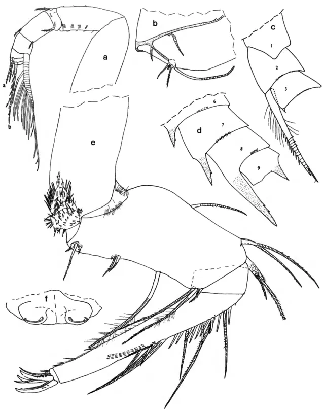
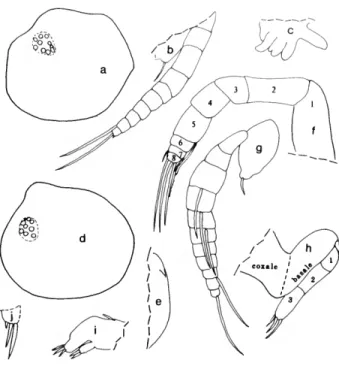
Paravargula digitata Kornicker, 1970
FIGURE 1
Paravargula digitata Komicker, 1970b:6, figs. 4-6.
HOLOTYPE.—USNM 125539,1 A-l male on 2 slides and in alcohol.
TYPE LOCALITY.—Tawi-Tawi, Sulu Archipelago, Philip- pines.
DISTRIBUTION.—Balimbing Point, Tawi-Tawi, Sulu Archi- pelago, Philippines, depth 1 or 2 m (type locality).
SUPPLEMENTARY DESCRIPTION OF A-l MALE HOLOTYPE
(Figure 1).—Infold of carapace with several minute bristles in
"pocket" of caudal process.
Second Antenna: Kornicker (1970a:9) stated that bristles of exopodial joints 2-8 have natatory hairs; it should have been joints 3-8.
Mandible: 3 claws of 3rd endopodial joint with marginal teeth.
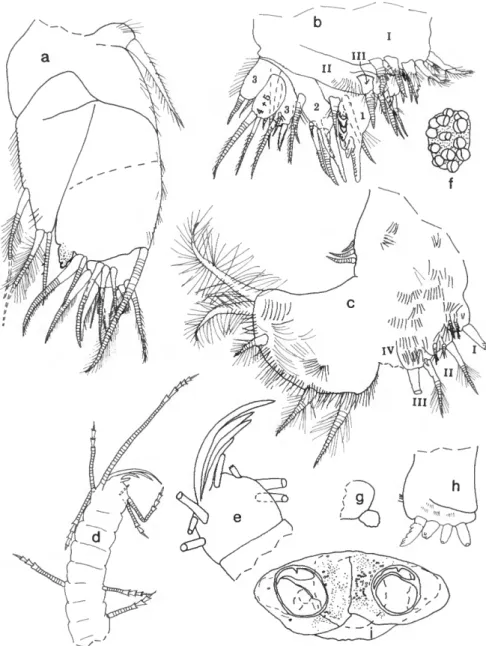
Sixth Limb (Figure la): 1 terminal bristle of endites II and III short. End joint having distal margin with spinules forming
lateral row along margin except in vicinity of 4 anterior and 2 posterior bristles; medial surface hirsute; distal margin with 13 or 14 bristles (both limbs with 2 bristles with bases on lateral surface near anterior end with short marginal spines, then margin with 2 long anterior bristles with long proximal and short distal spines, followed by 1 short bristle with short marginal spines; then left limb with 3 bristles with long proximal and short distal spines, and right limb with 1 long bristle with long proximal and short distal spines, 1 bristle with short marginal spines, 2 bristles with long proximal and short distal spines; then both limbs with short space followed by 3 bristles with long proximal and short distal spines, and 2 long hirsute posterior bristles).
Seventh Limb (Figure \b): Comb and jaw of limb illustrated.
Lateral Eye: With black pigment between ommatidia.
Upper Lip (Figure lc): Anterior unpaired part consisting of thin projecting rectangular process, transparent except for short posterior part along posterior edge that bears internal amber- colored granules (tranparent part of rectangular process with 4 truncate processes (glandular openings?); short granule-bearing part with similar truncate process). Paired posterior part with
SMITHSONIAN CONTRIBUTIONS TO ZOOLOGY
/lllll\W\\\
'ni\\\\\\\
l l
FIGURE 1.—Paravargula digitata Komicker, USNM 125539, hoiotype, A-l male: a, left 6th limb, lateral view (endites not shown); b, tip of 7th limb; c, upper lip, anterior to left.
long slender tusk (with 4 step-like proximal processes (glandular openings?) along anterior margin); then 2 short tusks (anterior of short tusks with about 4 glandular openings, posterior short tusk smaller and with 1 glandular opening).
Rounded surface between tusks and mouth hirsute.
Pigmentation: Carapace, body, and appendages without black pigment.
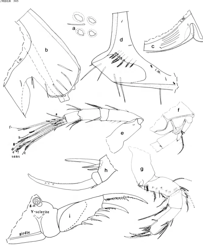
Paravargula trifax, new species
FIGURES 2 - 4
Paravargula sp. Komicker, 1987b:217.
ETYMOLOGY.—From the Latin trifax (kind of spear).
HOLOTYPE.—USNM 158322, adult female on slide and in alcohol (unique).
TYPE LOCALITY.—Enewetak lagoon (hoiotype from sta A, plankton trap 6, night, coral reef substrate).
DISTRIBUTION.—Enewetak Atoll.
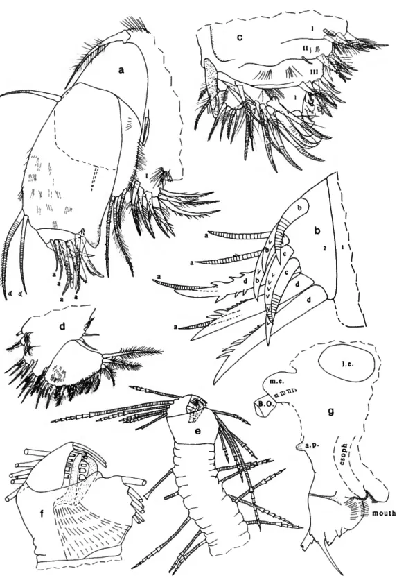
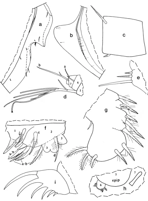
DESCRIPTION OF ADULT FEMALE (Figures 2-4).—Carapace
with slightly convex posterodorsal curvature, deep incisur, and small but projecting caudal process; surface smooth (Figure 2a-c).
Structures on Infold: Rostrum with oblique list intersecting dorsal edge of incisur (Figure 2d). Narrow list with anterior end near valve edge ventral to incisur (Figure 2d), continuing along ventral infold, then almost joining lip slightly overhanging inner margin of infold anterior to caudal process, and then continuing along caudal process and posterior infold close to inner margin of infold, finally terminating at dorsal end of caudal process (Figure 2e); lip overhanging inner margin of infold just anterior to caudal process, then continuing along caudal process as a line between list and inner margin of infold (Figure 2e). A 2nd line present between list and edge of ridge forming anterior boundary of "pocket" of caudal process (Figure 2e).
Bristles of Infold: Rostral infold with 14 bristles forming row (including 3 bristles forming ventral end of row longer and bifurcate, others shorter and unbranched), 2 long bristles and 1 short bristle forming row just dorsal to oblique list, and 4 minute bristles near middle of infold (Figure Id). 3 bristles on infold just posterior to inner end of incisur and pair of long bristles (longer of pair bifurcate) at inner end of incisur near valve edge. 2 small bristles on infold close to inner end of incisur. Anteroventral infold with short single bristle located inward from 4 long single bristles at valve edge anterior to list, and 7 minute divided bristles near midwidth of infold;
anteroventral list and anterior part of ventral list with 42 bristles (2 anterior bristles bare and single, next 3 bristles bare and bifurcate, remaining bristles bifurcate with spines on longer branch); ventral list (extending from posterior of bifurcate spinous bristle on anteroventral list to valve midlength) with 4 widely separated slender bare bristles (these could be pore canals). Inner edge of lip overhanging inner margin of ventral infold anterior to caudal process with 2 widely separated minute bristles (only posterior of these shown in Figure 2e). 1 small bristle present in triangular area at ventral end of caudal
FIGURE 2.—Paravargula trifax, new species, USNM 158322, holotype, adult female: a, complete specimen from left side, length 2.44 mm; b, c, inner views of right and left valves, respectively, showing location of central adductor muscle attachments, inner margin of infold anterior to caudal process, and "pocket" of caudal process;
d, inner view of anterior of left valve; e, inner view of caudal process of left valve;/, lateral view of left lamella of f urea; g, medial view of right lamella of f urea (only medial teeth of claws shown); h, upper lip, anterior to right;
i, part of body showing outline of left lateral eye, proximal part of furca, Y-sclerite and girdle;/ left lateral eye.
8 SMITHSONIAN CONTRIBUTIONS TO ZOOLOGY process between outer edge of lip and inner margin of infold. 1
fairly long bristle at ventral end of caudal process at about infold midwidth (Figure 2e). Line just posterior to list on ridge anterior to pocket of caudal process with 12 minute bristles.
Pocket of caudal process with about 13 minute bristles. Ridge forming anterior edge of pocket of caudal process with digitate processes forming 2 rows (Figure 2e): anterior row slightly medial to posterior row (latter on edge of ridge) and with about 17 digitate processes; posterior row with about 12 smaller digitate processes.
Selvage: Lamellar prolongation present along anterior and ventral margins, terminating posteriorly near ventral end of caudal process (Figure 2d); lamellar prolongation widest as well as narrowly striated in vicinity of incisur and divides at inner end of incisur. Double prolongation present along anterior half of ventral margin: inner layer about half width of outer and with serrate margin; outer layer minutely serrate near middle of ventral margin, smooth elsewhere.
Carapace Size: USNM 158322, length 2.44 mm, height 1.31 mm.
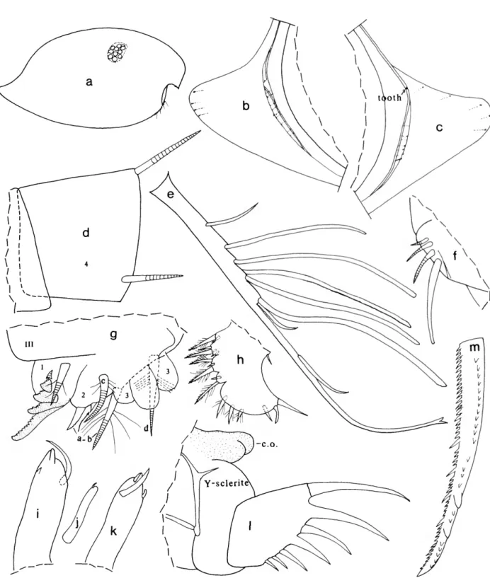
First Antenna (Figure 3a): 1st joint bare. 2nd joint with rows of spines. 3rd joint with short spinous ventral bristle and slightly longer subterminal dorsal bristle with few indistinct spines. 4th joint with short spinous ventral bristle and longer spinous dorsal bristle. 5th joint about half length of 4th joint;
stout sensory bristle with 9 long filaments followed by 3 shorter slender filaments and 1 very small subterminal filament. 6th joint with short spinous medial bristle. 7th joint a-bristle similar to bristle of 6th joint; b-bristle 3 times length of a-bristle, with 5 filaments (with marginal spines) followed by 2 spines; c-bristle about twice length of sensory bristle of 5th joint, with 9 filaments (with marginal spines) followed by 1 shorter subterminal filament. 8th joint: d- and e-bristles slightly shorter than sensory bristle of 5th joint, bare with blunt tips;
f-and g-bristles similar to c-bristle, small spine near base of some filaments.
Second Antenna: Protopodite with small spinous medial distal bristle (Figure 3b). Endopodite 1-jointed (could be interpreted as having small 2nd joint with indistinct suture separating it from 1st joint), with 4 proximal bristles and 1 long terminal filament (latter could be interpreted to be on small 2nd joint) (Figure 3b). Exopodite 9-jointed; 1st joint with few spines along concave margin (not shown); bristle of 2nd joint reaching 8th joint, with about 7 ventral and few dorsal proximal hairs followed by ventral spines and short subterminal ringed part (Figure 3c); bristles of joints 3 and 4 with ventral hairs near m id length spine -like and shorter than proximal and distal hairs;
bristles of joints 5-8 with natatory hairs, no spines; 9th joint with 4 bristles (short dorsal bristle with long hairs, and 3 long bristles with natatory hairs but no spines) and lateral spine about same length as joint; joints 3-8 with basal spines increasing in length on distal joints (spine of 8th joint more than twice length of 9th joint) (Figure 3d); joints 2-8 with minute spines forming row along distal margins.
Mandible (Figure 3e): Coxale endite spinous, with 2 stout
terminal spines with small peg between them; small bristle at base of endite (not shown); hairs at boundary between coxale and basale. Basale: dorsal margin with 3 ringed bristles (1 midlength and 2 subterminal); ventral margin with 3 a-bristles, 2 c-bristles with minute unringed peg just proximal to them, and 1 long d-bristle with long proximal and short distal spines (latter spines not shown). Exopodite about 2/3 length of dorsal margin of 1st endopodial joint, hirsute with 2 subterminal bristles (distal shorter). 1st endopodial joint with 4 ventral bristles; 2nd endopodial joint with distal bristles forming 3 groups with 1,1, and 2 unringed bristles (medial slightly longer than lateral); dorsal margin with 10 ringed bristles, 1 short unringed bristle with long marginal spines (base of bristle medial to 2nd ringed bristle), and 10 short unringed bare bristles; medial surface with indistinct spines forming rows (not shown). 3rd endopodial joint with 3 subequal claws (all with few teeth) and 4 ringed bristles.
Maxilla (Figure 4a,b): Endite I with 10 or 11 bristles; endite II with 7 bristles, endite III with 5 distal bristles and 1 short proximal bristle near base of exopodite. Coxale with dorsal fringe and 1 stout dorsal bristle with long spines along proximal
2/3 and short indistinct distal spines. Basale with 1 long anterior bristle and 1 shorter, ventral, medial bristle (dashed in Figure 4a). Exopodite with hairs along outer margin, 2 terminal bristles and 1 shorter subterminal bristle (subterminal and outer terminal bristle with long proximal and short distal spines, other with short spines). 1st endopodial joint spinous, with triangular cutting tooth with slightly undulate outer edge, 2 ringed alpha-bristles (medial spinous, lateral bare), and 2 ringed beta-bristles (inner medial bare, outer lateral slightly longer and pectinate). 2nd endopodial joint with 4 bare ringed a-bristles, 3 b-bristles (anterior ringed, others unringed, claw-like, pectinate), 2 ringed pectinate c-bristles, and 3 pectinate claw-like d-bristles (posterior ringed distally, others unringed).
Fifth Limb (Figure 4c): Epipodite with 48-51 hirsute bristles. Endite I with 5 bristles; endite II with 6 bristles; endite IN with 6 or 7 bristles (distal medial bristle stouter at base than other endite bristles and with long proximal spines and distal teeth). Protopodite with long slender anterior tooth with subterminal marginal tooth. 1st exopodial joint with 3 bristles in anterior group (all with long spines, inner stouter and pectinate) and 1 (with long spines) closer to tooth of protopodite; main tooth with 6 cusps, proximal peg (not shown), and 1 short bristle (with long proximal spines) proximal to peg (not shown). 2nd exopodial joint with 4 pectinate a-bristles, 3 pectinate ringed b'-bristles, and 4 pectinate ringed b*-bristles; posterior ringed c-bristle and anterior d-bristle with long proximal and short distal spines (d-bristle near 3rd joint). 3rd exopodial joint inner lobe with 1 proximal bristle with long proximal and short distal spines, and 2 terminal bristles with short marginal spines; outer lobe hirsute, with 2 terminal bristles with long proximal and short distal spines. Fused 4th and 5th exopodial joints hirsute, with 4 bristles (1 bare, 1 with long proximal and short distal spines, 2
FIGURE 3.—Paravargula trifax, new species, USNM 158322, holotype, adult female: a, medial view of right 1st antenna (c -, d -, e-, f-, and g -bri sties not shown); b, medial view of endopodite and part of protopodite of right 2nd antenna; c, d, lateral view of joints of left 2nd antenna (only bristle of 2nd joint shown); e, medial view of left mandible;/, genitalia.
10 SMITHSONIAN CONTRIBUTIONS TO ZOOLOGY
FIGURE 4.—Paravargula trifax, new species, USNM 158322, holotype, adult female: a, lateral view of left maxilla (only endue I shown); b, medial view of 2nd endopodial joint of right maxilla; c, anterior view of right 5th limb; d, medial view of right 6th limb; e, 7th limb;/, tip of 7th limb opposite that shown in e; g, anterior of body showing outline of left lateral eye, medial eye and Bellonci organ, anterior process, upper lip, mouth, and esophagus.
with short spines).
Sixth Limb (Figure Ad): Epipodite with 4 short bristles.
Endite I with 1 long terminal bristle, and 2 short medial bristles with long spines; endite II with 1 short and 1 long spinous terminal bristle, and 2 short medial bristles with long marginal spines; endite III with 1 short and 3 long spinous terminal bristles, and 1 long medial bristle with long proximal and short distal spines; endite IV with 1 short and 5 long spinous terminal bristles, and 1 long medial bristle with long proximal and short distal spines. End joint with 16 bristles: 2 short lateral near anterior end of joint (with short spines), 14 terminal (9 with long proximal and short distal spines, 3 with only short spines, and 2 posterior hirsute bristles); lateral spines along terminal edge of joint, and long hairs forming rows on medial surface.
Seventh Limb (Figure 4e,f): 9 or 10 scattered proximal bristles (4 or 5 on each side), each with 4-6 bells; edge of terminal segment (side bearing comb) with 6 bristles, each with 1-7 bells; edge of segment (side bearing jaw) with 5 bristles, each with 4-7 bells. Comb with total of 15 teeth comprising long middle tooth with 1 long slender tooth, 4 short narrow teeth, and 2 short broad square-tipped teeth on each side (Figure 4/); a sclerotized straight bar extending between teeth (Figure 4/). Jaw consisting of curved bar with distal end meeting straight bar (Figure 4/).
Furca (Figure 2f,g): Each lamella with 10 claws; claw 2 nonarticulated; claw 4 narrower but about same length as, or slightly shorter than, claw 5; medial teeth of claw 1 stouter than lateral teeth. Right lamella slightly anterior to left lamella.
Bellonci Organ (Figure 4g): Short, broad, with triangular tip.
Eyes: Medial eye light amber colored, bare (Figures 2i, 4g).
Lateral eyes well developed, larger than medial eye, with 36 brown ommatidia (in lateral view only about 30 ommatidia visible) (Figure 2/); without black pigment between ommat- idia.
Lips (Figures 2h, 4g): Anterior unpaired part consisting of thin projecting rectangular process, transparent except for short part along posterior edge, which bears internal amber-colored granules (tip of transparent part of rectangular process with 4 or 5 truncate processes (glandular openings?); short granule- bearing posterior part with similar projecting truncate terminal process). Posterior paired part with 5 step-like pointed processes (glandular openings?), a long slender tusk with narrow tip, and then 2 short tusks (anterior short tusk with 3 glandular openings, posterior short tusk smaller and with 1 glandular opening). Rounded surface between tusks and mouth hirsute, with 5 small processes (glandular openings?) on posterior surface adjacent to mouth (Figure 2h). Lower lip rounded, hirsute.
Genitalia (Figure 3/): Comprising 2 ovoid processes anterior to furca.
Anterior of Body (Figure 4g): Small rounded process about midway between medial eye and upper lip.
Posterior of Body (Figure 2i): Evenly rounded, bare.
Y-Sclerite (Figure 2i): Typical for subfamily.
Pigmentation (Figure 2i): Black pigmentation widespread in dorsal and posterior parts of body, with pigment more widely distributed on left side (area of pigmentation stippled in illustration); no pigment in appendages or carapace. (Pigment layer appears to be just beneath outer layer of integument.)
Eggs (Figure 2i): Spheres within body (observed only on left side) resemble eggs but anterior location suggests otherwise.
COMPARISONS.—Paravargula trifax differs from P. ensifera and P. digitata in having long proximal hairs on the bristle of the 2nd exopodial joint of the 2nd antenna (P. digitata is known only from the A-l male, and it is possible that the absence of proximal hairs is because of its age or sex; I have assumed that it is not). The carapace of P. trifax differs from P. hirsuta in having a deeper incisur, and from P. nanipollex in having a longer caudal process. The furca of P. trifax differs from that of P. arborea and P. nanipollex in having the 2nd claw nonarticulated. The upper lip of P. trifax differs from that of P.
ensifera in having a well-developed, rectangular, unpaired, anterior process. Parts of the body of the holotype of P. trifax have an underlayer of black pigment, but having only 1 specimen I am unable to determine its value as a specific character. The only other species of the genus having black pigmentation is P. arborea. The lateral eyes of P. digitata differ from those of P. trifax in having a dense black pigmentation between the ommatidia, but this could be the result of differences in preservation; however, the presence of black pigment in the body of P. trifax may indicate that the absence of eye pigment is not a matter of preservation.
Skogsbergia Kornicker, 1974
Skogsbergia Poulsen, 1962:162 [nocnen nudum|.
Skogsbergia Komicker, 1974:3 [designated type species].
TYPE SPECIES.—Skogsbergia minuta Poulsen, 1962:164, by subsequent designation (Kornicker, 1974:3).
COMPOSITION.—Ten species are referred to this genus including the new species described below (Poulsen, 1962:164;
Kornicker,1970a:10; 1974:8). A species referred to Skogsber- gia by Poulsen (1962:164), S. caudata (Cleve, 1905), has an upturned caudal process unlike those of other species of Skogsbergia, and I think it unlikely that it is a member of the genus. The species is insufficiently known to be able to refer it to another genus with a high degree of certainty; therefore, 5.
caudata is referred herewith to the category species inquirenda.
Two additional species referred to Skogsbergia by Poulsen (1962:164), S. mediterranea (Costa, 1845), and S. sarsi (Muller, 1912:11) are not identifiable from the original publication and are also referred to species inquirenda herewith (see discussion of S. mediterranea by Komicker, 1974:8).
DISTRIBUTION.—This genus is widespread between about 60°Nand 10°S in shallow to moderate depths.
12 SMITHSONIAN CONTRIBUTIONS TO ZOOLOGY Key to the Species of Skogsbergia
1. Furca with claws 2 and 3 nonarticulate S.squamosa Furca with no claws nonarticulate *•
Furca with only 2nd claw nonarticulate 4
2. Carapace with long projecting caudal process S. hesperida Carapace with rounded posterior or short caudal process 3 3. 5th furcal claw stouter than 4th S. costal 5th furcal claw not stouter than 4th S. megalops 4. Lateral eye small with 6-9 minute cells S. galapagensis Lateral eye large with 25-30 ommatidia 5 5. Carapace shorter than 1.3 mm S. minuta Carapace longer than 1.3 mm 6 6. Endopodite of 2nd antenna with 5 proximal bristles 7 Endopodite of 2nd antenna with 4 proximal bristles 8 7. 7th limb without teeth on jaw opposite comb S. curvata 7th limb with 4 small teeth (2 on each side) on jaw opposite comb . . . . S. lerneri 8. Tip of anterior process of male between medial eye and upper lip bifurcate
S. menezi Tip of anterior process of male between medial eye and upper lip rounded
S. strophinx, new species
Skogsbergia strophinx, new species
FIGURES 5 , 6
Skogsbergia sp. Komicker, 1987b:217.
ETYMOLOGY.—From the Greek strophinx (axle, pivot).
HOLOTYPE.—USNM 158307, adult male on slide and in alcohol.
TYPE LOCALITY.—Enewetak lagoon (holotype from sta B, plankton trap 7, night, coral rubble substrate).
PARATYPES.—Enewetak lagoon: Midwater trawl off Bokan- dretok Island: USNM 193626, 1 adult female on slide and in alcohol. Sta B, plankton trap 7, night, coral rubble substrate:
USNM 193660, 6 specimens. Sta B, plankton trap 10, night, coral rubble substrate: USNM 193654, 1 adult female in alcohol. Sta B, plankton trap 10, night, coral reef substrate:
USNM 193653, 3 adult females in alcohol.
DISTRIBUTION.—Enewetak Atoll.
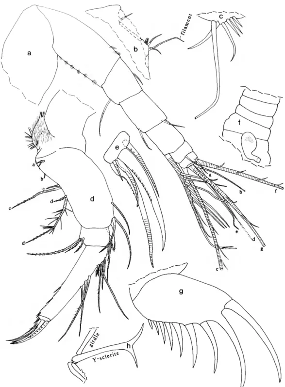
DESCRIPTION OF ADULT MALE (Figures 5, 6 a / ) . — Carapace with convex ventral and dorsal margins, broad rostrum, shallow incisur, and projecting caudal process delimited from posterior margin by slight angle (Figure 5a).
Structures on Infold: Narrow list with anterior end ventral to incisur, continuing close to ventral margin of valve, and broadening to form stout bar (with minute indistinct pustules along posterior edge) near anterior edge of caudal process (Figure 5c).
Bristles of Infold: Infold of rostrum with 7 or 8 bristles forming vertical row and 2 or 3 bristles forming horizontal row near incisur (on holotype these bristles with bases connected by
undulating line) (Figure 5b); 2 additional rostral bristles near infold mid width; a pair of bristles (longer bifurcate) and 1 small bristle present at inner end of incisur; 1 short bristle at infold mid width ventral to incisur (Figure 5b). Anteroventral infold and anterior third of ventral infold with 28 bristles (mostly divided and spinous); posterior half of ventral infold with 5 single bristles (these could be pore canals). Left valve only with single bristle near ventral end of broad bar of caudal process (Figure 5c). Broad bar of caudal process with transverse pore canals but no bristles; outer edge of infold of caudal process with about 11 minute pores (not shown).
Selvage: Lamellar prolongation with smooth outer edge present along anterior and ventral margins, terminating posteriorly near ventral end of caudal process; prolongation widest as well as narrowly striated in vicinity of incisur and divides at inner end of incisur (Figure 5b).
Carapace Size: USNM 158307, length 1.37 mm, height 0.81 mm.
First Antenna (Figure 5d): 1st and 2nd joints bare. 3rd joint with 2 small bristles (1 ventral, 1 dorsal); 4th joint elongate, with 2 small bristles (1 ventral, 1 dorsal); 5th joint short, triangular having sensory bristle with 8 or 9 long proximal filaments, 3 short distal filaments with marginal spines, and 1 very short bare filament near tip. 6th joint short, with long medial bristle (with few widely separated marginal spines) near dorsal margin (length of bristle equal to total length of joints 4-6 measured along dorsal margin). 7th joint a-bristle about
3A length of bristle of 6th joint, with few widely separated marginal spines; b-bristle about 3 times length of a-bristle, same length as sensory bristle of 5th joint, with proximal node
V
FIGURE 5.—Skogsbergia strophinx, new species, USNM 158307, holotype, adult male: a, complete specimen from left side, length 1.37 nun: b, inner view of anterior of right valve; c, inner view of caudal process of left valve; d, medial view of left 1st antenna (d-, e-, f-, and g-bristles not shown); e, medial view of endopodite and distal part of protopod ite of left 2nd antenna;/, medial view of right mandible; g, lateral view of right maxilla (part of 1st endopodial joint and all of 2nd not shown); h, lateral view of tip of right maxilla (only alpha-bristles and a -bristles shown); i, medial view of posterior distal comer of 1 st endopodial joint of right maxilla showing cutting tooth (stippled) and beta-bristles; y\ medial eye and Bellonci organ.
14 SMITHSONIAN CONTRIBUTIONS TO ZOOLOGY
and sucker on short stout proximal filament and 4 distal filaments (2 filaments with 3 and 2 small suckers, respectively, next 2 filaments with marginal spines), stem distal to last filament with 6 marginal spines; c-bristle almost twice length of b-bristle, with proximal node and sucker on short proximal filament (diameter of sucker 1.6-1.7 times diameter of large sucker of b-bristle) and 9 distal filaments (2 filaments with 2 small suckers, followed by 6 filaments with marginal spines, and 1 small bare filament near tip). 8th joint: d- and e-bristles bare, about 3A length of b-bristle; f- and g-bristles stout, about same length as c -bristle, with abundant long slender proximal filaments, followed by 8 stouter pectinate filaments and 1 small smooth subterminal filament (stem proximal to subterminal filament with spine).
Second Antenna (Figure 5e): Protopodite with short, bare, medial bristle. Endopodite 1-jointed (could be interpreted to have minute 2nd joint), with 4 proximal bristles (3 short and 1 about twice as long) and 1 long terminal filament (filament could be interpreted to be on minute 2nd joint). Exopodite: 1st joint with few indistinct spines along concave margin; bristle of 2nd joint reaching to about 8th joint, with proximal hairs (13 ventral, 11 dorsal) on unringed segment, 2 slender dorsal spines and 7 stout ventral spines on following ringed segments, and short terminal segment with closely spaced rings; bristles of joints 3 and 4 with natatory hairs and long slender spines (shorter than hairs) just proximal to bristle midlength; bristles of joints 5-8 with natatory hairs, no spines; 9th joint with 4 bristles (2 long, 1 medium, 1 short dorsal) with natatory hairs, no spines; joints 3-8 with basal spines increasing in length on distal joints; basal spine of 8th joint 3.6 times length of 9th joint; 9th joint with lateral spine about same length as joint;
joints 2-8 with indistinct minute spines forming row along distal margins.
Mandible (Figure 5/): Coxale endite spinous with 2 stouter terminal spines with small peg between them; with small basal bristle (not shown). Basale: dorsal margin with 1 bristle near midlength and 2 subterminal; ventral margin with 3 short medial a-bristles, 1 short lateral b-bristle, 2 small ventral c-bristles with small peg proximal to them, and 1 long d-bristle with long spines. Exopodite spinous, about 3A length of dorsal margin of 1st endopodial joint, with 2 bristles (base of proximal bristle near midlength of exopodite, distal bristle about 1h length proximal bristle). 1st endopodial joint with 4 ventral bristles. 2nd endopodial joint: medial surface with short spines forming rows (not shown); ventral margin with short spines, and 2 (weakly ringed) and 2 short slender pointed unringed bristles (paired bristles about same length and width); dorsal margin with 13 bristles (5 long bristles, 1 proximal, medium length bristle, 1 short ringed bristle (with long distal anterior spines) medial to 1st long bristle, 1 short ringed bristle medial to 3rd long bristle, and 5 short medial unringed bristles). 3rd endopodial joint with 3 claws (dorsal claw obscured, others with proximal ventral teeth) and 4 ringed bristles.
Maxilla (Figure 5g-i): Endile I with 11 bristles, endite II
with 7 bristles, endite III with 1 proximal and 6 distal bristles (Figure 5g). Precoxale with dorsal fringe. Coxale with plumose dorsal bristle. Basale with 1 short dorsal terminal bristle and 2 ventral terminal bristles (1 short, 1 long with long spines).
Exopodite hirsute, with 1 hirsute distal bristle and 2 terminal bristles (outer bristle hirsute). Endopodite: 1st joint with triangular cutting tooth, 2 slender bare ringed alpha-bristles, and 2 shorter ringed beta-bristles (inner with few marginal hairs, outer bare) (Figure 5h,i); 2nd joint with 4 bare ringed a-bristles, 3 pectinate claws plus about 5 additional obscured bristles (see description of adult female below).
Fifth Limb (Figure 6a): Epipodite with 50 plumose bristles.
Endite I with 7 spinose bristles; endite II with about 6 bristles;
endite III with about 7 bristles (not all endite bristles shown).
Protopodite with elongate sclerotized process. Exopodite: 1st joint: main tooth with smooth peg, 6 cuspate teeth, and spinous bristle proximal to peg; anterior side with 3 bristles forming row and 1 proximal bristle near protopodial process (not shown). 2nd joint with 4 stout pectinate unringed a-bristles, 2 pectinate ringed b'-bristles, 4 pectinate ringed b"-bristles, 1 spinous ringed posterior c -bristle, and 1 spinous anterior d-bristle (not shown). 3rd joint: inner lobe with short spinous proximal bristle and 2 bare terminal bristles (outer bristle unringed); outer lobe hirsute, with 2 terminal bristles with few long hairs. 4th and 5th joints hirsute, fused, each with paired bristles; indistinct small peg between pairs.
Sixth Limb (Figure 6b): Epipodite with 4 short bristles.
Endite I with 3 short, spinous, proximal, medial bristles and 1 or 2 terminal bristles; endite II with 2 short, spinous, proximal, medial bristles and 2 spinous terminal bristles (1 very small);
endite III with 1 long, spinous, proximal, medial bristle and 4 terminal bristles (1 short); endite IV with 1 short, spinous, proximal, medial bristle and 6 terminal bristles (1 short). End joint with 10-13 short spinous bristles followed by 2 longer plumose bristles; lateral edge with stiff spines; medial surface hirsute.
Seventh Limb: Proximal group with 7 bristles (3 or 4 on each side), each with 3-6 bells; distal group with 12 or 13 bristles (6
FIGURE 6 (opposite page).—Skogsbergia strophinx, new species. USNM 158307, holoty pe, adult male: a, posterior view of left 5th limb; b, medial view of right 6th limb; c, 7th limb; d, detail of tip of 7th limb shown in c; e, right lamella of furca and right copulatory organ;/, anterior of body showing joints 1 and 2 of right 1st antenna, anterior process, and upper lip. USNM 193626, paratype, adult female: g, complete specimen from left side showing location of central adductor muscle attachments and left lateral eye, length 1.64 mm; h, inner view of anterior of right valve; i, lateral view of left 1 st antenna (c-, d-, e-, f-, and g-bristles not shown); j , medial view of joints 6 and 7 of right 1st antenna (not all bristles shown); *, medial view of endopodite and distal part of protopodite of right 2nd antenna (ventral toward top of illustration); /, medial view of endopodial joints 1 and 2 of right maxilla (a-bristles not shown); m, posterior view of 1st and 2nd exopodial joints of left 5th limb (only a-bristles of 2nd joint shown); n, lateral view of left lamella of furca; o, anterior of body showing medial eye and Belkmci organ, anterior process, and upper lip; p, right lateral eye; q, genitalia with attached spermatophores.