Tyson belos, New Genus and Species of Western Pacific Fish (Gobiidae, Xenisthminae), with Discussions of Gobioid Osteology and Classification
VICTOR G. SPRINGER
SMITHSONIAN CONTRIBUTIONS TO ZOOLOGY • NUMBER 390
Emphasis upon publication as a means of "diffusing knowledge" was expressed by the first Secretary of the Smithsonian. In his formal plan for the Institution, Joseph Henry outlined a program that included the following statement: "It is proposed to publish a series of reports, giving an account of the new discoveries in science, and of the changes made from year to year in all branches of knowledge." This theme of basic research has been adhered to through the years by thousands of titles issued in series publications under the Smithsonian imprint, commencing with Smithsonian Contributions to Knowledge in 1848 and continuing with the following active series:
Smithsonian Contributions to Anthropo/ogy Smithsonian Contributions to Astrophysics
Smithsonian Contributions to Botany Smithsonian Contributions to the Earth Sciences
Smithsonian Contributions to Paleobiology Smithsonian Contributions to Zoology Smithsonian Studies in Air and Space Smithsonian Studies in History and Technology
In these series, the Institution publishes small papers and full-scale monographs that report the research and collections of its various museums and bureaux or of professional colleagues in the world cf science and scholarship. The publications are distributed by mailing lists to libraries, universities, and similar institutions throughout the world.
Papers or monographs submitted for series publication are received by the Smithsonian Institution Press, subject to its own review for format and style, only through departments of the various Smithsonian museums or bureaux, where the manuscripts are given substantive review. Press requirements for manuscript and art preparation are outlined on the inside back cover.
S. Dillon Ripley Secretary
Smithsonian Institution
S M I T H S O N I A N C O N T R I B U T I O N S T O Z O O L O G Y • N U M B E R 3 9 0
Tyson belos, New Genus and Species of Western Pacific Fish (Gobiidae, Xenisthminae), with Discussions of Gobioid Osteology and Classification
Victor G. Springer
SMITHSONIAN INSTITUTION PRESS City of Washington
1983
Springer, Victor G. Tyson belos, New Genus and Species of Western Pacific Fish (Gobiidae, Xenisthminae), with Discussions of Gobioid Osteology and Classification. Smithsonian Contributions to Zoology, number 390, 40 pages, 19 figures, 1983.—Tyson belos is described, based on specimens from the Great Barrier Reef, Trobriand Islands, and Lau Islands. This diminutive (gravid female 18.8 mm SL), coral-reef species differs conspicuously from all other gobioids in having the following combination of characters: pelvic fins sepa- rate, each comprising a single segmented ray and no spine; teeth on vomer;
anterior (spinous) dorsal fin lacking. Tyson appears to be closely related to Xenisthmus Snyder, Allomicrodesmus Schultz, and an undescribed genus (and species, reference D.F. Hoese). These genera share at least one synapomorphy within the Gobioidei: ventral lip with free ventral margin extending across dentary symphysis (margin interrupted across symphysis in other gobioids).
The subfamily Xenisthminae is recognized on this single character. There are, however, osteological specializations that are known only for Tyson and Xenisthmus and that are predicted to occur in the other two xenisthmine genera: premaxillary ascending process greatly reduced or absent (if present, lower than anterior maxillary articulating process of premaxilla); ascending processes replaced in position and function by a rostral bone (ossified rostral cartilage); basibranchials 2 to 4 absent. A rostral bone occurs in a few other gobioid genera, but in these the ascending premaxillary processes are well developed, basibranchials 2 to 4 are present, and not all the species in each genus where the rostral bone occurs have a rostral bone, indicating the probability that this bone is a homoplasy in those non-xenisthmine genera where it occurs.
The osteologies of Tyson and Xenisthmus are described and illustrated.
Synapomorphies are provided for the Gobioidei, Rhyacichthyidae, and Go- biidae, and the Rhyacichthyidae is proposed as the sister-group of all other gobioids.
OFFICIAL PUBLICATION DATE is handstamped in a limited number of initial copies and is recorded in the Institution's annual report, Smithsonian Year. SERIES COVER DESIGN: T h e coral Montastrea cavemosa (Linnaeus).
Library of Congress Cataloging in Publication Data Springer, Victor Gruschka, 1928-
Tyson belos, new genus and species of Western Pacific fish (Gobiidae, Xenisthminae) (Smithsonian contributions to zoology ; no. 390)
Bibliography: p.
Supt. of Docs. no. : SI 1.27:390
1. Tyson belos—Classification. 2. Gobiidae—Classification. 3. Bones. 4. Fishes—Classifi- cation. 5. Fishes—South Pacific Ocean—Classification. I. Title. II. Series
QL.S54 no. 390 [QL638.G7] 591s [597'.58] 83-600230
Contents
Page
Introduction 1 Methods 1 Materials 2 Acknowledgments 3
Family GOBIIDAE 4
Subfamily XENISTHMINAE 4
Tyson, new genus 5 Tyson belos, new species 6 Osteology of Xenisthmus and Tyson 7 Cranium 7 Jaws, Suspensorium, Superficial Bones of Head 12 Hyoid Arch 17 Branchial Apparatus 18 Pectoral and Pelvic Fins and Girdles 21 Vertebrae and Unpaired Fins 24 Characters and Classification of the Gobioidei 29 Notes 36 Literature Cited 39
in
Tyson belos, New Genus and Species of Western Pacific Fish (Gobiidae, Xenisthminae), with Discussions of Gobioid Osteology and Classification
Victor G. Springer
Introduction
In 1975, Tyson R. Roberts collected a single specimen of a fish from a coral reef in the Trob- riand Islands, off eastern New Guinea. He rec- ognized that it was distinctly different from any of which he had knowledge, but he was unable to assign it to a family. He asked my opinion of the specimen's affinities. Although the external anat- omy of the specimen did not allow one to assign it with confidence to any infraordinal taxon of fishes, I had the impression that the specimen was a gobioid. To confirm this impression would have required detailed information on the osteology of the specimen, and I was unwilling to sacrifice it for this purpose. Although Roberts generously offered to permit me to describe the species (and genus), I delayed description in the hope that additional specimens would become available. A second specimen, from the Great Barrier Reef, was obtained in 1981 by the Australian Museum, and I then made an osteological preparation of the first specimen. Shortly afterwards, in 1982, I collected two more specimens, from Fiji. It is now possible to confirm my impressions of the gobioid
Victor G. Springer, Department of Vertebrate Zoology, National Museum of Natural History, Smithsonian Institution, Washington, D.C. 20560.
affinities of the specimens. In addition, a sugges- tion by Douglass Hoese that the closest relation- ships of the new species (and genus) might be among those genera considered to be marine eleotridids (Larson and Hoese, 1980) opened up a fertile field of investigation into the family- group taxa of gobioids. I soon found myself im- mersed in the infamous and unwieldy morass of gobioid systematics, which was not my intention, nor did it fit my research priorities. I decided, therefore, to limit my study and present my find- ings to date.
In this study I describe the new genus and species, including its osteology and that of its sister genus {Xenisthmus Snyder), and assert the probable monophyly of the gobioid subfamily Xenisthminae, which contains only the new ge- nus, Xenisthmus, another undescribed genus (ref- erence D.F. Hoese, specimens unavailable to me), and Allomicrodesmus Schultz. I also discuss certain osteological characters used or useful in establish- ing the monophyly of the suborder Gobioidei and some included infrasubordinal groups.
METHODS.—Specimens (one each) of Xenisthmus clarus and Tyson belos and much of the other gobioid skeletal material used in this study were stained for cartilage with alcian blue, cleared with trypsin, and stained for bone with alizarin 1
red-s. When I discuss cartilaginous or bony struc- tures, I mean to imply only that the structures stained blue or red (pink), respectively. Some structures, notably fin-ray elements and portions of some skeletal tissues that otherwise stained blue, accepted little or no stain, and most are treated here as if they are bone (these unstained structures accept red stain in other gobioids). On the illustrations, blue-stained structures are indi- cated by a pattern of hatching; nonstained or red-stained structures are not differentiated; stip- pling is used sparingly to indicate depth or con- tours, and in Figure 12 a uniform fine screen was used to indicate depth (otherwise this screen was used for background contrast).
I prepared all the base drawings of the bones, the final renderings of which were executed by P.K. Hollingsworth.
The snout region of a large number of whole specimens of gobioids, including Xenisthmus and
Tyson and particularly genera usually considered to be eleotridids (because they have 6 branchio- stegals and separate pelvic fins), was examined to determine if the ventral lip had a complete free ventral margin, and the snout was partially dis- sected to determine whether premaxillary ascend- ing processes or a rostral bone were present. The osteologies of a number of gobioids have been discussed and illustrated in the literature (not all cited in the present study). I referred to much of this literature for relevant information. Where I encountered apparent discrepancies I attempt to rectify the differences in my discussions. (Bird- song, 1975, also encountered questionable osteo- logical characters in the literature, and I do not, in general, treat those errors that he corrected.)
Established procedures for making segmented dorsal- and anal-fin ray counts on gobioids call for enumerating the posteriormost two dorsal- or anal-fin segmented rays as a single ray. In contrast to the other segmented rays, which have a one- to-one relationship with pterygiophores in these fins, each of these ray pairs is supported by a single pterygiophore, and the two rays of each pair are closely approximate. The posterior mem- ber of each pair is often greatly reduced in size;
nevertheless, the structure of each element of each pair is that of a typical segmented ray. In my study I enumerate each element separately.
In referring to the commonly recognized fam- ily-group based on Eleotris Bloch, I use the family- level adjective "eleotridid." I do not recognize a family group based on Eleotris, however, but the adjective is useful when referring to earlier studies that do or when referring to gobioids with the following combination of characters: separate pelvic fins, 6 branchiostegals, fewer than 3 epur- als, no rostral bone, free margin of ventral lip not continuous across dentary symphysis. (All these characters, except the number of epurals, are plesiomorphic for gobioids; the number of epurals is plesiomorphic for all non-rhyacichthyid go- bioids.)
The following institutional abbreviations are used herein:
AMNH American Museum of Natural History, New York AMS Australian Museum, Sydney (catalog numbers be-
gin with I or IA)
CAS California Academy of Sciences, San Francisco GCRL Gulf Coast Research Laboratory, Ocean Springs,
Mississippi
USNM United States National Museum, the collections of which are housed in the National Museum of Natural History, Smithsonian Institution, Wash- ington, D.C. (fish specimens are in the Division of Fishes)
MATERIALS.—The osteological descriptions of Xenisthmus and Tyson are each based on a single specimen (counterstained for bone and cartilage):
Xenisthmus clarus (Jordan and Seale), USNM 235710, female, 23.6 mm SL (Figure 1); and Tyson belos, USNM 229985, female paratype, 18.5 mm SL (Figure 2). Most details of the skeletal anat- omy of X. clarus were compared for consistency with a counterstained specimen of a different (apparently undescribed) species of Xenisthmus:
USNM 247387, female, 25.3 mm SL. Differences exhibited by the comparative specimen are re- ported at appropriate points in the osteological descriptions (one of the prepublication reviewers of this study, D.F. Hoese, checked parts of the osteological description of Xenisthmus against a specimen of X. polyzonatus (Klunzinger)).
NUMBER 390
FIGURE 1.—Xenisthmus clams (from Snyder, 1982, pi. 68: fig. 3, Xenisthmus proriger [= X. clarus]).
Cleared and stained specimens (* = alizarin only; ** •• counterstained for cartilage) of other gobioids examined for this study include: Awaous tajasica (Lichtenstein), USNM 213491*; Butis am- boinensis (Bleeker), USNM 224964**; Callogobius species, USNM uncataloged (formerly CAS GVF
1956-28)**; Calumia godeffroyi (Gunther), USNM 224966**; Cerdale ionthas Jordan and Gilbert, GCRL V71:6563*; Clarkichthys bilineatus (Clark), GCRL V71:6019*; Eleotris amblyopsis (Cope), USNM 226200**; Eviota species, USNM 224539**; Gobiomorus dormitor Lacepede, USNM
Gobiosoma homochroma (Ginsburg), 121937*; Gunnellichthys pleurotaenia GCRL V82:19587*; Kraemeria bryani
USNM 143153*; K. species, AMS Microdesmus dipus Gunther, GCRL V69:361O*; M. longipinnis (Weymouth), GCRL V70:4613*; Mogurnda mogurnda (Richardson), USNM 217283**; Paragunnellichthys seychellensis Dawson, GCRL V82:19583*; Parioglossus taeniatus Regan, USNM 245268** and AMS 1.22138- 011*; Rhyacichthys aspro (Valenciennes), AMNH 48695 (complete)** and USNM 247300 (only gill arches and left jaws, suspensorium, and superfi- cial head bones)**; Valenciennea strigata (Brousso- nett), USNM uncataloged*.
A complete listing of whole specimens of go- bioids examined and partially dissected was not maintained, but those examined included a large number of genera and species, among which were the following: Bostrychus sinensis Lacepede, USNM 90325; Brachyamblyopus urolepsis (Bleeker), USNM 79070**
USNM Bleeker, Schultz, 1.20978*
217307; Bunaka canarensis (Day), USNM 164456;
Dormitator maculatus (Bloch), USNM 192189; Ero- telis species, USNM 192255; Gobiomorphus hubbsi (Stokell), USNM 198507; Gobiotrichonotus radiocu- laris Fowler, USNM 174949; Hetereleotris nebulofas- ciatus (J.L.B. Smith), USNM uncataloged (station no. VGS 69-28); H. vuigare (Klunzinger), USNM uncataloged (station no. VGS 69-23); H. zonatus (Fowler), USNM 210415; Leptophilypnus Jluviatilis Meek and Hildebrand, USNM 249722; Microper- cops daybri Fowler and Bean, USNM 83982; Ophio- cara porocephala (Valenciennes), USNM 243450;
Oxyeleotris marmorata (Bleeker), USNM 230328;
Percottus glehni Dybowski, USNM 77008; Per- iophthalmus species, USNM 249848; Rhyacichthys aspro, USNM 247300; Taenioides limicola C.L.
Smith, USNM 222998; Tridentiger obscurus (Tem- minck and Schlegel), USNM 214523.
I maintain a large and varied collection of cleared and stained specimens of teleost (mostly perciform) fishes that I use to evaluate skeletal characters. Many of these specimens have been listed in earlier studies of mine, and many of these specimens, as well as new additions to the collec- tion, were referred to during the course of the present study.
ACKNOWLEDGMENTS.—Appreciation for the
loan of specimens is extended to the following:
C.E. Dawson (GCRL), D.F. Hoese (AMS), and D.E. Rosen (AMNH). My colleagues at the Smithsonian Institution, E.A. Lachner and S.
Jewett, permitted me to examine skeletal material that they had prepared for use in their own
studies. Helpful discussions (some by mail) were carried on during the course of the work with R.S. Birdsong (Old Dominion University, Nor- folk, Virginia), D.F. Hoese, G.D. Johnson (Charleston, South Carolina), E. Murdy (Texas A&M University, College Station, Texas), D.E.
Rosen, and S.H. Weitzman. W.F. Smith-Vaniz (Academy of Natural Sciences, Philadelphia) pro- vided information on skeletal characters of several species of Opistognathidae. The manuscript was reviewed critically and substantially improved by suggestions from R.S. Birdsong, R. Winterbottom (Royal Ontario Museum, Ottawa), and D.F.
Hoese.
Grants from the Smithsonian Scholarly Studies Program and the Max and Victoria Dreyfus Foundation supported field work in Fiji that re- sulted in obtaining important material of the new genus and species.
Family GOBIIDAE
DIAGNOSIS.—Gobioid fishes (see "Characters and Classification of the Gobioidei," page 29) with the following synapomorphies: fewer than 3 epurals (3 only in variant specimens); no lateral line on body; no mandibular sensory canal; no more than 3 rows of ctenii on scales (only mar- ginal row may be tooth-like); dorsal end of inter- hyal articulating with hyomandibula, widely sep- arated from symplectic; large space separating shank of symplectic from preopercle.
Subfamily XENISTHMINAE
DIAGNOSIS.—Gobiid fishes with free ventral margin of ventral lip extending without interrup- tion across dentary symphysis; premaxillary as- cending process greatly reduced or absent (proc- ess, if present, lower than anterior maxillary ar- ticulating process of premaxilla; Figures 6 and 8b); rostral bone (ossified rostral cartilage, Figure 7) present, completely or almost completely re- placing premaxillary ascending processes in po- sition and function; and lacking: basibranchials 2 to 4, hypobranchials 3 (which may be repre-
sented vestigially as cartilaginous fragments), pterophenoids, and coronomeckelian bones.
All of the above characters, except the absence of the pterosphenoids, appear to be synapomor- phies of the Xenisthminae; however, the osteol- ogy is unknown for 2 of the 4 genera that I include in the Xenisthminae (based on the pres- ence of an uninterrupted ventral-lip margin; see
"Composition" below). The pterosphenoids are absent (homoplasiously?) in some microdesmin gobiids, for instance Clarkichthys bilineatus, but present in others, for instance Paragunnellichthys seychellensis. In the gobiin Parioglossus taeniatus, the pterosphenoids are represented on each side only by tiny fragments of bone and cartilage; the other species of Parioglossus examined had moderately well-developed pterosphenoids. It is conceivable that the pterosphenoids are absent in other spec- imens or species of Parioglossus. A rostral bone is present in Cerdale ionthas and Paragunnellichthys seychellensis, but absent in Microdesmus dipus, Clark- ichthys bilineatus, and Gunnellichthys pleurotaenia (all microdesmins). The rostral bone is also present in Parioglossus taeniatus (but not in the other species of Parioglossus examined), and in a species of Kraemeria (but not in K. bryani). In all these taxa the ascending premaxillary processes are well de- veloped, and all have basibranchials 2 to 4, hy- pobranchials 3, and coronomeckelian bones. The possibility exists, nevertheless, that some of these taxa have a sister-group relationship with the Xenisthminae.
The following xenisthmin characters also occur in various non-xenisthmin gobioids: 6 branchio- stegals, no postcleithra, no mesopterygoids, pelvic fins not united.
COMPOSITION.—Xenisthmus Snyder and Tyson, new genus, and probably an undescribed genus and species (based on characters furnished by D.F. Hoese, in litt., 1983), and Allomicrodesmus Schultz (monotypic). These four genera possess the uninterrupted free ventral-lip margin.
REMARKS.—Xenisthmus was first used as the basis of a family-group taxon by Miller (1973:426), who proposed the subfamily Xenisth- minae in the Gobiidae to include Xenisthmus
NUMBER 390
alone. None of Miller's diagnostic characters for the subfamily are synapomorphies. Birdsong (1975) questioned Miller's logic in establishing the taxon; with no more information available than that offered by Miller, I would have had to agree with Birdsong's doubts. My reaffirmation of the Xenisthminae stems from the number and extreme distinctiveness of the specializations ex- hibited by Xenisthmus and Tyson.
In recognizing the Xenisthminae as a subfam- ily of the Gobiidae, I am following essentially the gobioid classification scheme given by Miller (1973). Miller recognized only two families in the Gobioidei: Rhyacichthyidae and Gobiidae. Fam- ilies, such as Microdesmidae, Eleotrididae, Go- biodidae, and Kraemeriidae,1 were either rele- gated to subfamily status under the Gobiidae or were not recognized. I do not wish to comment on Miller's scheme except to say that I believe that several of his subfamily groups are probably paraphyletic or polyphyletic. In particular, I be- lieve this to be true of the Eleotrididae, which is usually considered to be the primitive sister group of all non-rhyacichthyid gobioids. My main rea- son for this belief is that there is no synapomorphy that defines the eleotridids.
Schultz (1966), based on inaccurately described characters, initially placed Allomicrodesmus in the Microdesmidae. Dawson (1974:409) gave a cor- rected description of the holotype (and only known specimen) and properly excluded the ge- nus from the Microdesmidae. Dawson was unable to assign Allomicrodesmus to a family, but Larson and Hoese (1980) assigned it to the Eleotrididae.
Allomicrodesmus dorotheae Schultz is known only from the holotype (USNM 113960), from the Marshall Islands, and a specimen from the Great Barrier Reef (AMS 1.18740-100). I have exam- ined the latter specimen and find it to be an adult male, 20.0 mm SL (measured from upper-jaw tip;
lower jaw protrudes). It is very delicate and slen- der (greatest depth 1.3 mm). The dorsal fin is single and continuous (the first 2 elements are spines, followed by 32 simple, segmented rays).
The anal fin comprises 25 simple elements, the first of which is probably a segmented ray, but I
was unable to satisfy myself of this. There are 3 simple rays in each pelvic fin (I cannot determine if a spine is also present) and the fins are closely approximate at their bases, but not connected by membrane. There are 6 branchiostegals on each side. The gill membranes form a continuous free fold across the isthmus.
Tyson, new genus
DIAGNOSIS.—A minute fish (less than 20 mm SL) of the subfamily Xenisthminae, lacking:
scales, extrascapular bones, lacrimal, gill rakers, an anterior spinous dorsal fin, exoccipital con- dyles (and, consequently, lateral condyles on the atlas), an uncinate process on epibranchial 1, an interarcual cartilage (Travers, 1981), and infra- pharyngobranchials 2 and 4 (including tooth- plate); and having: 13+13 vertebrae, an ossified basibranchial 1, teeth on vomer, fused anterior and posterior ceratohyals, and a single, simple, segmented ray (and no spine) in each pelvic fin (the pelvic-fin formula, 1, alone, is adequate to distinguish Tyson from all other gobioids).
Other characters, including many restricted to Tyson in the Xenisthminae, are those of the single included species, Tyson belos, new species (type- species by original designation and monotypy).
ETYMOLOGY.—The generic name is derived from the forename of Tyson R. Roberts, who collected the first specimen and recognized its distinctiveness. Gender, masculine. The stem for formation of family-group and higher level taxa is "tyson."
COMPARISONS.—Until more information is available on the other genera of the Xenisthmi- nae, I consider Tyson to be most closely related to Xenisthmus. In contrast to Tyson, Xenisthmus has scales, extrascapular bones, a lacrimal, gill rakers, an anterior spinous dorsal fin, exoccipital con- dyles, an uncinate process on epibranchial 1, an interarcual cartilage, and infrapharyngobranchi- als 2 and 4 (toothplate), only 10 precaudal ver- tebrae, a cartilaginous basibranchial 1, an eden- tate vomer, autogenous anterior and posterior ceratohyals, and pelvic fins 1,5. In addition, ex-
eluding the anteriormost anal-fin pterygiophore, Xenisthmus has only one dorsal- or anal-fin ptery- giophore inserted in each interneural or inter- hemal space, whereas Tyson has 2 or 3. It is possible, however, that Tyson is most closely re- lated to Allomicrodesmus, which, similar to Tyson, has greatly reduced the number of dorsal-fin spines (to 2) and segmented pelvic-fin rays (to 3 in each fin). The undescribed xenisthmine genus is unique among gobioids in having rows of pal- atine teeth (D.F. Hoese, in litt.).
Tyson belos, new species
FIGURE 2
This new species is so distinctive that only a diagnostic description is given here. The descrip- tion is supplemented by the generic diagnosis presented above and the osteological description (p. 7). Characters of the holotype are indicated by an asterisk.
HOLOTYPE.—AMS 1.22583-031; 19.0 ± 0 . 5 mm SL; sex unknown (immature); Australia, Queens-
FIGURE 2.—Tyson belos, USNM 229985, paratype, female, 18.8 mm SL, Kiriwina Island, Trobriand Islands, Papua-New Guinea (specimen now cleared, stained, and dissected): top, left lateral view; middle, enlarged ventral view of anus and urogenital papilla (drawings by J.R.
Schroeder); bottom, right lateral view (photograph by J.F. McKinney).
NUMBER 390
land, Escape Reef, outer barrier, coral bottom, depth 29 m; coll. 29 Oct 1981 by G. Allen, W.
Starck, and T. Ayling.
PARATYPES.—USNM 229985,18.8 mm SL, ripe female, Papua-New Guinea, Trobriand Islands, west side Kiriwina Island, coral reef, coll. 19 Sep
1975 by T.R. Roberts; specimen now cleared, stained, and dissected. USNM 233515, 12.9 and
16.2 mm SL, sexes unknown, Fiji, Lau Islands, Ono-ilau, outside barrier reef on northwest side of island, depth —13.7 to ~16.8 m, coll. 1 May
1982 by V.G. Springer et al.
DESCRIPTION.—Dorsal fin 1,9 to 1,10* (1,8 to 1,9* if last two rays are counted as one; see Figure 18). Anal fin 1,9 to 1,10* (1,8 to 1,9* if last two rays are counted as one). Pectoral fins 17*, 18, or 21. Pelvic fins not joined, each consisting of a single, unbranched, segmented ray (no spine).
Caudal fin emarginate, with 8* or 9 dorsal pro- current rays; 3 to 5* simple, segmented dorsal rays; 4* or 5 branched, segmented dorsal rays; 3*
or 4 branched, segmented ventral rays; 3 to 5*
simple, segmented ventral rays; and 9* ventral procurrent rays, for a total of 33 or 34* elements, of which 15 or 17* are segmented, and of these 7* to 9 are branched. Vertebrae 13* + 13* = 26*.
Branchiostegals 6*. Scales, gill rakers, sensory pores (except anterior and posterior nostrils) lack- ing.
Anterior nostril borne on short tube; tube, when depressed, reaches to upper lip; posterior nostril a simple pore just posterior to base of tube of anterior nostril.
Gill openings of holotype and largest paratype extend anteriorly to below level of middle of orbit; in smallest paratypes, to below level of posterior third of orbit.
Color in alcohol: ground color of body pale with few patches of melanophores. Holotype and smallest and largest paratypes each have patch of melanophores on midside of caudal peduncle.
Holotype and largest paratype have patches of melanophores posteriorly on dorsal and anal fins.
In holotype, dorsal- and anal-fin melanophore patches are extensive, intensive, and occupy ap- proximately same relative area as anal-fin melan-
ophore patch of largest paratype. Dorsal-fin me- lanophore patch of largest paratype much re- duced in area; patch completely absent in two smallest paratypes.
A color transparency photograph (in the USNM collections) taken of the freshly collected holotype is noteworthy mainly for the specimen's exhibiting an irregularly shaped, pinkish spot posteroventrally on the lateral surface of the head, and for a pale rust-colored perfusion about the melanophores on the caudal peduncle. Similar rust-colored areas are also present on the ventro- lateral portions of the head and abdomen.
REMARKS.—Although there are only four spec- imens of T. belos available, it appears that there may be geographic variation in fin-ray counts (asterisks indicate counts of holotype). Decision as to whether the differences noted are significant or the result of biased sampling will have to await the availability of more specimens.
Dorsal fin Anal fin Pectoral fins (Uft/right)
Locality
Trobriand Islands Great Barrier Reef Lau Islands
1,10 1,10*
1,9 1,9
1,10 1,10*
1,9 1,9
21/21 17/17*
18/18 18/17 ETYMOLOGY.—The specific name belos (arrow), here used as a noun in apposition, is from the Greek and refers to the arrow-like shape of the species.
Osteology of Xenisthmus and Tyson In the following descriptions, each portion of the skeleton is described first for Xenisthmus and then for Tyson.
CRANIUM
Xenisthmus (Figures 3 and 6): All the cranial bones are autogenous, with the possible exception of the pterosphenoids, which are either fused with the sphenotics or absent. The vomer (edentate) and median and lateral ethmoids are well ossified.
The lateral ethmoids are separated medially by the ethmoid cartilage, which fills the anterior
LATERAL EXTRASCAPULAE
POSTTEMPORAL EPIOCCIPITAL
EPIOCCIPITAL-1
| INTERCALAR PHENonc- P T E R 0 T I C
BAUDELOT'S LIGAMENT
POSTTEMPORAL
FIGURE 3.—Xenisthmus darns, cranium with some associated bones and ligaments (diagonal hatching represents cartilage): top, dorsal view; middle, lateral view; bottom, ventral view (medial aspect of interhyal illustrated; see also Figure 9).
NUMBER 390
interorbital region. Each lateral ethmoid bears a strong, cartilaginously tipped, laterally projecting process, to which its respective lacrimal attaches, and each is pierced by a foramen for the olfactory tract.
The frontals are two to three times as long as broad, and they are firmly joined to the ethmoids.
Each frontal bears a groove, which is perforated by three minute foramina (not visible in Figure 3), in the supraorbital region, along which the supraorbital canal courses. The groove (and canal) continues along the dorsolateral surfaces of the sphenotic and pterotic. In the postorbital region, each frontal bears a descending wing, which joins its respective sphenotic and prootic, and a parasphenoid ascending wing.
There are no parietals. The epioccipitals are joined along their dorsomedian margins (not vis- ible on external surface of skull) below the su- praoccipital, which bears a slight dorsomedian ridge posteriorly. There is no indication of carti- lage (blue stain) in the line of junction between the epioccipitals. A flattened area on the postero- lateral dorsal surface of each epioccipital serves as the area of attachment for the dorsal arm of its respective posttemporal.
The exoccipitals each bear a lateral condyle that articulates with the lateral condyle on its respective side of the atlas vertebra. The articu- lating surfaces of the exoccipital condyles are inclined ventroposteriorly. Each exoccipital is pierced by two foramina: the more anterior is the vagal foramen; the more posterior is probably for passage of a spinal nerve. Each exoccipital is excluded from meeting its respective prootic by an interosseous space (subtemporal fossa of Bird- song, 1975) that is bounded by the prootic, pter- otic, exoccipital, and basioccipital. Each interos- seous space is covered by a large intercalar, which broadly overlaps the four bones surrounding the space. A ligament extending from the ventral arm of each posttemporal attaches to the posterodorsal surface of its respective intercalar close to the junction of the intercalar with the exoccipital.
The exoccipitals form the dorsal and lateral walls of the foramen magnum, which is floored by the basioccipital.
The basioccipital articulates with the para- sphenoid anteriorly, the centrum of the atlas posteriorly, the exoccipitals dorsally, and the prootics dorsoanteriorly (most of each basioccip- ital-prootic joint is obscured from view by the overlying intercalar). Each Baudelot's ligament originates on its respective side from a minute process on the posterior midlateral surface of the basioccipital, extends posterolaterad, passing freely through a deep notch in the dorsal end of the cleithrum, and inserts on the posterointernal surface of its respective supracleithrum.
Each prootic articulates with the parasphenoid along the prootic's ventral and anterior margins;
with its respective frontal, sphenotic, and pterotic along the prootic's dorsal margin; with its respec- tive intercalar along the prootic's posterior sur- face; and with the basioccipital along the prootic's posteroventral margin. Each prootic forms only a minute part (ventral rim) of the large sphenotic socket on its respective side that articulates with the dorsoanterior condyle of its respective hyomandibula. (There is a relatively small socket, for reception of the posterodorsal condyle of each hyomandibula, in the anterolat- eral region of its respective pterotic.) Two large foramina, for passage of the trigemino-facial nerve complex, pierce the wall of each prootic.
On each side, ventral to the posterior of these two foramina, is the internal carotid foramen, formed by a notch in the margin of the parasphenoid.
Just anterolaterad of each of the internal carotid foramina, there is attached to the parasphenoid, a long, cord-like ligament that extends ventrolat- erally and attaches to a process on the medial surface of the respective interhyal (Figure 3).
The prootics lack internal shelves that might form a roof for the posterior myodome. There is, instead, a shallow, anteriorly opening pocket with a thin roof arising from the middorsal surface of the parasphenoid just posterior to the level of the anterior margins of the parasphenoid ascending processes. According to Birdsong (1975: 148, fig.
6B) the medial, inferior, and superior rectus mus- cles attach in this cavity.
The otoliths were not present, possibly having been lost during preservation and osteological
processing of the specimen. Otoliths are present in the comparative skull, but I did not attempt to extract them.
Tyson (Figure 4): Anteriorly there is a single, almost laminar, bone, which appears to comprise fused lateral and median ethmoids, which also appear to be fused to the dorsoanterior surface of the vomer. There is visible on the ventral surface of the cranium, however, a separation of the vomer from the fused ethmoid complex and the parasphenoid. The separation of the dorsoven- trally flattened shank of the vomer from the parasphenoid is also visible in a lateral view of the cranium, but no joint can be seen between the vomer and ethmoid complex when the cran- ium is viewed from in front. An opening for passage of the olfactory tract pierces the complex ethmoid bone on either side.
The vomer bears a single marginal row of nine teeth, comprising four large, recurved canines on each side, separated by a greatly reduced median tooth. These canines are clearly evident when the mouth of an intact specimen is open, and they form the posterior portion of a tooth patch in common with the anterior teeth of the premaxil- lae.
The anterior ends of the long (over four times longer than broad), paired frontals overlap the ethmoid bone posteriorly. Beginning ventral to the junction of the frontals and ethmoid complex, and extending anteriorly, is the mass of the eth- moid cartilage, which is unstained anteriorly.
The frontals, which bear no sensory canals, extend posteriorly for most of the dorsal surface of the skull, overlapping the anterior end of the supraoccipital, the dorsomedial margins of the fused sphenotic-pterotic complexes, and the an- terior ends of the epioccipitals; there are no pa- rietals. Each sphenotic-pterotic complex appears to be a single ossification, with no indication of a line of fusion. Even so, I treat the anterior portion bearing the facet for articulation with the dor- soanterior condyle of the hyomandibula as sphen- otic, and the remainder, which bears the facet for articulation with the dorsoposterior condyle of the hyomandibula, as pterotic. The separation of the pterotic portion from the dorsoposterior mar-
gin of the prootic is also unclear, but is marked by a narrow unstained area. I have illustrated (Figure 4) this unstained area as a joint line that is continuous with the joint line between the exoccipital and basioccipital.
On each side of the cranium, in the postero- medial portion of the orbital region, there is a prominent ventrally extending process that is con- tinuous with the sphenotic. Each process repre- sents either a flange of the sphenotic or a fused pterosphenoid (see discussion of prootic, p. 12).
Although the anterior margin of the supraoc- cipital is reasonably discernible through the trans- parent frontals, the remaining extent of the su- praoccipital and its joints with the epioccipitals are obscured by a granular-appearing area, pos- sibly representing the line of fusion of the supraoc- cipital with the epioccipitals. (The posterior mar- gin of the supraoccipital as delineated in Figure 4 is even less certain than indicated.) The pre- sumed posteriormost portion of the supraoccipital bears a very short, low, median ridge, which continues posteriorly as the slightly raised joint of the posterodorsally conjoined epioccipitals and the dorsally conjoined exoccipitals.
The relatively small epioccipitals each bear a flattened area dorsally, to which their respective posttemporal is attached. I was unable to deter- mine if the epioccipitals are conjoined synchon- drally beneath the supraoccipital (Birdsong, 1975, table 1).
The exoccipitals are most noteworthy for lack- ing condyles for articulation with the atlas (which, consequently, lacks lateral condyles and articulates only with the basioccipital). Each ex- occipital is excluded from making contact with its respective prootic by a meeting of the pterotic and basioccipital. The exoccipitals are each pierced by a large vagal foramen; no other foram- ina are present in these bones. The ligament extending from the posterior end of each posttem- poral attaches tightly to a minute process on the midanterolateral surface of its respective exoccip- ital just posterior to the joint between the exoc- cipital and pterotic and just below the level of the laterally widest expansion of the pterotic. There are no intercalars. The exoccipitals form the lat-
NUMBER 390 11
SPHENOTIC + PTEROTIC
SUPRAOCCIPITAL
MEDIAN + LATERAL ETHMOIDS
SPHENOTIC OR (?)PTEROSPHENOID
SPHENOTIC + PTEROTIC- FIGURE 4.—Tyson belos, cranium (diagonal hatching represents cartilage): top, dorsal view; middle,
lateral view (U = unstained portion of ethmoid cartilage); bottom, ventral view.
eral and dorsal walls of the foramen magnum, which is floored by the basioccipital.
The broadly Y-shaped basioccipital articulates with the atlas posteriorly and accepts the poste- rior end of the parasphenoid between its ante- riorly extending arms, each of which articulates anteriorly with the posterior ends of its respective prootic. Each Baudelot's ligament has a short bifurcation at its proximal end. The anterior branch of the bifurcation attaches to the postero- lateral surface of the basioccipital and the poste- rior branch attaches to a closely adjacent area on the anterolateral margin of the atlas vertebra.
The ligament extends posterolaterally and passes freely through a notch in the dorsal process of the cleithrum, then proceeds anteriorly and inserts on the dorsomedial surface of the supracleithrum.
Possibly, the bifurcation of Baudelot's ligament and its attachment to both the basioccipital and atlas, rather than only to the basioccipital (as in Xenisthmus), is an adaptation for strengthening the connection between the skull and vertebral column, which in other gobioids is shared by three skull bones rather than one.
Each prootic articulates with the parasphenoid along the prootic's ventral margin, with its re- spective fused sphenotic-pterotic along the prootic's dorsal margin, and impinges slightly (at most) on the ventrolateral margin of the ventrally extending process of the sphenotic in the post- orbital region. Each ventrally extending process appears to form the incomplete anterior border of an opening (anterior passageway for the trige- mino-facial nerve complex?) whose posterior bor- der is a notch on the anterior margin of the prootic. In most gobioids with pterosphenoids, each pterosphenoid forms the anterior margin, and the prootic the posterior margin, of the an- terior foramen for passage of the trigemino-facial nerve complex. Inasmuch as both foramina for passage of the trigemino-facial nerve complex are contained only in the prootic of Xenisthmnus, which lacks pterosphenoids, it would appear that the ventrally extending sphenoid processes of Ty- son represent fused pterosphenoids. Each prootic contains a single complete foramen for passage of the trigemino-facial nerve complex. The prootics
do not bear a shelf on their medial surface, and there is no bony roof over the posterior myodome.
The parasphenoid is perforated by a pair of carotid foramina near the anterior end of its posterior broad portion, and bears a low, median, anteriorly directed hook-like process on the dor- sal, interorbital surface of its slender anterior shank, which overlaps the shank of the vomer.
There is no bony pocket for reception of the rectus muscles of the eyes in the dorsal surface of the parasphenoid within the cranial vault, as there is
in Xenisthmus.
The otoliths were not present, possibly having been lost during preservation or osteological pro- cessing.
JAWS, SUSPENSORIUM, SUPERFICIAL BONES OF HEAD
Xenisthmus (Figures 5 and 6; for comparison with the snout region of a relatively unspecialized gobioid see Figure 7): The premaxillae are well- developed bones, each bearing an outer row of large recurved, conical teeth and an inner row of similar but much smaller teeth (in the compara- tive specimen, the outer row is only slightly larger than the inner teeth, which are scattered over the ventral surface of the premaxillae, but nowhere in more than three rows). Each premaxilla has anterior (articular) and posterior maxillary pro- cesses, but lacks an ascending process. The por- tion of the premaxilla that normally bears an ascending process is reduced to a flattened facet that articulates with a corresponding ventrolat- eral facet at the end of the median, unpaired, rostral bone, which has functionally replaced the ascending processes of both premaxillae. In the comparative specimen, the ascending processes are represented by facet-bearing knobs, which are, however, lower than the anterior articular processes. The rostral bone, which has a cartilag- inous dorsal end, is an ossified rostral cartilage.
The rostral bone bears thin anterodorsal and posteroventral flanges on each side. A band-like ligament connects each of the dorsoanterior flanges to the dorsoanterior margin of the pala- tine bone on its respective side (in other gobioids,
NUMBER 390 13
LATERAL EXTRASCAPULAE—i NASAL
MAXILLA
PREMAXILLA OPERCLE
QUADRATE
METAPTERYGOID HYOMANDIBULA
DENTARY
ANGULOARTICULAR
SUBOPERCLE- PREOPERCLE
I mm
FIGURE 5.—Xenisthmus clarus, lateral view of superficial bones, jaws, and suspensorium. (The limits of some bones that are obscured by others are indicated by dashed lines; see Figure 6 for dorsoanterior view of snout region.)
LATERAL ETHMOID NASAL
FRONTAL
MEDIAN ETHMOID
VOMER- PREMAXILLARY ARTICULAR PI
ROSTRAL CARTILAGE LATERAL ETHMOID
MEDIAN ETHMOID FRONTAL
PREMAXILLA
VOMER
PREMAXILLARY ARTICULAR PROCESS
^ M A X I L L A PREMAXILLARY
ASCENDING PROCESS
FIGURE 6.—Xenisthmus clarus, dorsoanterior view of snout region (frontals and palatines truncated posteriorly; nasal and lacrimal removed from one side; dotted lines indicate margins of maxillary palatine processes obscured by pala- tines).
FIGURE 7.—EUotris amblyopsis, dorsoanterior view of snout region (frontals and palatines truncated; lacrimal removed from one side; species lacks nasal bones). Structure of pre- maxillae and their relation to rostral cartilage represent generalized condition for gobioids.
this ligament attaches the palatine to the dorsal end of the ascending process, thus indicating a shift in "allegiance," but not in function, of the ligament). Another band-like ligament connects each of the posteroventral flanges to the dorsoan- terior portion of the palatine process of its respec- tive maxilla. In other gobioids, the same ligament exists, but it is attached to the rostral cartilage, which is, in turn, tightly attached to the posterior surface of the tips of the premaxillary ascending processes. Birdsong (1975:152, fig. 8) does not indicate the existence of a maxillorostral ligament in Microgobiusy but he does illustrate a ligament labelled "1," which he reports as attaching the base of the premaxillary process to the medial head of the maxilla. There is an unroofed nasal bone on each side of the rostral bone. A small foramen perforates the floor of each nasal bone.
Each maxilla is forked anteriorly where it clasps the articular process of its respective pre- maxilla. Just dorsal to each maxilla, and laterally overlapping each slightly, is a thin plate-like bone, the lacrimal, which has its strongest attach- ment with the laterally projecting process of its respective lateral ethmoid. The wall of each lac- rimal is pierced by a tiny foramen, but the lacri- mals do not appear to bear sensory canals. A suborbital (in agreement with Akihito, 1969) is lacking.
The dentary bones each bear a row of large outer teeth and, anteriorly, two inner rows of much smaller teeth, gradually reducing poste- riorly to one inner row (in the comparative spec- imen the outer teeth are only slightly larger than the inner teeth, which are more or less evenly distributed over the surface of the dentary, but nowhere in more than three rows). The long, club-like maxillodentary ligament on each side inserts in a groove on the anterolateral surface of its respective dentary and connects to the poste- rior end of its respective maxilla. The anguloar- ticular on each side inserts into a pocket at the posterior end of its respective dentary. Each long, rod-like Meckel's cartilage extends anteriorly from the endosteal process on the medial surface of its respective anguloarticular to a point in the
hollow end of its respective dentary, just anterior to the level of a foramen that completely perfo- rates the dentary. There is no sensory canal in the mandible. There are no coronomeckelian bones (sesamoid articulars). Each retroarticular is a small bone joined to the ventroposterior end of its respective anguloarticular and is connected by a ligament to the anterior end of its respective interopercle.
The quadrates are the pivotal bones of the suspensorium. Each bears a broad cartilaginous dorsal margin with which the anteroventral, car- tilaginous tip of its respective metapterygoid, the posterodorsalmost bony tip of its respective ectop- terygoid, and a minute portion of the anterior margin of its respective symplectic articulate.
There is a broad groove on the medial surface of each quadrate, to which the ventrolateral surface of its respective symplectic is tightly joined. Each preopercle narrowly overlaps the posterolateral margin of its respective quadrate, and the pos- terolateral margin of each ectopterygoid is nar- rowly overlapped by its respective quadrate.
The ectopterygoids are long, rod-like bones, each with a flattened ventrolateral surface along the anterior half of its length. The medial surface of each palatine articulates syndesmotically with a flattened surface of its respective ectopterygoid, but in the anterior region of this joint, the pala- tine bears a tiny rod of cartilage on its medial surface that participates in the ectopterygoid-pal- atine articulation.
The symplectics are relatively large. Each forms a synchondral joint with its respective hy- omandibula at the symplectic's dorsal end and has a cartilaginous ventral tip on the portion that attaches in the groove on the medial surface of its respective quadrate.
Posteriorly, each metapterygoid broadly over- laps and is tightly joined to the anterolateral surface of its respective symplectic and antero- dorsal surface of its respective hyomandibula.
Dorsoanteriorly, each metapterygoid is tightly joined to the anterolateral edge of its respective
sphenotic.
Each hyomandibula bears three convex con-
NUMBER 390 15 dyles, each of which articulates (anterior to pos-
terior) with a socket in its respective sphenotic, pterotic, and opercle. There is a thin, raised flange on the lateral surface of each hyomandibula, to which is tightly and conformably joined a thin, raised flange on its respective preopercle. The preopercular sensory canal courses ventrally along the posterior surface of the groove formed by the compound flange. Anteroventrally, on the medial surface of each hyomandibula, is a broad cartilaginous area. Posterodorsal to this area is the axil formed by the junction of the posterior convex condyle of the hyomandibula with the body of the hyomandibula. The medial surface of the preopercle lies lateral to this axillar area, and the dorsal end of the interhyal is ligamen- tously attached to this surface well dorsal to the cartilaginous ventral end of the hyomandibula.
There may also be weak membranous attach- ments of the interhyal to the hyomandibula in the axillar area, but I was unable to ascertain such with certainty. The ventral end of each interhyal joins the posterodorsal edge of its re- spective posterior ceratohyal. This joint forms a lateral bulge, which is accommodated by a deep rounded indentation in the ventral margin of the adjacent interopercle. Each interopercle is at- tached by ligament at its posterior tip to the ventroanterior margin of its respective suboper- cle.
The lateral extrascapulae (supratemporals of Akihito, 1971) are contained in the skin dorsal to the opercles. There are two, unroofed, lateral extrascapulae on the left side and three on the right (three on the left and two on the right in the comparative specimen). Akihito (1971) re- ported that Xenisthmus clams has two extrascapu- lae on each side, concluding that the anterior and median extrascapulae on each side, which are present in some gobioids, are fused. The presence of three extrascapulae on one side and two on the other in each of my specimens supports Akihito's conclusion; however, in one specimen it is the median and anterior elements that are fused, and in the other specimen, the median and posterior elements. Radiographs of several specimens of
Xenisthmus indicate that the presence of three or two extrascapulae on one or both sides are both common conditions. In the osteological prepara- tions, the floor of each of the three extrascapulae is pierced by a tiny foramen; the fused anterior plus median extrascapulae of the opposite side is pierced by two foramina, and the remaining ex- trascapula, by one foramen. Akihito (1971, figs.
1B, 2H) diagrammed the laterosensory canals and pores of X. darns.
Tyson (Figure 8): The premaxillae are slender bones bearing an outer row of small, fine, cani- niform teeth, with an incomplete second, inner, row of much larger canine teeth restricted to the anterior ends of the premaxillae. The largest tooth on each premaxilla is adjacent to the symphysis.
The premaxillae lack any vestige of ascending processes, but each has anterior (articular) and posterior maxillary processes. The anterior ends of the premaxillae are slightly separated at the symphysis, forming a space into which inserts a narrow process (not visible in Figure 8) at the ventroanterior end of the rostral bone. Each pre- maxilla articulates with the long, anteriorly forked maxilla on its respective side at the pre- maxilla's articular process, which is clasped by the maxillary fork. This articulation, together with the same articulation on the opposite side of the head, joins the ventrolateral end of the me- dian rostral bone, which functionally acts as a pair of long premaxillary ascending processes, riding over the dorsoanterior surface of the cran- ium. A pair of ligaments inserts on the lateral margin of each side of the rostral bone: one ligament of each pair attaches the lower third of the margin to the medial surface of the lateral fork at the anterior end of the maxilla, and the other attaches the middle of the margin to the dorsomedial process of the palatine. There is a process, which bears a cartilaginous cap, on the medial surface of each maxilla just posterior to the base of the maxillary fork.
Each dentary bone bears a single row of large, recurved, canine teeth, the largest of which is just posterolateral to the anteriormost tooth (near dentary symphysis), and just above the anterior
PREMAXILLA r~ MAXILLA
b
ROSTRAL r-ECTOPTERYGOID + CJMESOPTERYGOIDOPERCLE PALATINE / r - 0U A D R A T E
SUBOPERCLE PREOPERCLE FIGURE 8.—Tyson belos, superficial bones, jaws, and suspensorium: a, lateral view (limits of some bones that are obscured by others are indicated by dashed lines); b, dorsal view of rostral bone showing relationships with anterior ends of premaxillae and maxillae.
attachment of the long, large maxillodentary lig- ament, which connects the posterior end of the maxilla to the anterolateral surface (which is not grooved) of the dentary. Each dentary bears two or three small foramina, one anterolaterally and one or two along the midventral edge of the bone.
As there is no mandibular sensory canal, these foramina are probably for passage of nerves and/
or blood vessels. Each Meckel's cartilage is long and slender and extends for most of the length of its respective dentary, continuing posteriorly to its origin on the anguloarticular bone, whose anterior process inserts into a shallow pocket at the dentary's posterior end. There are no co- ronomeckelian or retroarticular bones, although the latter are probably fused indistinguishably to the anguloarticulars. A short, strong ligament attaches the ventralmost edge of the quadrate
articulating process of each anguloarticular to the anterior end of its respective interopercle.
Each palatine is a laminar bone with a two- tipped process at its anterior end. The ventral of the two tips bears a cartilaginous cap (not shown in Figure 8) and overlaps the lateral surface of the base of the lateral fork of its respective max- illa. The dorsal tip is the attachment point for a ligament that inserts on the midlateral margin of the rostral bone. The posterior end of each pala- tine broadly overlaps, on its respective side, the anterolateral surface of what appears to be a fusion of the ectopterygoid and mesopterygoid bones. As many gobioids, including Xenisthmus, either lack mesopterygoids or have them greatly reduced, it is possible that Tyson has no mesopter- ygoids and that the ectopterygoids and palatines have become broadly expanded relative to their
NUMBER 390 17 shape in other gobioids. The ventrolateral surface
of each ectopterygoid is joined to the dorsomedial surface of its respective quadrate.
Each quadrate is unusually long and shaped like a scimitar (blade tip directed posterodorsally) with a ledge coursing along its posterolateral margin. The dorsal margin of the ventroanter- iorly extending arm of its respective preopercle is tightly bound (fused?) to the ledge. On its medial surface, each quadrate attaches to the ventral half of its respective symplectic. The symplectics are long, rod-like bones, and ellipsoid in cross- section. The posterodorsal end of each symplectic is tightly attached (fused?) to a process on the ventroanterior margin of its respective hyoman- dibula. There is a broad gap between the sym- plectic and the preopercle, which is joined near its dorsal end to the ventral arm of the hyoman- dibula. There is no sensory canal in the preoper- cle.
Each interopercle, which is almost entirely in- ternal to its respective preopercle, is long and flat, except for a relatively heavy, cup-like process on its posteromedial surface. A knob-like process on the ventrolateral surface of the posterior end of the respective posterior ceratohyal articulates with the cup-like process. Each interopercle is well separated from the ventroanterior process of its respective subopercle and is attached to that process only weakly. The interhyals are tiny and completely ossified dorsally, and each is well sep- arated from its respective symplectic. Each inter- hyal is ligamentously attached at its dorsal end just dorsal to the ventral end of the medial surface of its respective hyomandibula and to the medial surface of its respective preopercle. Ventrally, each interhyal is attached to a dorsolateral proc- ess at the posterior end of its respective posterior ceratohyal.
Each opercle bears a strong, concave dorsoan- terior condyle that articulates with the posterior condyle of its respective hyomandibula, and the opercle overlaps and is tightly connected to the dorsolateral surface of its respective subopercle.
Each hyomandibula has two convex condyles dorsally and one posteriorly. The dorsoanterior
condyle articulates with a socket formed com- pletely by the sphenotic (no contribution from the prootic); the dorsoposterior condyle articu- lates with a socket formed by the pterotic; and the posterior articulates with a socket formed by the opercle.
There are no metapterygoid, lacrimal, subor- bital, nasal, or lateral extrascapular bones.
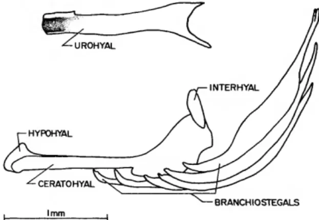
HYOID ARCH
Xenisthmus (Figure 9): Each side of the ven- troanterior end of the urohyal is connected by a short, strong ligament to a large process on the anteromedial surface of the hypohyal on its re- spective side. The dorsal and ventral hypohyals on each side are fused. (In the comparative spec- imen, autogenous dorsal and ventral hypohyals are present and the urohyal is attached to the ventral pair.) The dorsoanterior surface of the urohyal is directly beneath and slightly anterior to the median, cartilaginous basibranchial 1 (Fig- ure 11).
Each anterior and posterior ceratohyal pair is joined in a synarthrosis. Each anterior ceratohyal bears a cartilaginous tip ventroanteriorly where the anterior ceratohyal is capped by its respective hypohyal. Each hypohyal (dorsal hypohyal in comparative specimen) bears a rounded dorso- medial process that joins one of the two concave surfaces at the posterior end of the basihyal.
Somewhat lateral to the medialmost surface of each hypohyal rounded process is a small area of cartilage. The posterior, deeper portion of each hyoid arch, formed by the anterior and posterior ceratohyals on each side, is concave on its medial surface. Each anterior ceratohyal bears five bran- chiostegals, the two anteriormost, which are at- tached to the ventral side of the slender, anterior portion of the anterior ceratohyal, are the small- est; the next three branchiostegals are attached laterally on the expanded portion of the anterior ceratohyal, and the anteriormost of these is the largest of all the branchiostegals. The posterior- most branchiostegal is attached to the posterior surface of the posterior ceratohyal. The ventral
INTERHYAL LIGAMENT
ANTERIOR CERATOHYAL
I mm
end of each interhyal is syndesmotically joined to the dorsoposterior end of its respective posterior ceratohyal. Each interhyal bears a cartilaginously tipped dorsal process, a cartilaginously tipped ventroanterior process (which articulates with the posterior ceratohyal), and a prominent midanter- omedial process from which a strong cord-like ligament extends to the parasphenoid (Figure 3).
Tyson (Figure 10): The dorsoanterior surface of the urohyal is tightly joined to the ventral surface of the ossified basibranchial 1 (Figure 13).
There is a single hypohyal on each side (each probably representing a fusion of the dorsal and ventral hypohyals of each side), and a single ceratohyal on each side (each representing a fu- sion of the anterior and posterior ceratohyals of each side). Of the 6 branchiostegals on each side, the 2 smallest are attached to the narrow anterior process of their respective ceratohyal, and the remaining four to the broad posterior process.
Each interhyal is attached on its ventromedial surface to a process on the dorsolateral surface of the posterior portion of its respective ceratohyal.
There is a tiny area of cartilage on the medial surface of the interhyal at its point of attachment to the ceratohyal. The presence of a ligament extending from the interhyal to the parasphenoid was not noticed (and was probably not present), but may have been removed early in the dissec- tion before I became aware of the existence of
FIGURE 9.—Xenisthmus clarus, lateral view of urohyal and left hyoid arch, rotated ~45°
clockwise about long axis (the limits of some bones that are obscured by others are indi- cated by dashed lines; two small areas of cartilage on the interhyal and one at the anterior end of the anterior ceratohyal are indicated by diagonal hatching; ligament attaching to interhyal extends to parasphen- oid, Figure 3).
such a ligament in Xenisthmus. The right cerato- hyal exhibits an incomplete ventral separation, with cartilage present between the attachment of the fourth and fifth posteriormost branchioste- gals. This separation is probably a landmark for separation of the anterior and posterior cerato- hyals that are normally present in fishes.
BRANCHIOSTEGALS
FIGURE 10.—Tyson belos, lateral view of urohyal (isolated) and left hyoid arch, rotated —45° clockwise about long axis.
B R A N C H I A L A P P A R A T U S
Xenisthmus (Figure 11; for comparison with a relatively unspecialized gobioid branchial appa- ratus, see Figure 12): The basihyal is a relatively thin, dorsally concave, ventrally convex bone that
NUMBER 390 19
-INFRAPHARYNGOBRANCHIAL I INTERARCUAL CARTILAGE
^INFRAPHARYNGOBRANCHIALS 2 3 4(TOOTHPLATE-NO CARTILAGE)
BASIHYAL
,'N-HYPOBRANCHIALS 2
GILL RAKERS
CERATOBRANCHIALS
FIGURE 11.—Xenisthmus clanis, dorsal view of gill arches and basihyal (right-side dorsal elements removed; left-side dorsal elements illustrated both in place and separately; arches spread slightly for clarity).
attaches to the hypohyals at its posterior end and bears a cartilaginous margin anteriorly. The ba- sihyal is slightly removed anteriorly from the small cartilaginous basibranchial 1, which is at-
tached posteriorly to the anterior, cartilaginous ends of the hypobranchials 1. There are no other basibranchials present, either as cartilage or bone.
Of the hypobranchials, only 1 and 2 are present
INFRAPHARYNGOBRANCHIAL I INTERARCUAL CARTILAGE
INFRAPHARYNGOBRANCHIALS
5 ^ (CARTI LAGE+TOOTHPLATE) BASIHYAL
2 V-BASIBRANCHIALS
V-HYPOBRANCHIALS 3
CERATOBRANCHIALS
FIGURE 12.—Eleotris amblyopsis, dorsal view of gill arches and basihyal (right-side dorsal elements removed and illustrated from ventral aspect; left-side dorsal elements illustrated both in place and separately). Structure represents generalized condition for gobioids.