STRUCTURAL CHARACTERIZATION OF THE RECEPTOR FOR ADVANCED GLYCATION END PRODUCTS REVEALS A TWO DOMAIN MODULAR
ARCHITECTURE
By
Brian Matthew Dattilo
Dissertation
Submitted to the Faculty of the Graduate School of Vanderbilt University
for the degree of
DOCTOR OF PHILOSOPHY in
Biochemistry August, 2007
Nashville, Tennessee
Approved:
Professor Walter J. Chazin Professor Charles R. Sanders, II
Professor Martin Egli Professor Billy G. Hudson
ACKNOWLEDGEMENTS
First and foremost I thank my advisor, Dr. Walter Chazin. Not only did he provide the opportunity to freely perform exciting research in his laboratory, but he stuck with me during the low points of my project and development as a scientist.
I also thank the other members of my continuing committee, Drs. Billy Hudson, Charles (Chuck) Sanders, Martin Egli, and Matthew Breyer, for advice and discussion.
But mostly I thank them for one particular meeting that helped me make the leap to the problem solver I have become today.
I thank the other three founding members of the CLDT. They know who they are and what the acronym means.
Writing this dissertation would not have been as enjoyable without the presence of two great friends, Kelly and Mike. Thank you for being great distractions from the many hours of sitting, reading, and writing.
All of the work in this dissertation has been performed in close collaboration with Dr. Guenter Fritz from the University of Konstanz. Thanks for provided stimulating discussion throughout the development of this project in addition to his contributions as an expert protein crystallographer. In addition, I thank Dr. Estelle Leclerc for her valuable Biacore expertise.
Of all the past and present members of the Chazin I especially thank Dr. Craig Vanderkooi and Susan Meyn. Craig helped develop the initial ideas for this dissertation, taught me everything I know about NMR, and provided incredible discussion throughout.
I thank Susan, of course, for putting up with my shenanigans in the lab but also for helping me develop as a person and fight through the tough times during grad school.
Funding was provided by the molecular biophysics training grant and NIH research grants. The Diabetes Research Training Center provided initial pilot funding which helped launch this work.
Finally I thank my family. Thanks for the example that provided me with all the tools needed to achieve success. And thanks for the love and support that helped me maintain enough focus to climb out of the low points of my training.
TABLE OF CONTENTS
Page
ACKNOWLEDGEMENTS... ii
LIST OF TABLES... vii
LIST OF FIGURES ... viii
LIST OF ABBREVIATIONS... xi
Chapter I. INTRODUCTION ...1
The Multiligand Receptor for Advanced Glycation End Products...1
RAGE Biology...3
Advanced Glycation End Products (AGEs)...3
AGE Chemistry...3
AGEs in Normal Aging and Diabetes...4
AGE Signaling Induces Oxidative Stress Though RAGE...6
Signaling Pathways Triggered by AGEs ...7
RAGE Activation by Amphoterin: a Physiological Role for RAGE...7
RAGE Binding to Amyloid β: Implications in Neurotoxicity...10
S100 Proteins are RAGE Ligands...11
Divergent Signaling Pathways and Essential Signaling Elements ...14
Kinase Pathways ...14
Dominant-Negative RAGE...14
Positive Feedback Loop...15
Potential for RAGE-Based Therapeutics ...15
Diabetes ...16
Cancer ...16
Alzheimer’s Disease ...17
Chronic Inflammation...17
Concluding Remarks...17
S100 Calcium-binding Proteins ...18
Structure of the EF-hand Motif...18
Calcium Sensing by EF-Hand Proteins...19
Architecture of the S100 Protein...19
S100 Proteins and Zinc ...22
Higher Order Oligomeric States of S100 Proteins...22
Target Recognition by S100 Proteins ...24
Cytokine Receptor Classification ...27
Scissor-like Activation of EPOR ...28
Transmembrane Rotation Model for Activation of GHR ...29
Structural Studies on sRAGE and Ligand Interactions...32
II. THE EXTRACELLULAR REGION OF THE RECEPTOR FOR ADVANCED GLYCATION END PRODUCTS IS COMPOSED OF TWO INDEPENDENT STRUCTURAL UNITS ...33
Introduction...33
Production of sRAGE and Domain Constructs ...34
Stability of sRAGE Domains...37
sRAGE is Composed of Coupled V and C1 Domains With an Independent C2...45
Homology Modeling of sRAGE ...56
Summary ...59
III. HIGH RESOLUTION CRYSTAL STRUCTURE OF VC1 ...61
Introduction...61
Crystallization and Structure Determination ...62
Structural Characteristics of VC1 ...64
The V-C1 Interface ...72
Summary ...76
IV. LIGAND BINDING TO SRAGE...77
Introduction...77
S100B Binding Induces Structural Change in sRAGE...88
NMR Characterization of S100B Binding to sRAGE ...83
S100B Binds to RAGE with nM Affinity...87
CML Binding to RAGE ...89
Summary ...93
V. CONCLUSIONS AND FUTURE DIRECTIONS...94
Implications and Conclusions ...94
The Modular Structure of sRAGE ...94
Implications for Ligand Binding...101
Implications for RAGE Signaling...103
Future Directions ...109
High Resolution Structure of the C2 domain...109
The Oligomeric State of Intact RAGE...112
Variable RAGE Surfaces Used in Ligand Recognition...114
Stoichiometries of RAGE-Ligand Complexes...117
Potential Mechanisms for RAGE Autoinhibition ...117
Functional Role of the C2 Domain ...121
Prospects for sRAGE Based Therapeutics...122
Closing Remarks...123
VI. MATERIALS AND METHODS...124
Molecular Biology ...124
Protein Expression and Purification...126
Limited Proteolysis/Protease Protection ...127
Mass Spectrometry...128
Circular Dichroism...128
Dynamic Light Scattering ...129
Differential Scanning Calorimetry...129
Surface Plasmon Resonance ...130
Nuclear Magnetic Resonance Spectroscopy...130
Homology Modeling and Structure Analysis ...131
CML Synthesis...132
Synthesis of N(alpha)-Boc-N(epsilon)-(carboxymethyl)lysine...132
Synthesis of N(epsilon)-(carboxymethyl)lysine ...133
Appendix A. MULTIPLE SEQUENCE ALIGNMENT OF SRAGE PROTEINS...135
B. SUMMARY OF CRYSTALLIZATION STATISTICS FOR VC1 ...137
C. 15N-1H HSQC SPECTRA OF OPTIMIZED SRAGE CONSTRUCTS ...139
D. TABLE OF BACKBONE ATOM CHEMICAL SHIFTS FOR C2 ...144
BIBLIOGRAPHY...147
LIST OF TABLES
Table Page
6.1 Summary of expression vectors with expression and purification strategies
of recombinant proteins ...125
B.1 Statistics of data collection and Zn MAD phasing ...137
B.2 Statistics for highest resolution dataset...138
D.1 1H, 15N, 13C backbone chemical shifts for C2...144
LIST OF FIGURES
Figure Page
1.1 Schematic of RAGE domain organization...2
1.2 Chemical pathway towards AGE formation ...4
1.3 Structure of rat amphoterin ...9
1.4 Solution structure of Ca2+-loaded human S100B...13
1.5 Calcium induced conformational change in S100A6 ...21
1.6 Variability in target recognition by S100 proteins...26
1.7 Two models for cytokine receptor auto-inhibition and activation...31
2.1 Multiple sequence alignment of RAGE proteins ...35
2.2 The effect of reducing agent on Ig domain tertiary structure ...36
2.3 Limited proteolysis of sRAGE reveals two stable domains ...38
2.4 Resistance to proteolysis of V and C1 is enhanced by covalent attachment ...39
2.5 The C2 domain is stable to trypsin proteolysis...40
2.6 VC1 undergoes a complex thermal transition...41
2.7 CD thermal melt of VC1...43
2.8 Thermal stability of C1C2, V and C1 constructs...44
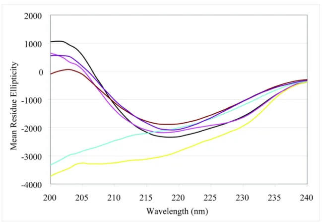
2.9 Circular dichroic spectra of sRAGE and domain constructs ...46
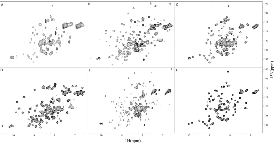
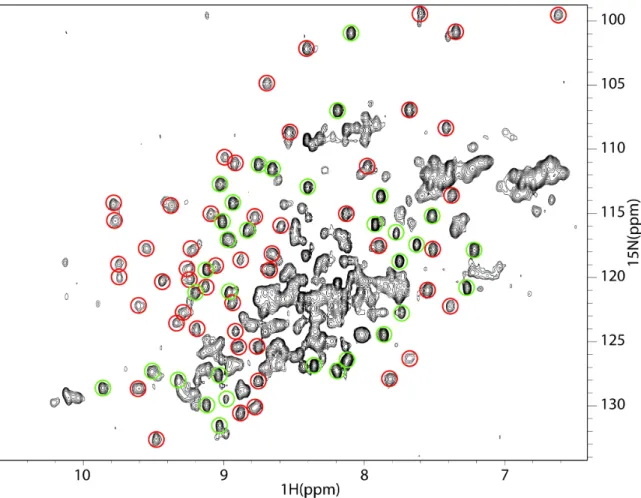
2.10 Heteronuclear NMR spectra of RAGE constructs...50
2.11 VC1 and C2 are separate structural units...52
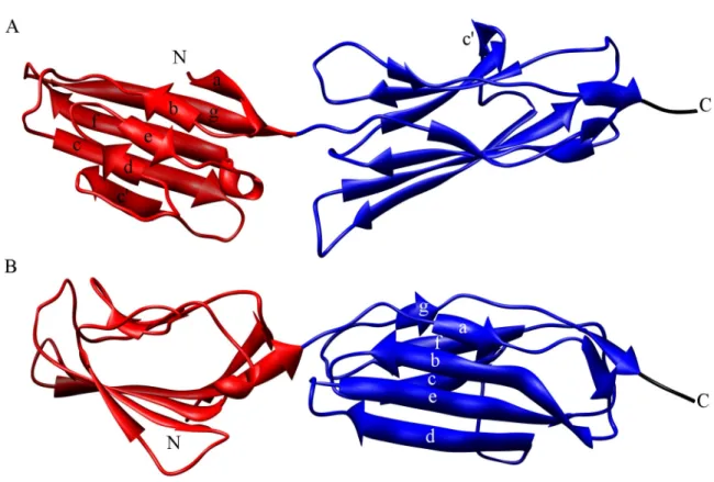
2.12 The V and C1 domains form a discrete structural interface ...55
2.13 Homology modeling of VC1 ...58
3.1 The x-ray crystal structure of VC1 ...63
3.2 Topology diagram of the x-ray crystal structure of VC1...65
3.3 Side chain hydroxyl stabilizes a bulge in a C1 β-strand...66
3.4 Strand swap at the C-terminus of the C1 domain ...68
3.5 Comparison of the homology model and x-ray crystal structure of VC1...71
3.6 The V-C1 interdomain angle is different in the x-ray crystal structure...73
3.7 Interdomain contacts in the VC1 x-ray crystal structure ...75
4.1 Cys84 of S100B covalently attacks the V domain disulfide bond...79
4.2 S100B binding causes a conformational change in sRAGE...81
4.3 S100B binding is localized to the V domain ...82
4.4 C2 retains its structural independence when in complex with Ca2+-S100B ...84
4.5 Both domains of VC1 are broadened by S100B...86
4.6 SPR binding of S100B to sRAGE domains...88
4.7 CML titration into 15N-enriched V ...90
4.8 CML is unperturbed in the presence of the isolated V domain ...92
5.1 Homology model of C2 suggests S-type Ig fold...97
5.2 Model for the structure of sRAGE...100
5.3 Structural characterization of the intracellular domain (IC) of RAGE...106
5.4 Models for ligand-induced activation of RAGE through oligomerization ...108
5.5 15N-1H HSQC spectrum of C2 with backbone assignments ...111
5.6 Regions of ligand recognition by RAGE ...116
5.7 Solvent accessible side chains as a mechanism for autoinhibition ...120
6.1 Reaction scheme for synthesis of N(epsilon)-(carboxymethyl)lysine ...134
A.1 Multiple sequence alignment of sRAGE from four species ...136
C.1 15N-1H HSQC spectra of VC1(23-233)...140
C.2 15N-1H HSQC spectra of V(23-119) ...141
C.3 15N-1H HSQC spectra of C1(119-233) ...142
C.4 Optimized V shows structural changes in the context of VC1 ...143
LIST OF ABBREVIATIONS
Aβ Amyloid β Peptide
AD Alzheimer’s Disease
AGE Advanced Glycation End products
APP Amyloid Precursor Protein
BBB Blood Brain Barrier
BSA Bovine Serum Albumin
CD Circular Dichroism
CML N-(epsilon)-(CarboxyMethyl)Lysine
DLS Dynamic Light Scattering
DSC Differential Scanning Calorimetry dnRAGE dominant negative RAGE
DTT DiThioThreitol
EBP Erythryopoietin Binding Protein EMP Erythryopoietin Mimetic Peptide
EPO ErythroPOietin
EPOR ErythroPOietin Receptor
ERK Extracellular signal-Regulated Kinase FRET Fluorescence Resonance Energy Transfer
GH Growth Hormone
GHR Growth Hormone Receptor
GHbp Growth Hormone binding protein
GST Glutathione S-Transferase
HMG High Mobility Group
HSA Human Serum Albumin
HSQC Heteronuclear Single Quantum Coherence
IC IntraCellular domain
Ig Immunoglobulin
IPTG IsoPropyl β-d-1-ThioGalactopyranoside IL-4R InterLeukin-4 Receptor
JNK JaNus Kinase
MAPK Mitogen Activated Protein Kinase
MALDI-MS Matrix Assisted Laser Desorption Ionizing Mass Spectrometry NCAM Neural Cell Adhesion Molecule
NF-κB Nuclear Factor kappa B
NMR Nuclear Magnetic Resonance
NOESY Nuclear Overhauser Effect SpectroscopY
PDB Protein DataBank
RAGE Receptor for Advanced Glycation End products RMSD Root Mean Square Deviation
SDS-PAGE Sodium Dodecyl Sulfate-PolyAcrylamide Gel Electrophoresis
SPR Surface Plasmon Resonance
sRAGE soluble RAGE (extracellular ligand binding region)
TM TransMembrane helix
TOCSY TOtal COrrelated SpectroscopY
CHAPTER I
INTRODUCTION
RAGE: A Multiligand Receptor for Advanced Glycation End Products
RAGE is a cell surface receptor initially discovered and named for its ability to bind proteins modified by glucose metabolites termed advanced glycation end products (AGEs) (1, 2). AGE signaling through RAGE is widely thought to play a role in the progression of diabetic complications and chronic inflammatory processes (3). In the past 15 years, RAGE has been shown to interact with a range of structurally and functionally diverse ligands including AGEs, amphoterin, amyloid-β peptides, and members of the S100 protein superfamily (4). Interaction with these ligands implicates RAGE in several physiological processes (neural growth, acute inflammation) and disease pathologies (diabetes, tumor growth/metastasis, Alzheimer’s disease, chronic inflammation) implicating RAGE as a promising lead for therapeutic intervention (4, 5).
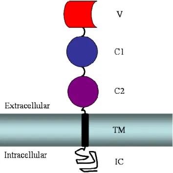
RAGE is a member of the immunoglobulin (Ig) superfamily with three predicted Ig-like domains in its N-terminal extracellular region, in addition to a single transmembrane helix and small intracellular domain (Figure 1.1). The extracellular Ig- like domains have been classified as an N-terminal V-type domain and two subsequent C-type domains (1). However, no direct structural data existed prior to the work in this dissertation to confirm or refute this assignment, specify RAGE quaternary structure, elucidate the basis for interactions with structurally diverse ligands, or generate hypotheses about receptor autoinhibition and activation. The issue of sRAGE quaternary
is important for understanding how an initial binding event causes a signal to be transferred across the cell membrane. The basis for interactions with structurally diverse ligands is critical when addressing the issue of pharmaceutical intervention. And lastly, addressing both of these issues can provide insight into mechanisms of receptor autoinhibition and activation. Thus, the desire to obtain a broader knowledge of how RAGE functions in the cell and malfunctions in disease set the stage for the structural, biochemical, and biophysical work presented in this dissertation.
Figure 1.1: Schematic of RAGE domain organization. The membrane is shown as a horizontal rectangle with the extracellular and intracellular regions marked appropriately.
The intracellular domain (IC) is shown as a random black sketch. The single transmembrane helix (TM) is a black vertical rectangle. The three predicted Ig-like domains of sRAGE are shown above the membrane. The predicted C-type Ig domains are depicted in dark blue (C1) and violet (C2). The predicted V-type Ig domain is shown in red.
RAGE Biology
In this section, I describe the current knowledge of RAGE function associated with normal physiology and cellular dysfunction. There is a wealth of information on the biology associated with RAGE as a signaling receptor. One fascinating aspect of RAGE biology is its recognition of structurally diverse ligands, which can be grouped into four classes: (1) AGEs, (2) amphoterin, (3) amyloid-β peptides (Aβ), and (4) S100 proteins.
While multiligand recognition is not directly addressed, the conclusions and future directions in this dissertation are discussed with respect to signaling mechanisms specifically implicated by multiligand recognition. For scientific completeness, I also delve into the diverse biology associated with these ligands and some of their non-RAGE functions.
Advanced Glycation End Products (AGEs) AGE Chemistry
Protein glycation is a non-enzymatic process initially discovered by Louis Maillard who observed chemical modifications to amino acids when heated in the presence of reducing sugars, a process often referred to as the Maillard reaction. During the early stages of glycation, sugars such as glucose form a Schiff base with free amino groups on proteins at the N-terminus or on lysine and arginine side chains (Figure 1.2) (6). The Schiff base undergoes a rearrangement involving proton transfer to the more stable and reversible Amadori Product (Figure 1.2). The late stages of glycation are irreversible and consist of a variety of chemical modifications. The best studied AGE moiety is N(epsilon)-(carboxymethyl)lysine (CML) which results in a charge reversal
(Figure 1.2) that could disrupt intra- or intermolecular salt bridges. Overall, the consequences of AGE formation can be as diverse as their chemistry.
Figure 1.2: Chemical pathway towards AGE formation. In the first reversible step, D- glucose reacts with free amino groups on a protein forming a Schiff base (circle with letter “P” represents the protein). Reversible proton transfer leads to Amadori product formation in the second step. Finally, a series of irreversible non-enzymatic chemical reactions leads to AGE-modified protein (example shows CML modification to lysine side chain). Figure adapted from (6).
AGEs in Normal Aging and Diabetes
The accumulation of advanced glycation end products is dependent on two primary factors, the concentration of aldose sugars such as glucose and the half-life of the modified protein. Prolonged hyperglycemia associated with diabetes increases the concentration of glucose, a precursor to many AGEs. Observations in the late 1980s suggested AGEs played a role in the pathogenesis of cardiovascular disorders (7). These authors took advantage of the yellow-brown fluorescent pigmentation with characteristic UV absorption of AGEs. They compared the levels of an Amadori product to those of the highly fluorescent advanced glycation end product in rat aorta. Amadori product levels reached a maximum between 50 and 70 weeks whereas levels of the fluorescent
observation implied a correlation between AGE formation and age. A similar study on humans showed increased Amadori levels between Type 1 diabetic patients versus control patients, but no correlation between duration or severity of diabetes (8). However, there was a direct correlation between the duration and severity of diabetic complications and the levels of fluorescent protein adducts, assumed to be AGEs as a result of prolonged hyperglycemia (9). Thus, it appeared that the irreversible AGE moieties and not the reversible Amadori products were associated with diabetes.
AGE formation has been directly observed on cellular components associated with complications of diabetes. Human cataract eye lens show characteristics of AGE formation compared to lens from non-cataract patients (10). Similar observations were made on insoluble fractions of collagen comparing both old to young patients and normal patients to those age-matched with Type 1 diabetes (11, 12). Additional work correlates AGE-β2 microglobulin in patients on long-term hemodialysis suggesting AGE-related pathogenesis in patients being treated for renal failure (13). Lastly, in vitro formation of amyloid-β peptide (Aβ) aggregates, a major component of senile plaques, occurs rather slowly but is markedly enhanced when seeded with AGE-modified Aβ suggesting AGE modification may be a catalyst for aggregate formation in Alzheimer’s brain (14). In a similar example, antibodies positively stain two AGEs, pentosidine and pyrraline, on senile plaques and neurofibrillary tangles (15). Thus, the high insolubility and resistance to denaturant of neurofibrillary tangles could be a byproduct of AGE induced covalent aggregation (16).
Initial work to discover potential receptors for AGEs characterized a 90 kDa protein from murine macrophages that bound a synthetic AGE compound with
nanomolar affinity (17, 18). Concurrent with work on macrophages were experiments on endothelium, tissue that lines the interior of blood vessels and whose dysfunction is connected with diabetic vascular complications. AGE-bovine serum albumin (AGE- BSA) specifically binds to cultured endothelial cells (19). Interestingly, monolayers of endothelial cells show increased vascular permeability, in addition to modulated coagulant properties, when in contact with AGE-BSA (19). Thus, the aggregate evidence suggests AGEs accumulate in long-lived proteins such as collagen and their localization leads to interaction with cell surface molecules causing cellular dysfunction. Studies of these diverse aspects of AGE biology lead to the search for and eventual discovery, isolation, cloning, and characterization of RAGE.
AGE Signaling Induces Oxidative Stress Though RAGE
The progression of diabetic hyperglycemia to cardiovascular disease, renal failure, etc. may arise from tissue damage or altered signaling pathways caused by increased oxidative stress in cells. Cultured capillary endothelial cells (a cell type present in the vasculature that is damaged in diabetes) show increased oxidative stress in response to treatment with AGE-BSA, which was blocked by antibodies to RAGE (20).
In addition, electrophoretic mobility shift assays show AGE-BSA binding to RAGE induces specific translocation of nuclear factor kappa-B (NF-κB) to the nucleus (20). NF- κB is a well characterized transcription factor best studied for its role in gene expression in response to inflammatory stimuli in the immune system (21). In fact, oxidative stress alone in the form of hydrogen peroxide and superoxide radical induces NF-κB activation on T cells (22). Therefore one possible interpretation of the data is that AGE-BSA interacts with RAGE leading to increased oxidative stress which in turn induces
activation of NF-κB by a secondary mechanism. Another possibility is that AGE-BSA binding to RAGE directly leads to activation of signaling pathways causing oxidative stress on the target cell. Subsequent work has confirmed that activation of signaling pathways, and not binding alone, is necessary thereby establishing RAGE as a signaling receptor (23).
Signaling Pathways Triggered by AGEs
Many of the kinases involved in signaling between ligand binding and NF-κB activation have also been identified. In pulmonary artery smooth muscle cells treated with AGE-BSA, activation of NF-κB occurs through p21ras and the extracellular signal regulated kinase 1/2 (ERK1/2) (24). In human monocyte cells, both ERK and p38 mitogen activated protein kinase (MAPK) were activated by CML modified human serum albumin (HSA) (25). This study also showed no activation of the third kinase family, janus kinase. Thus, RAGE signaling in response to AGE exposure is a link to cell stress associated with diabetic vascular complications and stimulated interest in understanding RAGE biology.
RAGE Activation by Amphoterin: a Physiological Role for RAGE
Analysis of RAGE tissue distribution shows higher protein expression in developing tissue than in adult tissue (26). This observation suggests endogenous ligands may be present to activate RAGE during development. Amphoterin was identified as the first “natural” ligand for RAGE (AGE formation being an “unnatural” ligand due to the non-enzymatic chemistry). In situ hybridization and immunohistochemistry showed high expression and colocalization of RAGE and amphoterin in developing cerebral cortex,
hippocampus, and cerebellum of rat brains (27). In vitro, amphoterin binds to RAGE with the highest affinity of all ligands (KD = 6.4 nM) and induces neurite outgrowth on cultured neurons (27). Receptor-dependent neurite outgrowth, therefore, was the first established native function for RAGE in the cell.
Amphoterin is one of several diffusible factors that induce neurite outgrowth on cultured neurons, a function vitally important during nervous system development (28, 29). Prior to its discovery as a neurite outgrowth factor described above involving RAGE, amphoterin (high mobility group 1, HMG1) had been characterized for its DNA binding activity and association with chromatin as a non-histone component (30, 31). In addition to DNA binding activity, HMG1 interacts with DNA-binding proteins and may modulate DNA processing events such as transcription (32, 33).
Amphoterin is described as containing two ~10 kDa HMG box motifs identified by sequence homology and shown to be structurally independent by limited proteolysis (34, 35). In addition, a highly acidic C-terminal extension exists adjacent to the two structured domains (34). Solution structures are available for each of the two HMG box motifs of rat amphoterin (Figure 1.3). The two domains are highly homologous with backbone RMSD of 2.0 Å (36, 37). The fold is predominately α-helical with helices 1 and 2 forming an approximate right angle with respect with helix 3 (Figure 1.3). This L- shaped α-helical structure is quite different from the small chemical modifications that make up AGEs. In addition, the name amphoterin is derived from the primary sequence showing two regions with completely different electrostatic properties; both HMG box domains are quite basic while the C-terminal extension is quite acidic. In fact, it appears that the basic region (pI ~10.0), part of which is α-helical in the HMG box B structure
(Figure 1.3), is capable of binding to RAGE (38). Thus, it is possible that molecules with different electrostatic properties are recognized by RAGE.
Figure 1.3: Structure of rat amphoterin. Ribbon diagrams representing one model of solution structures of the two HMG box domains of rat amphoterin (rat and human are
>99% identical). The N-terminal “A” domain corresponding to residues 1-83 of native amphoterin (PDB 1AAB) is shown in green. The C-terminal “B” domain corresponding to residues 88-164 of native amphoterin (PDB 1HME) is shown in blue. N- and C-termini of each domain are marked accordingly. A portion of the amphoterin peptide known to bind and antagonize RAGE is colored in pink. The remainder of this peptide was not included in the construct used in structure determination. All protein structure images in this dissertation were made using Chimera (39).
RAGE Binding to Amyloid β: Implications in Neurotoxicity
Alzheimer’s disease (AD) is a progressive neurodegenerative disorder characterized by lesions in regions of the brain important for intelligence and memory.
Two primary morphologies are observed, neurofibrillary tangles and senile plaques. The major protein component of neurofibrillary tangles are tau protein aggregates while that for senile plaques are amyloid fibrils composed largely of amyloid β (Aβ) peptides from the amyloid precursor protein (APP). RAGE has been identified as a protein capable of binding Aβ peptides and does so with low nanomolar affinity (40).
APP is an integral membrane protein with a large extracellular domain, a single transmembrane helix, and a small intracellular domain. Normal cellular processing of APP in lysozomal compartments leads to proteolytic fragments that are secreted from various cells in culture (41). However, neither the function of APP nor the role of normal protein turnover is well understood. In senile plaques, Aβ peptides are heterogeneous in length but average around 40 amino acids. Nonetheless, isolated Aβ peptides offer functional and structural features unique from AGEs and amphoterin.
The interaction of soluble Aβ peptide with RAGE induces migration of microglial cells and oxidative stress in endothelial cells, both of which are RAGE dependent (40).
Thus, RAGE may enhance neurotoxicity directly through Aβ peptide interaction or indirectly by inducing microglial infiltration. The role of RAGE as a signaling receptor in neurodegenerative disorders was strengthened by studying a transgenic mouse model for AD expressing a mutant form of APP (42). Transgenic mice also expressing a signaling deficient form of RAGE (dnRAGE, see below) show markers such as enhanced NF-κB
type RAGE, consistent with its role as a signaling receptor in Aβ induced neuropathy (42).
S100 Proteins are RAGE Ligands
The continued search for native ligands of RAGE lead to the identification of a gene termed EN-RAGE (endogenous RAGE) (43). Analysis of the sequence revealed EN-RAGE was a member of the S100 superfamily of EF-hand proteins, S100A12. The precise function of S100 proteins is not well understood. Nonetheless, they are involved in diverse biological processes.
S100A12 (EN-RAGE) is one of the calgranulins, which are exclusively expressed and secreted from granulocytes of the immune system. Radioligand binding assays show S100A12 binds with KD ~90 nM to both purified RAGE and RAGE on the endothelial cell surface (43). RAGE activation by S100A12 on endothelial cells induces nuclear translocation of NF-κB and the subsequent expression of vascular cell adhesion molecule-1 and intercellular adhesion molecule-1, both mediators of the inflammatory response. Furthermore, S100A12 clearly promotes cell migration of mononuclear phagocytes in a chemotaxis assay strongly suggesting a RAGE-S100 protein role in acute inflammation (43).
A second S100 protein, S100B, was shown to activate RAGE (44). S100B is known to regulate the extension of neurites in vitro (45, 46). At nanomolar concentrations of S100B, neurite outgrowth is promoted. This function appears to be coregulated in an additive manner by S100B, S100A1 and amphoterin up to 1 µM. At micromolar concentrations, however, both S100 proteins lead to cellular toxicity and apoptosis
through the caspase cascade (44). For all three proteins at neurotrophic concentrations, RAGE activation led to NF-κB upregulation (44).
S100A12 and S100B are the best studied RAGE ligands in the S100 superfamily.
However, other S100 proteins are implicated in RAGE biology. S100P is overexpressed in many cancers. In both NIH3T3 and pancreatic cancer cells, S100P is coimmunoprecipitated with RAGE and activation of NF-κB is blocked with a signaling deficient RAGE (dnRAGE, see below) (47, 48). S100A4 leads to RAGE dependent activation of p38 MAPK, nuclear translocation of NF-κB, and overproduction of matrix metalloprotease 13 in cultured human chondrocytes (49). And lastly, S100A11, overexpressed in osteoarthritic cartilage, appears to activate chondrocytes by signaling through RAGE and p38 MAPK (50). S100 protein activation of RAGE is likely regulated by the subcellular localization and tissue-specific nature of S100 proteins since many in the superfamily bind to and activate RAGE.
S100s are small, acidic, α-helical proteins with high structural similarity across the family. A ribbon diagram of human S100B is shown in Figure 1.4. While S100 proteins share secondary structure elements with amphoterin, there is no shared tertiary structure nor is there any similarity to AGEs or Aβ. In addition, S100 proteins can be quite acidic with S100B having an isoelectric point of 4.6. The biochemistry of S100B as a RAGE ligand is studied in this dissertation. Therefore, a more detailed discussion of S100 protein structure is warranted and included in the next section.
Figure 1.4: Solution structure of Ca2+-loaded human S100B. The ribbon diagram (PDB 1UWO) shows one subunit of the symmetric homodimer in red and the other in orange.
All S100 protein structures in this dissertation are shown in this orientation with the two HIV helices in front and the two HI helices in back.
Divergent Signaling Pathways and Essential Signaling Elements Kinase Pathways
AGE binding to RAGE induces the activation of NF-κB through p21ras and ERK1/2. Much like AGEs, amphoterin induces the translocation of NF-κB to the nucleus in cultured neuroblastoma cells (23). However, rac and Cdc42 were used to signal to NF- κB instead of ras and ERK1/2. Working in a fibrosarcoma cell line (HT1080), Ishihara and coworkers showed amphoterin activates ERK1/2 (51). This cell line is more similar to the endothelial cells used in AGE studies than the neural cells used by Huttunen and coworkers. Thus, it is possible that the signaling pathways used by RAGE are cell type dependent but not ligand dependent. Interestingly, ERK1/2 coimmunoprecipitated with RAGE from HT1080 cells after activation with amphoterin. The interaction with ERK2 (ERK1 was not tested) was specifically localized to the first 18 amino acids of the intracellular domain in glutathione S-transferase (GST) pull downs from rat lung extracts (51).
Dominant-Negative RAGE
A very important observation was made during initial studies with amphoterin. A truncation mutant of RAGE was transfected into neuroblastoma cells that had endogenous expression of RAGE. This truncation mutant lacked the ~40 amino acid intracellular domain (IC). Translocation to the membrane was not prevented nor was amphoterin or AGE-BSA binding. Cell signaling, however, was deficient in the construct lacking the IC (23). Since wild-type RAGE is also present at the cell surface and ligand binding is not diminished, a dominant-negative form of RAGE (dnRAGE) implies that RAGE functions in some sort of oligomeric state. The likely role of RAGE in neural
growth and development and in progression of diabetes and chronic inflammatory disorders, coupled with utilization of divergent signaling pathways described in this section, has greatly stimulated interest in RAGE as a signaling molecule.
Positive Feedback Loop
Additional insights into RAGE signaling arose from analysis of its promoter. Two functional NF-κB-like binding sites have been characterized, both of which are necessary for basal RAGE expression and increased RAGE expression in response to stimuli (52).
RAGE expression is shown to increase in diabetic versus normal vasculature, as well as other pathologic settings (53). Therefore, an interesting scenario has arisen in RAGE biology. Binding of ligand to RAGE activates NF-κB which in turn enhances RAGE expression increasing potential cell surface binding sites. This positive feedback loop has been hypothesized to be responsible for the RAGE induced amplification of chronic disorders such as diabetes and inflammation (54).
Potential for RAGE-Based Therapeutics
As described above, RAGE participates in a diverse array of biological and pathological processes each defined by the particular class of ligand activating the receptor. Due to its implied role in multiple disease states, RAGE is considered a therapeutic target (55). In fact, ligand binding in a variety of culture systems leads to activation of signaling pathways consistent with the associated disease states (5). This suggests blockade of the RAGE signaling axis could provide an avenue for pharmaceutical intervention. Exogenous sRAGE has been used to block ligand binding
and to prevent receptor activation in cell culture. These observations have been translated into mouse models for various diseases.
Diabetes
Apolipoprotein E-null mice naturally develop accelerated atherosclerosis and show a greater than 2 fold increase in plasma AGEs (56). Intraperitoneal administration of sRAGE significantly blocked the development of atherosclerotic lesions in the aorta (57). The mechanism is presumably through blockade of AGE-RAGE interaction since neither hyperglycemia nor insulinemia were affected.
Cancer
Transplanting C6 glioma cells into immunocompromised mice causes rapid tumor growth that is significantly suppressed by intraperitoneal administration of either sRAGE or antibodies against RAGE or amphoterin (58). Furthermore, stably transfected C6 glioma cells either expressing dnRAGE or secreting sRAGE showed neither tumor growth nor tumor metastasis. Thus, the leading edge colocalization of amphoterin and RAGE observed in culture (27) implies a mechanism for promoting (and blocking) tumor invasion.
In a similar study, B16-F1 melanoma cells were injected into the tail veins of immunocompromised mice and assessed for colonies indicative of lung metastasis (38).
Two strategies suggested an amphoterin-RAGE signaling axis is important for formation of metastatic colonies in the lung. One, injecting cells transfected with dnRAGE showed 72% fewer colonies than injection of parental B16-F1 cells (38). The second involved coinjection of B16-F1 cells with an amphoterin peptide antagonist (amphoterin 150-183),
which caused a 64% reduction in lung metastatic colonies (38). Thus, multiple lines of evidence point to an amphoterin-RAGE axis important in tumor metastasis.
Alzheimer’s Disease
The vascular theory of AD states that accumulation of Aβ in Alzheimer’s brain may arise from circulating Aβ and dysfunction of the equilibrium of peptides across the blood brain barrier (BBB) (59). BBB transport would presumably require specific cell surface receptors for Aβ. Using an in vitro model for BBB, anti-RAGE antibodies prevented transport of peptides suggesting a role for RAGE (60). Transgenic mice expressing mutant APP function as a model for AD since Aβ containing plaques accumulate rapidly in the brain and correlate with impaired learning and memory (61).
Treatment with sRAGE results in high levels of sRAGE-Aβ coimmunoprecipitates from plasma in addition to decreased levels of Aβ plaque formation (62).
Chronic Inflammation
Another potential therapeutic approach is based on the interaction of RAGE with S100A12 which is highly associated with chronic inflammation such as inflammatory bowel disease. Interleukin-10 deficient mice serve as a model for enterocolitis characterized by hyperactive bowel inflammation (63). In this model, administration of sRAGE prevented inflammatory infiltrates into the colon likely by blocking S100-RAGE interaction and signaling (43).
Concluding Remarks
There is a wealth of information on the biology associated with RAGE as a signaling receptor. Comparing the signaling pathways utilized by all ligands suggests that
RAGE universally signals through NF-κB. The biological processes involved range from neural growth and development and acute inflammatory responses to complications associated with diabetes and AD. Thus, knowledge gained by studying one aspect of RAGE function has the potential to transcend all aspects of RAGE biology.
S100 EF-Hand Calcium-Binding Proteins
S100 proteins, specifically S100B, are a major focus of this dissertation. These proteins have been well characterized structurally and biochemically (64) and reagents were readily available to pursue structural studies on S100-RAGE interaction. In this section I take a broader look at calcium signaling involving S100 proteins in addition to the current knowledge about S100 protein structure and function.
Structure of the EF-hand Motif
The calcium ion is known to function as a second messenger in many signaling cascades. Signal transduction from a physiological change in calcium concentration to a biochemical response in the cell is mediated by EF-hand calcium-binding proteins. The EF-hand is a helix-loop-helix motif in which the protein chelates calcium ions using a series of side chain and backbone oxygen atoms (65). The prototypical EF-hand protein is calmodulin, a single polypeptide with four EF-hand motifs organized into two structural domains separated by a flexible linker. Each domain consists of paired EF-hand motifs that fold into a stable four helix bundle. Each EF-hand is able to bind one ion of calcium.
Thus, calmodulin binds four calcium ions, two in each structural domain.
Calcium Sensing by EF-Hand Proteins
The functional role of EF-hand proteins in calcium signal transduction is mediated by structural and biophysical responses to changes in intracellular calcium concentration (66). The prototypical EF-hand calcium sensor, calmodulin, binds calcium ions with KDs of ~10-6 and ~10-5 for the C- and N-domains, respectively, in the range of the intracellular calcium signal (67). Thus, at basal levels of calcium, calmodulin is in a magnesium-bound or the apo- (calcium-free) state. The release of calcium from intracellular stores during response to a stimulus leads to micromolar concentrations of calcium, with possible subcellular compartments exposed to millimolar concentrations.
At mid-micromolar or higher concentrations of calcium, all four binding sites are filled and calmodulin is activated. The structural basis for calmodulin signaling activity is revealed by comparative analyses of apo- and calcium-bound structures. Upon binding calcium, both domains of calmodulin undergo large structural rearrangements exposing deep hydrophobic pockets frequently used to bind to α-helical target peptides (66).
Architecture of the S100 Protein
S100 proteins are EF-hand containing calcium binding proteins named for solubility in 100% saturated ammonium sulfate. The architecture of an S100 protein is quite different from calmodulin. The minimal structural and functional S100 unit is a highly integrated symmetric homodimer first seen in the structure of S100A6 (calcyclin) determined in our laboratory (68). Each subunit of the S100 dimer consists of two EF- hands connected by a hinge region similar to each of the domains of calmodulin. The C- terminal EF-hand motif is canonical (calmodulin-like) whereas the N-terminal EF-hand is
S100-specific (69). Unlike the canonical 12 residue EF-hand, the S100-specific EF-hand is 14 residues and displays weaker affinity for calcium. Much like calmodulin, affinities for calcium are in the range of the intracellular calcium signal classifying most S100 proteins as calcium sensors.
Calcium binding to S100 proteins induces a similar yet different conformational change than calcium binding to calmodulin for two reasons. One, helices HI and HIV form the tightly integrated dimer interface and are therefore restricted. Two, the S100-specific EF-hand resembles the calcium bound state even when calcium is absent (70, 71). It has been proposed that the S100-specific EF-hand is stabilized in the absence of calcium by the presence of a water molecule in the binding site. Such a water molecule showing similarity to the calcium coordination geometry was observed in the S100-specific binding loop of apo-S100A3 (72). Regardless of the origin of the effect, the major conformational change in S100 proteins occurs in the C-terminal EF-hand, specifically in helix HIII (Figure 1.5). Calcium binding to S100 proteins creates a shallow hydrophobic surface much different from the deep hydrophobic pocket formed by calcium binding to calmodulin.
Due to the high sequence homology among the S100 protein family, it is not surprising that they adopt similar structures in both the absence and presence of calcium (73). However, many subtleties exist. For example, S100A7 and S100A10 have amino acid substitutions or deletions that inactivate calcium binding at key glutamate/aspartate residues in the EF-hands (74). Remarkably, an x-ray crystal structure of S100A10 bound to a target peptide reveals that calcium is not required to stabilize the open conformation since it is sufficiently stabilized by the binding of target (75). It has yet to be determined
whether S100A10 adopts the calcium-bound state in the absence of target binding or is in dynamic exchange between the apo- and calcium-bound states.
Figure 1.5: Calcium induced conformational change in S100A6. Solution structures of apo-S100A6 (left, PDB 1CNP) and Ca2+-loaded S100A6 (right, PDB 1A03). For each homodimer, one subunit is shown as a light blue Cα trace with a dark blue ribbon diagram showing the 4 helix bundle. The two molecules have been aligned by helices HI
and HIV at the dimer interface. Colored in pink is helix HIII emphasizing the calcium induced structural change.
S100 Proteins and Zinc
S100 proteins exhibit the ability to bind other metals, a characteristic that distinguishes them among EF-hand proteins. For example, S100A2 has at least two high affinity sites for zinc (II) that are independent from the calcium binding sites (76).
Binding to zinc (II) occurs with nanomolar KDs, near the physiological levels of intracellular zinc (II). A solution structure of S100B bound to zinc (II) reveals utilization of sites separate from the calcium binding sites (77). Unlike calcium binding which induces a large conformational change in helix HIII, binding of zinc (II) to S100B causes very little structural perturbation (77).
The S100A8/S100A9 heterodimer has also been characterized as a zinc (II) binding protein (78). S100A8/S100A9 is highly expressed in monocytic cells of the immune system and is thought to play a role in the hummoral response to pathogen invasion (79). Addition of the heterodimer but not the individual homodimers inhibits growth of cultured Candida albicans (80). Furthermore, growth inhibition by the S100A8/S100A9 heterodimer is reversed by addition of excess zinc (II). Thus, the antimicrobial activity of the S100A8/S100A9 heterodimer may occur through chelation of essential zinc (II). A newly described human disorder characterized by aberrant zinc metabolism, called hyperzincaemia, is correlated with highly elevated levels of S100A8/S100A9 heterodimer (81).
Higher Order Oligomeric States of S100 Proteins
Although S100 proteins form highly integrated dimers, evidence has been gathering in support of S100 proteins functioning in vivo as higher order oligomers. Early
reports on the neurotrophic effects of S100B suggested disulfide-linked higher order oligomeric species were essential for function (45, 46, 82). More recent evidence arises primarily from crystal structures and other biophysical techniques. S100A12 has been crystallized in numerous forms, including a hexameric structure (83, 84). This hexamer was apparently stabilized by additional calcium binding sites between individual dimers (85). There is some biophysical support for higher order oligomer formation in the absence of lattice forces from crystal packing. Dynamic light scattering (DLS) reveals an increase in the hydrodynamic volume of S100A12 when calcium concentrations reach extracellular physiological levels (~1 mM) (85).
Gel filtration of S100A4 revealed multiple oligomeric states, which were correlated with potential to induce neurite outgrowth on cultured hippocampal cells.
Fractions containing S100A4 tetramer or higher were necessary to induce neurite outgrowth (86). However, it is not clear whether the higher order oligomeric species are in equilibrium with the dimeric species. Thus, further studies involving mutations at key residues involved specifically in higher order oligomer formation are definitely required to test this proposal.
The x-ray crystal structure of S100A8/S100A9 heterodimer has three tetramers in the asymmetric unit (PDB 1XK4). Significant contacts are made between helices HIV of one dimer and the calcium binding loops of the second dimer. This tetramer interface places the traditional S100 target interacting surfaces of opposite dimers in proximity which could generate novel target recognition surfaces. Chemical crosslinking and mass spectrometry suggest calcium dependent tetramer formation of S100A8/S100A9 (87, 88).
The crystal structure of S100A10 in complex with a peptide from annexin II also reveals
disulfide linked tetramers (75). In addition, a recently described crystal form of S100B shows the presence of four tetramers (89). It is intriguing to consider S100 oligomerization in the context of receptor activation. Not only can a S100 dimer potentially dimerize a receptor (or alter the organization of a pre-formed receptor dimer), but higher order oligomers can multimerize or create novel binding surfaces for receptor recognition.
Target Recognition by S100 Proteins
Multiple structures of S100 proteins bound to peptide fragments of cellular targets are available and reveal differences in the details of target recognition (64). Crystal structures of S100A10 and S100A11 bound to peptides from annexin II and annexin I, respectively, provided early insight into S100-target recognition (75, 90). In both cases, the annexin peptides adopt α-helical secondary structure and are bound to the S100 protein in a calcium-dependent manner with a stoichiometry of two peptides per S100 dimer (Figure 1.6). Thus, S100 proteins could function by oligomerizing target proteins, by bridging two different molecules, or by inducing conformational changes in proteins by binding to two different regions of one polypeptide.
The interaction between annexin peptides and their respective S100 proteins is stabilized by a combination of hydrophobic and hydrophilic interactions primarily in the shallow pocket formed between helices HIII and HIV. Unique to these two structures are contacts between the peptide and helix HI of the opposite subunit. This type of contact is absent in structures of S100B bound to target peptides from p53 and NDR kinase (Figure 1.6) (91, 92). Nonetheless, the peptides adopt α-helical secondary structure and
contribute a combination of hydrophobic and hydrophilic side chains to stabilize the interaction. Earlier in this chapter I discussed multiligand recognition of structurally diverse molecules by RAGE. Ig domains do not contain a significant amount of α-helical secondary structure. Typically, 3-4 helical turns are required for efficient binding to S100 proteins (Figure 1.6). Thus, the work described in this dissertation sets the stage to study the potentially novel interface used by S100 proteins to bind and activate RAGE.
Figure 1.6: Variability in target recognition by S100 proteins. Displayed are the complexes of S100A10 with annexin II peptide (PDB 1BT6), S100B bound to a p53 peptide (PDB 1DT7), and S100B bound to a NDR kinase peptide (PDB 1PSB). Ribbon diagrams shown with both subunits of the S100 dimer in black and the target peptides in red. Figure adapted from (64).
Mechanisms of Cytokine Receptor Signaling and Autoinhibition
RAGE functions as a signaling receptor. The structural work in this dissertation allows me to propose hypotheses about receptor activation taking advantage of the current knowledge available in the literature. A common theme within receptor activation is the concept of ligand induced oligomerization. An increasing level of sophistication in autoinhibition and activation models evolved as more information became available suggesting oligomerization is necessary but not sufficient for activity. In this section, I examine the available data on two well studied systems, growth hormone receptor (GHR) and erythropoietin receptor (EPOR), summarizing structural mechanisms of receptor autoinhibition and activation.
Cytokine Receptor Classification
Cytokine receptors are integral membrane proteins sharing common features such as a large extracellular domain responsible for ligand binding, a single transmembrane helix, and an intracellular domain containing either intrinsic kinase activity or adaptor function to interact with kinases (93). The receptors are classified according to the size and complexity of their extracellular ligand binding domains. Traditionally, “simple”
receptors contain only extracellular domains necessary for ligand binding and include GHR, EPOR, and the interleukin-4 receptor (IL-4R). “Complex” receptors contain structured domains not involved in ligand recognition but play a critical role in signal transduction, the best studied of which is the insulin receptor.
Scissor-like Activation of EPOR
Early studies in cytokine receptor function hypothesized the initial step towards activation required ligand-induced dimerization. For example, cysteine mutants in the extracellular region (containing two Ig-like domains D1 and D2) of EPOR (EPO binding protein, EBP) lead to disulfide linked dimers and constitutive activation (94). Production of a peptide that mimics receptor activation (EPO-mimetic peptide, EMP) and induces dimerization of EBP supported ligand-induced dimerization (95, 96). Additional EMPs that efficiently promote dimerization but lack the ability to activate the receptor have been characterized (97). Thus, the basic hypothesis was refined with the discovery that dimerization is necessary but not sufficient for activation.
The structure of unliganded dimeric EBP provides insight into the autoinhibition mechanism of EPOR shown schematically in Figure 1.7 (98). Analysis of this structure placed the membrane proximal D2 domains > 70 Å apart compared to the direct contacts between D2 domains observed in the x-ray crystal structure of erythropoietin bound to EBP (EPO-EBP) (99). Thus, the physical separation of the intracellular domains and bound kinases functions as the autoinhibited state. Ligand binding physically reorganizes the receptor bringing the bound kinases into proximity for trans-phosphorylation and signal activation. This scissor-like model is supported by a fluorescence complementation assay where two fragments of dihydrofolate reductase are fused to EPOR and transfected into Chinese hamster ovary cells. Intracellular binding of fluorescently conjugated methotrexate (which requires proximity of the two fragments) was only possible when the receptor was activated by ligand or when a 30 amino acid linker was present
providing extreme flexibility (the observations with this linker are also consistent preformed EPOR dimers) (100).
Comparisons of the x-ray crystal structures of EMP-EBP and EPO-EBP complexes revealed insights into subtle details of activation (96). The two EBP molecules show ~120° rotation in the crystal structure of the EPO-EBP complex (99). Two EMP- EBP structures show relative rotations different from 120°. Since one EMP was inactive and the other was partially active, a logical conclusion is that a specific receptor orientation is required for efficient activation. Therefore, both proximity of the bound kinases and a specific orientation are required for signal transduction.
Transmembrane Rotation Model for Activation of GHR
GHR was also thought to function through ligand-induced dimerization when a combination of titration calorimetry and gel filtration suggested a stoichiometry of 1:2 between growth hormone (GH) and the ligand binding region of GHR (GHbp) (101).
This stoichiometry was confirmed in the crystal structure of GH-GHbp (102). Studies using a variety of monoclonal antibodies that target the GHR extracellular domain show that dimerization is not sufficient for activation but that specific structural orientation of the receptor is necessary (103). Unlike EBP, GHbp exists as a monomer despite a combination of coimmunoprecipitation and FRET studies showing unliganded GHR dimers in cell culture (104). A mechanism for GHR signal activation has also been proposed and is depicted schematically in Figure 1.7. A series of alanine insertions were made between the transmembrane helix and the intracellular domain. Constitutive activation was achieved when exactly four alanines were introduced, which is predicted
to produce an ~40° net rotation (total of ~400°) of the intracellular domain if they continue α-helical secondary structure (104). Thus, unlike EPOR, which dimerizes through the extracellular domain in an inactive conformation physically separating the kinase adapter intracellular domain, GHR appears to dimerize through the transmembrane helix with its kinase adapter domains rotated in an orientation that autoinhibits trans-phosphorylation.
Figure 1.7: Two models for cytokine receptor autoinhibition and activation. (A) Scissor- like model for EPOR as described in the text. D1 and D2 domains of EPObp are shown in red and blue, respectively. JAK2 is shown as a blue circle associated with the intracellular domain. EPO is shown as a violet circle. Figure adapted from (99). (B) Rotation model for activation of GHR as described in the text. The two extracellular domains of GHbp are shown as red and blue 3D rectangles. One associated JAK2 is a blue circle and the other is a blue ellipse. GH binding (violet circle) induces rotation aligning the JAK2 molecules for activation. Figure adapted from (104).
Structural Studies on sRAGE and Ligand Interactions
The central focus of this dissertation is to understand the structure of RAGE and its function as a signaling receptor. Specifically, I describe the first detailed structural and biophysical characterization of sRAGE (Chapter II). The techniques used include limited proteolysis, a powerful approach for analyzing flexible regions of a protein (105). A combination of CD and NMR spectroscopy are used to determine the secondary and tertiary structure elements of sRAGE and single and tandem domain constructs. The combination of these experimental procedures revealed unique features of sRAGE quaternary structure. Further insights into sRAGE structure and function were obtained from a high resolution x-ray crystal structure of the tandem VC1 construct of sRAGE (Chapter III). Lastly, the interaction of sRAGE with two of its physiological ligands, S100B and CML, is characterized using a combination of protease protection, surface plasmon resonance, and NMR (Chapter IV). The combination of these results provided insight into sRAGE structure and RAGE-ligand interactions, the broader implications of which are discussed along with ideas about future directions for structure-based research on RAGE (Chapter V).
CHAPTER II
THE EXTRACELLULAR REGION OF THE RECEPTOR FOR ADVANCED GLYCATION END PRODUCTS IS COMPOSED OF TWO INDEPENDENT
STRUCTURAL UNITS
Introduction
RAGE is a member of the immunoglobulin (Ig) superfamily containing three predicted Ig-like domains in its N-terminal ligand binding region (1). Further classification to an N-terminal V-type Ig domain and two subsequent C-type Ig domains was proposed based on sequence analysis; however, this assignment was based largely on homology to NCAM, whose β-strand topology has since been shown to differ slightly from V-type and C-type domains (106).
As described in Chapter I, a critical aspect to understanding mechanisms of receptor signaling is obtaining information on the natural or ligand-induced quaternary structure of the receptor. In fact, while the general tertiary structure of the domains in sRAGE is assumed to be a two-sheet β-sandwich stabilized by an internal disulfide bond, little information is known about sRAGE quaternary structure. In this chapter, production protocols for sRAGE and a series of domain constructs are developed to establish a foundation for structural studies. Subsequently, the issue of sRAGE quaternary structure is addressed using limited proteolysis, which can identify variability in structured vs.
unstructured regions of the protein (105). Lastly, the relationship of the three Ig domains is assayed by characterizing their thermal denaturation midpoints and analyzing multidimensional NMR spectra and a homology model for the tandem VC1 domains.
Production of sRAGE and Domain Constructs
High level production and purification protocols were developed for sRAGE and the five single and tandem domain constructs (VC1, C1C2, V, C1, C2; see Figure 1.1).
While the production of recombinant sRAGE has been described for both eukaryotic and prokaryotic expression systems (43, 107-109), the strategy described here provided consistent high level production of all six constructs without the need to refold protein from inclusion bodies. Bacterial expression vectors were constructed that enable purification via an N-terminal His6-tag and nickel affinity chromatography (see Chapter VI). Use of cells providing an oxidizing environment and optimization of expression parameters (temperature, duration, IPTG concentration) produced soluble protein for each of the constructs. Protein yields after purification were similar for sRAGE, VC1, V, and C2 (15-20 mg/L) and lower for C1C2 and C1 (5-10 mg/L) as a result of their lower expression levels.
When performing structural analysis on isolated domains of a protein, it is important to properly define construct boundaries. Initial boundaries were based on sequence alignment with known Ig family sequences and secondary structure prediction.
Ala23 represents the first amino acid of the native receptor after loss of the signaling peptide and was selected as the N-terminus for our sRAGE construct. The C-terminus of sRAGE, Gly327, represents the end of the C2 structural region, which is linked to the transmembrane helix. The N-terminus for the V and VC1 constructs and the C-terminus for the C1C2 and C2 constructs were the same as for sRAGE. The predicted linker region separating C1 and C2 was identified by multiple sequence alignments of four RAGE genes (human, bovine, rat, and mouse; Figure 2.1). The eleven amino acid insert in
bovine RAGE after Trp230 aided identification since regions of low identity and homology often represent loop and linker regions. The intervening boundaries between V and C1 proved to be more challenging to precisely identify and had to be manually optimized based on expression level, solubility, and stability. This analysis suggests there may be unique features relating V to C1.
Figure 2.1: Multiple sequence alignment of RAGE proteins. From top to bottom, human (Q15109), bovine (Q28173), rat (Q63495) and mouse (Q62151) RAGE were aligned using ClustalX run on a Windows PC. The numbering on the top represents the residue number of the human sequence. The symbol (*) denotes 100% conservation while the symbols (:) and (.) represent high and moderate conservation, respectively. See Appendix A for full sequence alignment.
Analysis of purified constructs under reducing and non-reducing SDS-PAGE conditions showed a mobility shift consistent with formation of disulfide bonds.
Observing a single band under non-reducing conditions suggested homogeneity in disulfide formation. Interestingly, 15N-1H HSQC spectra of V acquired in the presence of reducing agent showed a collapse of the chemical shift dispersion in the 1H dimension, which is characteristic of an unfolded protein (Figure 2.2). This suggests any experiments in the presence of reducing agents could lead to artifacts due to protein unfolding.
Figure 2.2: The effect of reducing agent on Ig domain tertiary structure. (A) 15N-1H HSQC spectrum of isolated V in the absence of DTT acquired according to Chapter VI.
(B) 15N-1H HSQC spectrum of V in the presence of 1 mM DTT.
Stability of sRAGE Domains
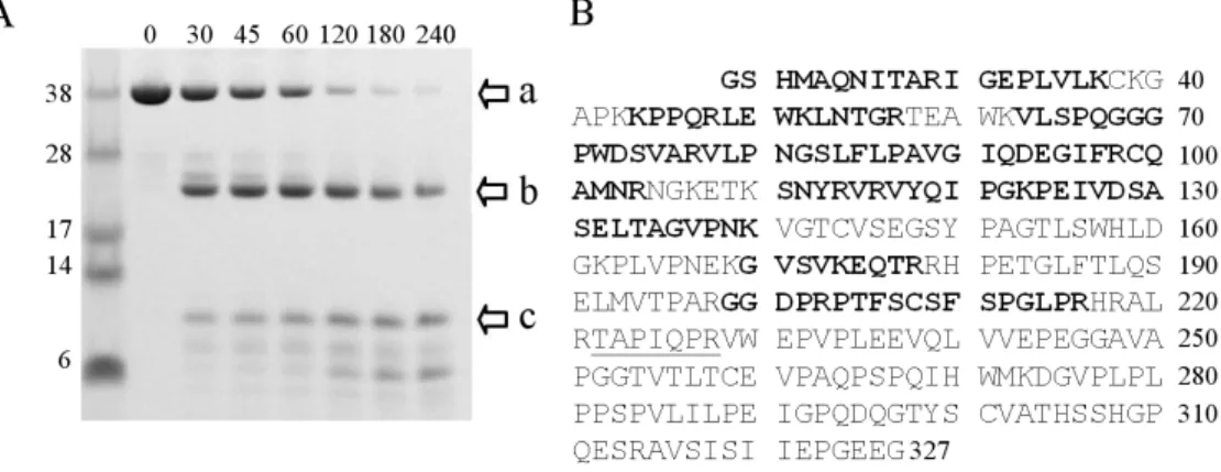
A series of limited proteolysis experiments were performed in order to further characterize the domain structure of sRAGE. A time course for digestion of the protein by trypsin shows that sRAGE is completely digested within four hours (Figure 2.3A).
Two major species are formed during early time points, one migrating at ~25 kDa (Figure 2.3A, arrow b) and another at ~12 kDa (Figure 2.3A, arrow c). In-gel digestion of the 25 kDa band with trypsin followed by MALDI-MS analysis showed this band contained peptides unique to the V and C1 domains. Figure 2.3B shows the sequence coverage obtained for both intact sRAGE (Figure 2.3A, arrow a) and the ~25 kDa fragment.
Notably, coverage obtained for the V and C1 domains was nearly complete in the 25 kDa band (Figure 2.3A, arrow b), ranging from a peptide encoding a region of the N-terminus (23-29) to a peptide near the C-terminus of C1 (199-216).
Figure 2.3: Limited proteolysis of sRAGE reveals two stable domains. (A) Reducing SDS-PAGE of sRAGE digested by trypsin (500:1, w/w) at room temperature. Arrows at right highlight the three main fragments. Each of these bands shift in mobility when gels are run under non-reducing conditions indicating they contain one or more disulfide bonds. Time points are given in minutes. (B) Sequence of recombinant sRAGE after removal of His6-tag. The first four amino acids (GSHM) remain after thrombin cleavage.
Ala23 is the first amino acid of the mature protein after the signal sequence is removed.
Highlighted in bold are peptides observed in mass spectrometry analysis of the trypsinized 25 kDa band (band “b”) in panel (B). The only peptide observed in the mass spectrometry analysis of band “a” that is not seen in band “b” is underlined. The numbering on the right corresponds to the last amino acid in each row.
To further confirm the origin of the 25 kDa band, limited proteolysis of VC1 (Figure 2.4) was performed under conditions identical to those used for sRAGE.
Digestion of VC1 produced a small truncation to a band with the same mobility as the 25 kDa band generated from sRAGE digestion (arrow d, Figure 2.4). Both the intact VC1 and the 25 kDa fragment were digested at the same rates as those observed in the experiment with sRAGE. Notably, the 25 kDa fragment produced from either sRAGE or VC1 digestion was largely stable to further proteolysis despite the presence of both lysine and arginine residues in the expected linker region between the two domains. To further understand the apparent stability of the V-C1 linker we performed limited proteolysis experiments on isolated V and C1. Figure 2.4 shows that both intact V and especially C1 were much more susceptible to trypsin proteolysis than the 25 kDa fragment that contained both V and C1. Furthermore, we failed to observe a truncation to a stable fragment for either construct that would indicate the presence of a single stable V or C1 domain.
Figure 2.4: Resistance to proteolysis of V and C1 is enhanced by covalent attachment.
Reducing SDS-PAGE analysis of VC1, V, and C1 proteolyses by trypsin (500:1, w/w).
The mobility of band “d” and that for the intact protein shift when run under non- reducing conditions. Time points are given in minutes.
Unlike sRAGE and VC1, the C1C2 construct was completely digested by trypsin in 5 minutes, and only a single 12 kDa band was observed thereafter (Figure 2.5, arrow e). This stable band had identical mobility to the 12 kDa band seen in the sRAGE digestion (Figure 2.3, arrow c). The presence of this band in sRAGE and C1C2 digestions, but not for VC1, implies the band belongs to the C2 domain. Exposure of isolated C2 to trypsin showed it is completely stable to digestion by this protease (Figure 2.5). This observation is identical to that made for the 12 kDa band in the sRAGE (Figure 2.3, arrow c) and C1C2 digestions. The assignment of the 12 kDa band to C2 and the absence of other bands in C1C2 digestions indicate that the C1 domain on its own is not stable.
Figure 2.5: The C2 domain is stable to trypsin proteolysis. Reducing SDS-PAGE analysis of C1C2 and C2 by trypsin (500:1, w/w) at room temperature. The mobility of band “e” corresponds to the ~12 kDa band “c” in Figure 2.3A. Time points are given in minutes.
Differential scanning calorimetry (DSC) experiments were performed to better understand the stability and interdependence of sRAGE domains. Intact sRAGE showed a single thermal transition with a Tm at 55.1 °C, which was surprising given the much higher Tm value of isolated C2 (vide infra). Unfortunately, heating beyond 60 °C leads to aggregation and precipitation (Figure 2.6, solid line), which precludes further analysis.
VC1 responded in a very similar manner, with a single transition corresponding to Tm at 55.1 °C followed by aggregation and precipitation (Figure 2.6, dashed line).
Figure 2.6: VC1 undergoes a complex thermal transition. DSC thermograms are shown for sRAGE (−), VC1 (--) and C2 (··). Experimental details are described in Chapter VI.
In an attempt to determine if the thermal transition derives specifically from one domain, DSC experiments were performed on isolated V and C1. However, although the proteins remained soluble, no discrete transitions were observed for either of the isolated domains (Figure 2.8A). These observations are consistent with the limited proteolysis experiments, which suggested that isolated V and C1 are not stably folded independent domains. Based on the evidence from the thermal denaturation and limited proteolysis data, we attribute the unfolding event at 55 °C to uncoupling of the V and C1 domains.
Remarkable parallels are found in a very thorough study of the unfolding of a tandem (one V-type and one C-type domain) Ig protein from a multiple myeloma κI light chain, from which the authors concluded that the phenomena observed did not correspond to unfolding of independent structural domains (110). As an alternative to the DSC approach, the temperature dependence of the CD spectrum of VC1 was examined. The rationale is that the degree of unfolding of the protein would be reflected in the degree of loss of CD ellipticity. Since the protein precipitates at higher temperatures, the maximum temperature was restricted to 55 °C. Figure 2.7 shows a reversible (and incomplete) loss of CD ellipticity is observed as the temperature is raised. The reversibility of the thermal melt rules out precipitation as the only contributing factor to the CD transition. In addition, the retention of significant secondary structure (~60%) is consistent with uncoupling of V and C1, and not global unfolding, as interpreted for the DSC results.
Figure 2.7: CD thermal melt of VC1. Shown are the melting (−) and subsequent cooling (··) of VC1 from 25 °C to 55 °C. Raw data were converted to mean residue ellipticity according to Chapter VI. The difference in mean residue ellipticity at 25 °C is consistent with a small amount of protein precipitation during the experiment.