S M I T H S O N I A N C O N T R I B U T I O N S T O B O T A N Y 0 N U M B E R 65
The Phareae and Streptogyneae (Poaceae) of Sri Lanka:
A Morphological-Anatomical Study
Thomas R. Soderstrom, Roger P. Ellis and Emmet J . Judziewicz
SMITHSONIAN I N S T I T U T I O N PRESS Washington, D.C.
1987
A B S T R A C T
Soderstrom, Thomas R., Roger P. Ellis, and Emmet J. Judziewicz. T h e Phareae and Streptogyneae (Poaceae) of Sri Lanka: A Morphological-Anatomical Study.
Smithsonian Contributions to Botany, Number 65, 27 pages, 8 figures, 1987.-A morphological-anatomical study of the herbaceous bamboos (Poaceae: Bambusoideae) of Sri Lanka is presented. T h e occurrence of Scrotochloa urceolata and Leptaspis zeylanica of the Phareae and Streptogyna crinita of the Streptogyneae on the island represents the easternmost region in which these tribes of animal-dispersed, rain forest grasses are found together, a fact of phytogeographic significance, since both groups are the only herbaceous bamboo tribes found in both the Old World and the New World. Morphologically, Leptaspis and Scrotochloa possess strongly pseudo- petiolate, inverted, oblique1 -veined, usually broad leaves with an inner ligule and flowered spikelets; the mature female florets are in ated and covered with uncinate macrohairs that facilitate epizoochory, and the male spikelets have six stamens.
Streptogyna has narrower, non-inverted, parallel-veined leaves that possess inner and outer ligules. T h e perfect, several-flowered spikelets have two or three stigmas and two stamens and are borne in a spikelike raceme; at maturity the florets may detach and adhere to animals by means of a complex of awns, retrorsely barbed stigmas, and rachilla internodes. Details of leaf anatomy indicate that both tribes are peripheral members of the Bambusoideae.
Transverse sections of the blades reveal typically bambusoid features such as a complex midrib vasculature, prominent fusoid cells, at least rudimentary arm cells, a multi-layered inner vascular bundle sheath composed of small, thick-walled cells, and an outer bundle sheath of large, colorless cells. T h e epidermides of the Phareae and Streptogyneae are quite distinct from each other and from typical bamboos. T h e pharoid grasses, unlike bamboos, possess intercostal fibrous bands, dumbbell-shaped, horizontal silica bodies, and poorly developed bulliform cells. Streptogyna, while less anomalous, also possesses somewhat fiber-like intercostal cells. Bicellular microhairs and well-developed, minute epidermal papillae, both typically bambusoid features, are absent from the leaf-blades of both tribes.
transverse veinlets. T h e in
K
orescence is an open anicle bearing unisexual, one-ff
OFFICIAL PUBLICATION DATE is handstamped in a limited number of initial copies and is recorded in the Institution’s annual report, Smithsonian Year. SERIES COVER DESIGN: Leaf clearing from the katsura tree Cercidiphyllum japonicum Siebold & Zuccarini.
Library of Congress Cataloging in Publication Data Soderstrom, Thomas R.
T h e Phareae and Streptogyneae (Poaceae) of Sri Lanka.
(Smithsonian contributions to botany ; no. 65 Bibliography: p.
Includes index.
Supt. of Docs. no. : SI 1.29 :65
1. Grasses-Sri Lanka. 2. Grasses-Morphology. 3. Grasses-Anatomy. 4. Botany-Sri Lanka. 5.
Botany-Morphology. 6 . Botany-Anatomy. I. Ellis, Roger P. 11. Judziewicz, Emmet J. 111. Title.
IV. Title: Streptogyneae (Poaceae) of Sri Lanka.
Q K 1 .S2747 no. 65 581 s 86-600284 [QK495.G74] [584’.909549’3]
V. Series.
Contents
Page
Preface
. . .
ivIntroduction
. . .
1TribePHAREAE
. . .
2Genus Scrotochloa
. . .
33 Scrotochloa urceolata
. . .
3Key to the Genera of the Phareae
. . .
3Key to the Species of Scrotochloa
. . .
Morphology. . .
4Anatomy
. . .
7Genus Leptaspis
. . .
9Key to the Species of Leptaspis
. . .
10Leptaspis zeylanica
. . .
10Morphology
. . .
10Anatomy
. . .
12T ~ ~ ~ ~ S T R E P T O G Y N E A E
. . .
12Genus Streptogyna
. . .
13Key to the Species of Streptogyna
. . .
13Streptogyna crinita
. . .
13Morphology
. . .
13Anatomy
. . .
15Phylogenetic Considerations
. . .
1717 18 21 ListofTaxa
. . .
25Literature Cited
. . .
26 Morphological and Anatomical Diagnosis of the Bambusoideae. . .
Relationships of the Phareae to the Bambusoideae
. . .
Relationships of the Streptogyneae to the Bambusoideae
. . .
111 ...
Preface
T h e impetus for this paper came through an invitation from Dr. F. Raymond Fosberg to prepare an extensive treatment of the grasses of Ceylon to be included in “A Revised Handbook to the Flora of Ceylon.” I gladly consented to do so, especially in view of the fact that Fosberg’s Flora of Ceylon Project, headquartered at the Smithsonian Institution, would provide me with round-trip air transportation to that far-away island and cover all expenses of a three-month stay collecting grasses.
It occurred to me that it would be ideal if I could enlist the cooperation of other agrostologists, each visiting the island for a different three-month period to make general collections and be responsible for writing the treatments of a particular set of genera, especially in his field of expertise. This plan worked out admirably, and three leading agrostologists seized upon this opportunity. Dr. W. Derek Clayton of Kew travelled to Ceylon from January through March 1970, overlapping somewhat the period of March through May when Dr. Frank W. Gould of Texas A & M University and his wife, Lucile, visited the island. From July through September Dr.
Michael Lazarides of C.S.I.R.O. in Australia collected in Ceylon. Dr. Norman Bor, who had published widely on the grasses of Southern Asia (1960) had kindly consented to identify the first set of specimens from Ceylon as they were assembled at Kew.
My own trip to Ceylon had been made the year before, from October through December, when I went to make collections of the bamboos. Mrs. Gesina Berendina Threlkeld, who had made many illustrations for me previously, accompanied me to Ceylon to work with fresh material. When flowering material was not available, dissections of herbarium specimens were made later in Washington and illustrations of these rendered by Alice R. Tangerini. T h e world’s expert on bamboos and my mentor at the Smithsonian was Dr. F.A. McClure, who had agreed to prepare the bamboo treatment and also to go to Ceylon to collect. But shortly after our preliminary discussions in early 1969 he told me that he did not feel that his state of health would permit an arduous trip to Ceylon and he would not be able to go;
furthermore he asked that I d o the treatment with the assurance that he would help me along the way. Shortly after my return from Ceylon, my dear friend and mentor passed away on April 15, 1970.
T h e unexpected death of Dr. Bor on December 22, 1972, was a great loss to the field of agrostology and to our project in particular. I was fortunate to find another collaborator, Dr. Gerrit Davidse of the Missouri Botanical Garden, to write up the genera for which Bor was responsible. O u r newest collaborator travelled to Ceylon in 1974, where he made a large and impressive collection of grasses and other plants during his stay there from October to mid-December.
My own assignment, the Bambusoideae, proved difficult from the beginning, as my previous studies had been confined to herbaceous bambusoid grasses, with little attention paid to the bamboos themselves during the time McClure was active. After McClure’s death, I was thrust into the study of bamboos by editing his manuscript entitled, “Genera of Bamboos Native to the New World (Gramineae: Bambu- soideae),” which appeared in 1973. Almost a whole year was devoted to this task and that of sorting out his office, in which I had the wonderful assistance of his wife, Ruth Drury McClure.
T h e state of taxonomy of the woody bamboos in general, not just those of Sri iv
NUMBER 65 V
Lanka, provided the impetus to undertake a broad survey of the leaf anatomy of the Bambusoideae; toward this end I began a collaborative effort in 1980 with Dr. Roger P. Ellis of the Botanical Research Institute in Pretoria, South Africa. This survey was a necessary step in understanding better the generic limits of the bamboos and in interpreting the genera of Sri Lanka. T h e results of this anatomical study are now being written up, as is the study of the Ceylonese bamboos themselves; and the Grasses of Ceylon volume is now being edited.
T h e present paper, which is a small portion of the entire study, covers only the herbaceous bamboos of Sri Lanka. When w e wrote the first draft of this paper there were two described genera: Streptogyna and Leptaspis. During the past two years, a graduate student at the University of Wisconsin (Madison), Emmet J. Judziewicz, has made a study of the genus Pharus, which is confined to the New World. He is now continuing his investigations on its Old World sister genus, Leptaspis and a further genus, which he segregated from it and called Scrotochloa. Judziewicz is presently at the Smithsonian Institution as a pre-doctoral fellow (September 1985-August 1986) to carry out research on several herbaceous tribes of Bambusoideae, including the Anomochloeae, Phareae, Streptochaeteae, and Streptogyneae. Because of the exper- tise that he has acquired in the Phareae and his present studies on the Streptogyneae, w e have asked him to contribute to this paper.
This publication is thus the result of a long series of events and, we hope, will be a worthy contribution to our understanding of these unusual herbaceous bamboos.
We have attempted to present details of the morphology, along with those of leaf anatomy, of the two herbaceous tribes represented in Sri Lanka. More interesting, perhaps, than such details of the plants’ structure is the distribution of the genera and their Singhalese connection. As the reader will learn, we have not denied ourselves the opportunity to speculate on this intriguing question.
T . R . Soderstrom 19 September 1985
The Phareae and Streptogyneae (Poaceae) of Sri Lanka:
A Morphological-Anatomical Study
Thomas R. Soderstrom, Roger P. Ellis and Emmet J . Judziewicx
Introduction
In the shaded understories of the world’s tropical forests occur seven or eight tribes and perhaps 150 species of herbaceous, broad-leaved grasses that, based on details of leaf anatomy and spikelet structure, are now generally considered to be allied to the woody Bambusoideae (Cald- er6n and Soderstrom, 1980). Three tribes of these herba- ceous bamboos, the Streptochaeteae, Streptogyneae, and Phareae, have mature spikelets that are variously adapted for dispersal via the fur and feathers of animals. These epizoochorous adaptations may be an important reason why these probably ancient, relictual grasses have been able to survive to the present day (Soderstrom, 198 l ) , while distant cousins that lacked such adaptations have become extinct or nearly so. For example, Anomochloa marantoidea of the monotypic tribe Anomochloeae is now known only from a single forest in Brazil.
Sri Lanka marks the easternmost outpost in which two of these epizoochorous tribes co-occur. Scrotochloa urceolata and Leptaspis zeylanica (both Phareae) and Streptogyna crinita (Streptogyneae) are present on the island, and it is note- worthy that these groups are the only tribes of herbaceous bamboos that are native to both hemispheres. T h e Phareae are found in the tropical regions of Latin America, Africa, Southeast Asia, Australia, and Oceania; the Streptogyneae grow in tropical Latin America, Africa, southern India, and Sri Lanka. T h e occurrence of these grasses on Ceylon poses a question of great phytogeographical interest, and two explanations may be advanced for their pantropical distri- butions.
Thomas R. Soderstrom, Department of Botany, National Museum of Natural History, Smithsonian Institution, Washington, D.C., 20560. Roger P. Ellis, Botanical Research Institute, Private Bag X l O l , Pretoria, South Africa. Emmet J . Judziewicz, Department of Botany, University of Wisconsin, Madison, Wiscon-
sin, 53706.
Based on their resemblance to the woody bamboos, both the Phareae and the Streptogyneae are probably quite an- cient taxa, and so it is tempting to explain their amphi- Atlantic ranges on the basis of continental drift. In this scenario, the proto-pharoid and proto-streptogynoid stocks were present throughout the Gondwanaland super-conti- nent, and upon the separation of South America from Africa during the Cretaceous (Raven and Axelrod, 19’74) new climatic and biological parameters, aided by genetic isolation, allowed natural selection to proceed in different directions on both sides of the newly-formed Atlantic Ocean. Phylogenetic differentiation proceeded to the genus level in the Phareae and to the species level in the Strepto- gyneae. At the beginning of the Paleocene, South America and Africa were separated by about 800 km, or about ’h of their present distance, while to the east the Indian subcon- tinent, including Sri Lanka, had completed its separation from Madagascar. In succeeding eras the original Afro- Indian plants could then have been rafted northeastward;
they probably migrated to Asia when that plate collided with the Indian plate in about the Middle Eocene. During this rafting period it is believed that India and Sri Lanka retained a warm, wet climate (R.O. Whyte, pers. comm.), and upon “impact” with Asia the pharoids could have spread eastward to Southeast Asia before the late Tertiary cooling and drying trend in world climate reduced their ranges to their present isolated sites at the southern tip of the Indian subcontinent. T h e streptogynoids either did not reach Southeast Asia or have since become extinct in the region.
One difficulty with this hypothesis is that there is no evi- dence that the Poaceae were in existence during the Meso- zoic; the earliest fossil grass pollen known is from the Paleocene and occurred in South America, Africa, and Australia (Muller, 198 1).
A second explanation for the present distribution of the pharoid and streptogynoid grasses might invoke relatively 1
2 SMITHSONIAN CONTRIBUTIONS TO BOTANY
recent (late Tertiary), long-distance dispersal by migrating o r stray birds (Carlquist, 1974) as the means by which these taxa attained their present wide ranges. This process might have been facilitated by the manifestly epizoochorous na- ture of the disseminules in both tribes. T h e pharoid female floret is a compact, uncinate-covered structure, generally less than 1 cm long, that detaches singly from the branches of the inflorescence. T h a t the Phareae can certainly travel is evidenced by their presence on many isolated islands.
Pharus lappulaceus grows on many of the Lesser Antilles and also on the tiny Swan Islands north of Honduras.
Leptaspis angustqolia, a clearly specialized taxon, has n the Pacific Ocean, and L. zeylanica grows on islands in three oceans: Sao Tome in the Atlantic, the Comoro Islands, Madagascar, and Sri Lanka in the Indian Ocean, and New Britain, New Ireland, and Bougainville in the Pacific. T h e spikelets of Streptogyna are larger (2-3 cm long) and usually disarticulate as a group via a complex of awns and coiling stigmas, making the possibility less likely that a bird would complete a trans-oceanic journey with such a mass of large, foreign objects adhering to its feathers.
T h e relatively awkward nature of this type of epizoochory may be reflected in the more continental distribution of the genus: the only islands from which Streptogyna is recorded are Trinidad and San Jose Island (Panama) in the New M’orld and Fernando Po (Hubbard, 1956) and Sri Lanka in the Old World, all of which are less than 100 km from a continent .
T h e more likely method of dispersal of these grasses is continental drift, probably followed by over-water dispersal by birds when the American and African land masses were only a few hundred kilometers apart or bridged by island chains during the early Tertiary. T h e possibility remains that Leptaspis, Scrotochloa, and especially Streptogyna have continuously resided o n Sri Lanka for tens of millions of years. T o understand the full phytogeographic significance of the distribution of these extraordinary Singhalese grasses will require elucidation of the intra- and inter-tribal taxon- omic relationships of all the tribes of herbaceous bamboos, based on careful anatomical and morphological studies.
This study is a contribution toward that goal.
the leaf-blade in cross-section, dried (herbarium) material was hydrated by boiling o r treatment with Contrad 70.
Both dried and field-preserved (in a formalin-acetic acid solution) material was then treated with hydrofluoric acid to remove silica, dehydrated Lvith 2-2-dimethoxypropane, embedded in paraffin, and sectioned on a rotary microtome at 10 o r I 3 pni. Staining was effected by either the safranin- fast green series o r by chlorozol black E, and photographs were taken through a Leitz Ortholux compound micro- scope. Epidermal peels were prepared following the method of Tomlinson ( 1 96 l ) , then stained in safranin 0 o r chlarozol black E and mounted in clear resin under weighted cover XIATERIALS A N D METHODS-For anatomical studies O f
slips. Pressed specimens were examined from the National Herbarium of Sri Lanka in Peradeniya (PDA), from the United States Yational Herbarium (US), and, for specimens of the pharoid genera from B, BR, GH, L, LE, N Y , and P.
ACKNOWLEDGMENTS.-The first author is grateful to Dr.
F. Raymond Fosberg for the opportunity afforded him to carry out fieldwork in Sri Lanka as a participant in the Flora of Ceylon project; and to the Smithsonian Research Opportunity Fund that made it possible to travel to South Africa in 1983 to ivork in the laboratory of Dr. Ellis. We thank Stanley Yankowski of the Smithsonian for preparing many of the leaf anatomical slides used in this study; and Alice R. Tangerini (ART) of the same institution for the several fine and detailed line drawings that she prepared for this paper. Thanks are also due to Mrs. Gesina Beren- dina Threlkeld (GBT), who prepared illustrations in Ceylon for all the bambusoid species.
Tribe PHAREAE PHAREAE Stapf, 1898:319
TYPE GENus.-Pharus P. Browne.
DIAGsosrs.-Herbaceous grasses of shaded forest un- derstories. Leaves with a prominent pseudopetiole twisted 180 O at the summit, thus bringing the morphologically abaxial surface into an upward-facing, functionally adaxial position; outer ligule absent; inner ligule small, membra- nous; blades with the lateral nerves diverging obliquely from the midnerve. Znjorescence: an open, terminal panicle, the rachis and branches covered with minute, uncinate hairs.
Spikelets: unisexual, 1 -flowered, mostly paired, the female spikelets large, subsessile to short-pedicelled, the male spike- let small, short- to long-pedicelled. Female spikelets: glumes 2, usually purple, shorter than the floret; lemmas indurate, covered wholly o r in part by uncinate macrohairs; palea 2- nerved; lodicules apparently absent; ovary with a single style and 3 stigmas; fruit a caryopsis, the narrow hilum extending its entire length, the embryo small, basal. Male spikelets:
membranous; glumes 2 , shorter than the floret; lodicules 0-3; stamens 6; basic chromosome number n = 12 (Hun- ziker, Wulff, and Soderstrom, 1982).
T h e Phareae are an anomalous tribe of three well-defined genera and about a dozen species of tropical forest grasses.
T h e genera are differentiated principally by the morphol- ogy of the mature female florets, which are covered with hooked hairs as an adaptation for external animal dispersal, and by the mode of disarticulation (if any) of the branches of the panicle. T w o genera, Leptaspis and Scrotochloa, occur in Sri Lanka. Leptaspis may be the most primitive genus in the tribe, based on its persistent, non-disarticulating panicle branches and the presence of glume-like bracts subtending the 1st female glunie and also the branchlet on which the female/niale spikelet pair is borne. Scrotochloa is clearly advanced in its umbelliform panicles that detach from the
NUMBER 65 3 summit of the peduncle. A third genus, Pharus, occurs in
the New World and appears to be intermediate in terms of specialization.
DISTRIBUTION.-Three genera in the tropics of both hemispheres.
Key to the Genera of the Phareae
1 . Mature female floret cylindrical, with strongly inrolled, usually free margins; axis of inflorescence prolonged into a naked or few-flowered bristle; male floret persistent; New World (7 species, Florida, Mexico to Argentina, Uruguay)
. . . .
. . .
Pharus Mature female floret globose, inflated, with fused margins; axis of inflorescence not prolonged into a bristle; male floret deciduous; Old World. . .
2 2 . Mature female lemma urceolate, 13-nerved (at least at summit). the pore through which the style exits appearing terminal; some female spikelets solitary; main branches of inflorescence in verticils of 4-8, these and the associated node disarticulating as a unit from the summit of the peduncle at maturity; principal nodes of inflorescence 1 (rarely 2); female and male glumes blunt, caducous (2 species, Sri Lanka, India to Australia, Solomon Islands).. . .
Scrotochloa Mature female lemma cochleate, strongly 7-nerved, the pore through which the style exits appearing lateral; female and male spikelets all paired; main branches of inflorescence borne singly, in pairs, o r in verticils of 3, not disarticulating from the rachis at maturity; nodes of inflorescence 3-8; female and male glumes cuspidate, persistent (3 species, Guinea, Angola to Taiwan, Australia, Fiji). . .
Leptaspis 1.2 .
Genus Scrotochloa
Scrotochloa Judiiewicz, 1984:299.
TYPE SPECIES.-Scrotochloa urceolata (Roxburgh) Jud- ziewicz.
DIAGNOSIS.-P~ants with hollow culms (or apparently solid in S. tararaensis), erect or with age becoming decum- bent and rooting at the nodes. Leaves: sheaths somewhat compressed; blades narrow to broad. InJlorescence: long- pedunculate, unibelliform, with one principal node and 4- 8 primary branches, the entire structure at maturity disar- ticulating at the summit of the peduncle, the rachis not prolonged into a bristle. Spikelets: paired o r the female
solitar) ; female spikelet short-pedicelled, the male spikelet long-pedicelled. Female spikelets: borne on slightly clavate pedicels; glunies caducous, blunt to acute, 5-7-nerved; lem- nias urceolate, lvith connate margins and a terminal pore through which the style exits, indurate and densely covered with uncinate macrohairs at maturity; stigmas subplumose.
Male spikelets: caducous; lemmas more o r less tubular but with free margins; lodicules apparently lacking; basic chro- mosome number unknown.
DISTRIBUTION.-sri Lanka and southern India to Viet- nam, Indonesia, New Guinea, Australia, a n d the Solomon Islands.
SPECIES OF Scrotochloa IN SRI LANKA.- Scrotochloa urceo- lata.
Key to the Species of Scrotochloa
Leaf blades oblong, 4-8 cm wide, glabrous; female spikelets 4-6 mm long; range of the genus
. . .
S . urceolata Leaf blades linear, about 1 cm wide, usually puberulent beneath (adaxially); female spikelets 3-4 mm long; rare, Papua, New Guinea and Australia. . .
S . tararaensisScrotochloa urceolata Leptaspis urceolata (Roxburgh) R. Brown in Bennett, 1838:23, pl. 6;
Thwaites 1864:357; Hooker f., 1900:190-191; Senaratna, 1956:21- 22; Bor, 1960:617,619.
Leplaspis manillensis Steudel, [ 1855]:8. [Type: Philippines, Leyte, Cumzng 1739. Holotype, P!, isotypes BM!, L!, LE!, P!.]
Scrotochloa urceolata (Roxburgh) Judziewicz, 1984:299.
Pharus urceolatus Roxburgh, 1 8 3 2 6 1 1-6 12. [Type: Malaysia, Pulo Pinang, u?ithout collector, Herbarium Roxburgh. Holotype, BM!]
4 SMITHSONIAN CONTRIBUTIONS TO BOTANY
MORPHOLOGY
FIGURES 1 , 2
DESCRIPTION.-Phnts: with shallow rhizomes, the strongly ribbed internodes 1-4 cm long, the sheathed nodes each bearing 1 o r 2 thick, hard, long primary (prop) roots, these 1-2 mm in diameter, naked for several cm before bearing secondary roots; rhizome continuous into the aerial axis (culm) of the plant, further culms often produced singly from the 2 or 3 rhizome nodes proximal to the main culm, resulting in formation of a clump of few individuals. Culms:
to about 50 cni tall, covered by a thick, keeled layer of overlapping sheaths, the lowermost without blades, the succeeding ones with gradually larger blades. Leaves:
sheaths elongate, glabrous, strongly ribbed, narrowed into a pseudopetiole about ' / i the length of the blade, manifestly tessellate on the inner (adaxial) surface, glabrous except at the narrowed and irregular transition zone between sheath and pseudopetiole where shortly appressed-pubescent; ribs of the sheath continuous with those of the outer (abaxial) side of the pseudopetiole and lower (morphologically adax- ial) face of the blade; blades broadly obovate, 15-25 cm long, 4-6 cm wide, flexuous, tapering at the base, acuminate at the apex; upper (abaxial) surface glabrous, darker green than the lower, the midvein prominent, elevated, green, the primary veins emanating from it along the lower half; lower (adaxial) surface glabrous, pale green, strongly tessellate, the broad midvein level o r slightly depressed, stramineous, smooth, shiny; ligule about 0.5 mm long, glabrous on the inner (adaxial) surface, densely ciliate on the outer surface.
Injlorescence: terminal on an exserted peduncle; rachis about 12 cni long, strongly angled and densely covered by short uncinate hairs, bearing whorls of branches at the single principal node; primary branches of principal node about 6 to 14 cni long, at first ascending, at maturity stiffly spread- ing o r some reflexed, densely covered with short uncinate hairs; upper nodes of inflorescence greatly reduced, the second node bearing about 3 short primary branches, suc- cessive nodes indistinct. Inflorescence at maturity falling entire, disarticulating just below the principal node, the whorled branches persistent o r occasionally disarticulating from the rachis. Primary inflorescence branches bearing short, thin secondary branches, these terminating in a male spikelet; belou this a pedicelled female spikelet. Female spzkelets: 4-6 mm long, when young obovate and symmet- rical, purplish-brown; glumes caducous, slightly coriaceous, greatly wrinkled; 1st glume shorter than the 2nd, about 4/i its length, more or less obovate, indistinctly 7-9-nerved, glabrous; 2nd glume the length of the spikelet at anthesis, broadly obovate and with inflexed margins, 2-keeled, con- cave between the keels, indistinctly 7-nerved, with some transverse veinlets, glabrous; lemmas at anthesis shorter than the 2nd glume, more or less fusiform, narrowed above into a short neck with an opening through which the stigmas
and tip of the palea emerge, densely covered with delicate uncinate hairs, obscurely nerved; lemmas at maturity ex- panding into a hardened, inflated, urceolate structure with a narrowed, stipe-like base and a coarse abaxial groove, densely covered with stout uncinate macrohairs except at the flattened, slightly beaked summit, evidently 13-nerved only a t the summit; palea longer than the lemma, tongue- like, 2-keeled, the sides parallel, glabrous below, the emer- gent tip densely covered on both surfaces by uncinate hairs.
Gynecium: ovary gradually narrowed into an elongate style, dividing above into 3 subplumose stigmas; vestigial andre- cium present as 6 minute staminodes situated asymmetri- cally around the base of the ovary; caryopsis not well- developed in the material examined but apparently about 'LJ the length, of the lemma, in front view more o r less obtriangular, in lateral view strongly arched on the hilum (palea) side, deeply grooved, the broad hilum lying in the center of the groove; embryo about Ih the length of the fruit. Male spikelets: overtopping the female and terminating the entire inflorescence, 3-4 mm long, ovoid, stramineous;
1st glume about 'h the length of the spikelet, triangular with a broad apex, glabrous, nerveless; 2nd glume twice as long as the first, broadly obovate with an irregular dentate margin, 3-nerved; lemma more o r less tubular and com- pletely enclosing the andrecium, narrowing at the summit, covered by tiny appressed uncinate hairs, 7-nerved with some transverse veinlets; palea slightly shorter than the lemma, narrow with parallel sides and bifid at the apex;
lodicules lacking. Andrecium: anthers orange-yellow, about 3 mm long, basifixed, the filament attached to the connec- tive at the lower 'h of the anther; vestigial gynecium present as a tiny, rudimentary, 3-pronged structure in the center of the andrecium.
SPECIMENS EXAMINED.-KANDY DISTRICT: Peradeniya Botanical Garden [cultivated?], without collector, anno 1903 (NSW). KURUNEGALA DISTRICT: Kurunegala [as Kuruna- galle], Aug 1847, G a r d n e r s.n., C.P. 972 (PDA); Dolukanda, Soderstrom 2562 (US); [as Doluwa Kande], Dec 1883, Trimen s.n. (PDA); H i n a d u r e [as Himidoor], M a y 18[5]5, without collector, C.P. 972 (PDA). KEGALLA DISTRICT: Ca. 40 miles SE of Kandy, across river from Kitugala, C o u l d and N . B a l a k r i s h n a n 13874 (PDA, US); Kelaniya River, near Kitul- galle, K o s t e r m a n s 28395 (AAU). R ATNAPURA DISTRICT: Sri Palabaddala, Waas 419 (NY, PDA, US); Gilimale, N . Bala- k r i s h n a n 620 (PDA, US); Horagalkanda, Sinharaja, Waas
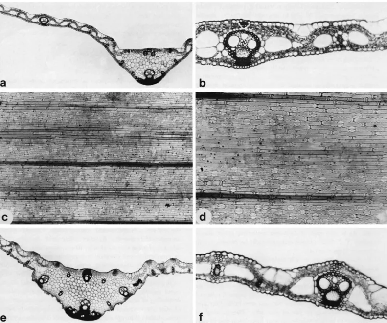
FIGURE I.-Scrotochloa urceolata: a, habit of basal part of plant, showing
decumbent culm with thick prop roots ( X 0.6); 6, habit of upper part of plant ( X 0.6); c, enlargement of section of blade t o show transverse veinlets;
d , ligule, with twisted pseudopetiole ( X 4); e, pseudopetiole showing twisting at summit ( X 2 ) ; inflorescence showing point of disarticulation from peduncle ( X 0.6); g, enlargement of surface of female glumes to show uncinate macrohairs. All drawings based on Bahkrzshnan 620, from Sri Lallkd.
NUMBER 65 5
6 SMITHSONIAN CONTRIBUTIONS T O BOTANY
N U M B E R 65 7
2016 (NY, PDA, US). G A L L E D I S T R I C T : Hinidun-kanda, Jayasuriya et al. 1795 (PDA, US). D I S T R I C T UNKNOWN: Kek- una Ethula Mukalane, 3-2-[ 19124, de Silva s.n. (PDA); Kopa Kande, Marawa Kale, 28 Feb. 188 1, without collector (PDA);
Kalugammana, J . M . Silva 46 (BM, NSW, NY). LOCALITY UNKNOWN: Walker [as W a l k ] 1404 (PDA); Walker s.n. (GH);
C.P. 972 (BR, G H , LE, MEL, P, US).
DIscussIoN.-Scrotochloa urceolata, the only widespread species in this small, recent segregate of Leptaspis, reaches the western limits of its range in the wet forests of south- western Sri Lanka and southern India. Both this species and its relative, Leptaspis zeylanica, occur in shaded forests, where individuals may form clones by decumbent culms that root at the lower and middle nodes. Because of the paucity of collections from Sri Lanka it is not possible to give the blooming phenology of this species for the island, but based on African and Asian collections it appears that both pharoid genera exhibit a flowering peak in the local rainy season.
T h e dispersal mechanism of Scrotochloa urceolata is quite specialized. T h e number of conspicuous o r principal nodes in the rachis of the inflorescence is reduced to one, although often one o r two naked, knob-like swellings a r e found on the peduncle between the uppermost foliage leaf and the whorl of panicle branches, indicating where additional pan- icle branches have been reduced and lost. At maturity the principal node of the inflorescence with its whorl of branches disarticulates from the summit of the peduncle (Figure Ij), and since the branches and female florets are covered with a dense indumentum of hooked macrohairs the entire structure is easily capable of adhering to animals.
Unlike Leptaspis zeylanica, the inflated female florets are neither conspicuously ribbed nor brightly colored at matu- rity.
T h e Singhalese specimens of S. urceolata at o u r disposal did not include young inflorescences o r mature fruits, so some material from Sumatra (Bartlett 8223) and New
FIGL'RE 2.--ScrotochIoa urceolata, dissection of spikelets: a, pair of spikelets:
inale left, female right, on clavate pedicel ( X 12); b, glumes, 1st (left) and 2nd (right) of male spikelet (X 12); c, lemma of male spikelet ( X 12); d, palea of male spikelet ( X 18): e, andrecium with 6 stamens a n d pistillodium i n center ( X 12);f; 1 st glunie of female spikelet ( X 12); g, 1st glume removed on female spikelet to show I o u n g fusiform anthecium with stigmas begin- ning to exit through apical pore (x 12); h, 2nd glume of female spikelet, inner view ( X 12): t , 2nd glunie of female spikelet, side view ( X 12); J ,
lemma of young female spikelet covered with uncinate hairs and with stigmas and palea exiting through apical pore ( X 12); k , tip of female palea enlarged ( X 48): I, mature female spikelet, side view ( X 6); m, mature female spikelet as viewed from above (X 6); n, gynecium showing the 3 subplumose stigmas (x 12); o, pistilodium ( X 32); p , 6 staminodes at base of ovary ( X 3'2); 4, caryopsis showing broad linear hilum in groove (X 12): r, caryopsis showing basal embryo ( X 12); s, palea of female spikelet ( X 16). Drawings a-e a n d o based on Bartktt 8223, Sumatra; f-k,n,p, and s on Brass 32709, Kew Guinea: 1 and m on Balakrishnan 620; 9 a n d r on Gardner s.n. from Sri Lanka.
Guinea (Brass 32709) was used to prepare portions of the species description and illustration.
ANATOMY
FIGURE 3 (cf. Figures 7 and 8)
DEsCRIPTIoN.-Leaf-blade i n transverse section. Outline:
blade flat, expanded, the margins somewhat reflexed; lam- ina thickness 175 pm. Ribs and furrows: absent, and no undulations associated with vascular bundles. Midrib outline:
distinct abaxially projecting keel (Figure 3 a ) , this represent- ing the functionally adaxial side of blade as the pseudo- petiole is twisted through 180" at its summit; symmetrical and semicircular o r rounded in shape; no lacunae in par- enchymatous ground tissue of keel. Midrib abaxial vascular bundles: 1 median and 2 minor vascular bundles present;
median bundle with well-developed abaxial cap of scleren- chynia present, but with n o girder linking it to the epidermis (Figure 3 a ) , the fibrous tissue surrounding the phloem separated from the epidermis by a continuous layer of niesophyll cells; minor bundles also lacking girders. Midrib adaxial vascular bundles: 1 minor bundle, partially em- bedded in a thin, broad plate of sclerenchyma extending the width of the midrib. Vascular bundle arrangement i n lamina: 9 first-order bundles present in leaf section, includ- ing single first-order bundle in keel. Number of third-order bundles between successive first-order bundles variable from midrib to margin: 5-7 present between midrib and first lateral first-order bundle, 10-1 2 separating central first-order vascular bundles and 5 o r 6 between first-order bundles near margin. All bundles located in center of blade.
Vascular bundle description: first-order bundles ovate, with broader side uppermost (Figure 36); lysigenous cavity pres- ent; metaxylem vessels circular, with unthickened walls, of about the same diameter as the outer bundle sheath cells.
Third-order vascular bundles elongated vertically, tall, an- gular, and narrow; xylem and phloem tissue distinguishable.
Vascular bundle sheaths of primary bundles: double sheath present. Outer sheath square, composed of 16-1 8 cells with straight, vertical sides (Figure 3b), entire, although adaxial and abaxial cells much smaller than lateral cells; no bundle sheath extensions present. Cells achlorophyllous, regular in shape and distinct from chlorenchyma cells, although vir- tually same size, square-shaped with straight radial and outer tangential walls, the walls without secondary thick- ening. Inner niestome sheath entire, consisting of at least 2 layers of cells with uniformly thickened walls (Figure 3b).
Sclerenchyma: girders not associated with vascular bundles but adaxial strands developed adjacent to all bundles (Fig- ure 3 b ) ; minute abaxial strands sometimes associated with bundles. Thickened, lignified pairs of epidermal cells oc- curring throughout both epidermides, in surface view ap-
8 SMITHSONIAN CONTRIBUTIONS T O BOTANY
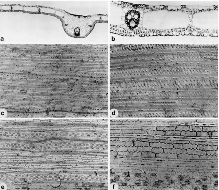
FIGURE 3.-Leaf anatomy of Scrotochloa urceolata: a and 6, leaf-blade anatomy as seen in transverse section, the adaxial (downward-facing) surface facing up i n these views; a, outline of midrib showing shape and vasculature
(X 55): b, anatomical detail with fusoid cell shape, multi-layered mestome sheath and epidermal fibers in the intercostal zones visible (X 225); c and d, abaxial (upper) epidermis in surface view, the long axis of the leaf hori7ontal; c, general cellular arrangement sholcing little differentiation into costal and intercostal zones ( X 90); a vein in located near the bottom,
pearing as fibers (Figure 3 c 4 , each pair enclosing a short or long cell; no sclerenchyma present between bundles except for these fibrous bands scattered throughout both epiderniides. Leaf margin with a small cap of fibrous tissue projecting into short, narrow point. Mesophyll: chloren- chyma not radiate, consisting of single layers of cells located immediately subjacent to both upper and lower epider- mides; remainder of leaf thickness occupied by fusoid cell
and many fibrous bands are visible in the upper portion; d, detailed structure of the intercostal fibrous bands showing their tapering and inter- connecting ends and associated silica bodies, interference contrast, (X 225);
e andf; adaxial (lower) epidermal structure. Note the strongly sinuous long cells; e, distinct stomata1 bands flanking two veins, the intercostal zones composed of alternating areas of files of fibrous bands and long cells (X 9O);f; stomata1 band above vein (lower), fibrous bands and long cells (upper)
(X 225). Based on Soderstrorn 2562 from Sri Lanka.
cavities. Uniseriate columns of chlorenchyma connecting adaxial and abaxial layers of chlorenchyma cells between adjacent fusoid cavities. Cells of abaxial (upper) chloren- chyma layer somewhat palisade-like, vertically oriented, tightly packed, larger than adaxial cells; arm cells poorly- developed, with only relatively short and indistinct inward- facing invaginations. Colorless parenchyma cells not pres- ent. Bullqorm cells: absent on both epidermides. Macrohairs,
NUMBER 65 9 prickles, minute papillae: absent o n both epidermides. Abaxial
epidermal cells: Long cells small, inflated, each with a n individual cuticle on their outer tangential walls. Groups of fibrous bands occurring a t regular intervals among files of typical long cells (Figure 3b); stomata not observed. Adaxial epidermal cells: intercostal long cells small, uniform in size, thin-walled, without distinct cuticle; groups of lignified fi- brous bands interspersed among these normal long cells.
Stomata occurring in bands on either side of vascular bun- dles, serving to indicate position of costal zones overlying the vascular bundles.
Abaxial epidermis: Costal and intercostal zones sometimes difficult to distinguish in surface view, as vascular bundles often not connected to epidermis by sclerenchyma girders.
Epidermal long cells: consisting of two intergrading types.
Normal long cells elongate-rectangular (length/width ratio 5:l; Figure 3 d ) with thin, often very sinuous lateral anti- clinal walls and straight end walls, these grading into thick- walled, very elongate (length/width ratio over 100: I), lig- nified fibers with tapering ends, the latter occurring lat- erally in files of three contiguous cells, the central file containing costal-type silica bodies and small, short, thin- walled long cells, the two sandwiching lateral files of each group quite fibrous, exceedingly long (1.5-4 mm) and ta- pering, and either interlocking with another fiber or di- verging into a V configuration, and making contact with normal intercostal long cells. Fibrous bands typically 6- 10 between each costal zone, not continuous along leaf length but ending abruptly among normal intercostal long cells.
Bulliform cells: absent. Stomata: infrequent in short, inter- rupted files (Figure 3 c ) ; subsidiary cells dome-shaped, some- times with slightly flattened tops. Intercostal short cells: pres- ent only as interrupted files of silica cells interposed between the files of fibers in the fibrous bands; normal long cells not separated by short cells. Papillae: absent. Prickles: not pres- ent on specimens examined. Microhairs: none seen. Macro- hairs: absent. Silica bodies: much narrower than other epi- dermal cells; small, horizontally elongated, round to oblong o r somewhat dumbbell-shaped, usually single but sometimes paired and alternating with short to long cells between epidermal fibers; granules o r vesicles present in all bodies.
Costal cells: very narrow, with silica bodies present only as single files between epidermal fibers; of short to medium length and often appearing to be silicified. Files of fibers located below vascular bundles probably continuous along the whole length of associated bundle.
Adaxial epidermis: Similar to abaxial epidermis, but with costal and intercostal zones well-demarcated, Stomata: oc- curring in bands of 3-4 cells on both sides of the costal zones (Figure 3e); subsidiary cells dome-shaped but appear- ing to be flat-topped, as stomata1 complexes are located below the level of the adjacent long cells (Figure 3J; sto- niatal rows adjacent to one another, a single interstomatal long cell separating successive stomata in a file. Intercostal fibrous bands: present, fewer in number (5-7) than on
abaxial epidermis, and with n o fibrous cells transitional to normal long cells. Fibers tapering to long, sharp points, occasionally branching. Intercostal long cells: of normal type, shorter, wider, and slightly less sinuous than their abaxial counterparts (Figure 3c-j).
SPECIMENS EXAMINED.-S. urceolata: Soderstrom 2562, Sri Lanka (US); Brass 29119, New Guinea (US).
DIscussIoN.-The prominent fusoid cells and complex midrib vasculature observed in leaf cross-sections of this species are familiar bambusoid features, but the intercostal fibrous bands of Scrotochloa (Rao and Naidu, 1981; Ren- voize, 1985; Figure 3) and its relatives, Leptaspis (Metcalfe, 1960; Clifford and Watson, 1977; Palmer and Tucker, 198 1) and Pharus (Metcalfe, 1960; Renvoize, 1985; Figure 8) are without exact parallel in the family. Each band consists of lateral files of elongate, thick-walled, tapering fibers that invariably enclose a single central file of silica cells, prickles, o r modified, small long cells. O n the abaxial surface of Scrotochloa urceolata (Figure 3 c ) there appears to be a complete gradient between these fibers and typical thin-walled, sinuous long cells. On the adaxial surface of the species, however, as well as on the abaxial epidermides of all the Pharus species examined for this study (Figure 8), there is a fairly sharp demarcation between the two types.
Thus, preliminary evidence indicates that Scrotochloa, al- though advanced morphologically, may be primitive with respect to this character.
Genus Leptaspis
Leptaspis R. Brown, 1810:211.
TYPE SPECIES.-L. banksii R. Brown.
DIAcNOSIS.-P~ants cespitose o r spreading by decum- bent, rooting culms; culms solid. Leaves: sheaths com- pressed; blades narrow and linear to broadly elliptic. Injlo- rescence: a long-pedunculate panicle bearing appressed o r spreading, single or whorled, persistent branches. Spikelets:
paired, the female large, subsessile, the male small, pedi- celled. Female spikelets: sometimes subtended by a glume- like bract; true glumes 2, persistent, cuspidate; lemmas with fused margins, at maturity becoming bladder-like and form- ing a shell-shaped (cochleate) utricle, strongly 7-ribbed, covered with uncinate macrohairs, somewhat indurate, the style exiting through a lateral pore; palea narrow, longer than the lemma and exiting through the pore; stigmas subplumose. Male spikelets: glumes persistent, shorter than the deciduous floret; lemmas more or less tubular; lodicules 0-3; basic chromosome number, n = 12 (Hunziker, Wulff, and Soderstrom, 1982).
DISTRIBUTION.-Tropics of the Old World, from Guinea eastward to Madagascar, Sri Lanka, Taiwan, Australia, New Caledonia, and the Fiji Islands.
SPECIES OF Leptaspis I N SRI LmKA.-Leptaspis reylanica.
10 SMITHSONIAN CONTRIBUTIONS T O BOTANY
Key to the Species of Leptaspis
1. Main inflorescence branches in whorls of 3; leaf-blades 2.5-6 cm wide, glabrous;
bract subtending 1st female glume present; Guinea, Sri Lanka, to New Guinea, Bougainville
. . .
L. xeylanica 1. Main inflorescence branches borne singly at the nodes; leaf-blades 1-2.5 (-3.5) cm wide, glabrous o r puberulent beneath (adaxially); bract subtending 1st female glume typically absent. . .
2 2. Leaf-blades lanceolate, (1.5-) 2-3 (-3.5) cm wide, puberulent beneath; Taiwan, Indonesia to New Caledonia, Australia. . .
L. banksii 2. Leaf-blades linear, about 1 cm wide, glabrous; Fiji Islands. . .
L. angustyolia Leptaspis zeylanicaLeptaspis zeylanica Nees ex Steudel, 1853:8. [Type: Sri Lanka. Neotype, here designated: Sri Lanka, without locality or collector, C.P. 896, B!]
Leptaspis cochleata Thwaites, 1864:357. [Type: Sri Lanka, Central Prov- inces. Lectotype [chosen by C.C. Mez in 19221: Sri Lanka, without locality or collector, C.P. 896, B!] Hooker f., 1900:191; Senaratna, 1956:22;
Bor, 1960:617, fig. 7 3 .
Leptaspis conchzjera Hackel, 1887:211-212, pl. G , fig. A . [Type: Sao Tome,
“prope IMorros d e Monte Cafe a d 850 m (Moller) et prope Mongo ad 480 m (Moller); Angolares (F. Quintas).” Holotype, Moller and Quintas 148, M’, not seen: isotype, K , not seen; fragment of holotype, US! sheet no. 81844.1
Leptaspis comorensis A. Camus, 1924:513. [Type: Comoro Islands, Humblot 321. Holotype, P!; isotypes, BM!, K, not seen.]
MORPHOLOGY
FIGURE 4
DESCRIPTION.-PlantS: with shallow rhizomes, the strongly ribbed internodes 1-6 cm long, the sheathed nodes each bearing a thick, hard, long, adventitious (prop) root, this bearing fibrous roots: rhizome continuous into the aerial axis (culm) of the plant, o r new culms sometimes borne from a decumbent node. Culms: up to 1 m tall, covered by overlapping sheaths. Leaves: sheaths elongate, glabrous, strongly ribbed, narrowed to the ligule and continuous with the pseudopetiole, sparingly tessellate on the outer surface, glabrous, the ribs of the sheath continuous with those of the abaxial face of the blade; blades oblong-lanceolate, 10- 20 cm long, 2.5-5 cm wide, flat, narrowed at the base into a pseudopetiole 1 / ~ ~ ~ - 1 / ~ - ) the length of the blade, the apex acute: upper (morphologically abaxial) surface glabrous ex- cept for the pilose adaxial surface of the pseudopetiole, the midvein slightly elevated, green, strongly ribbed, the pri- mary veins emanating from it along the lower half, tessel- late; lower (morphologically adaxial) surface glabrous, strongly tessellate, the broad midvein flush with the epider- mis, stramineous, smooth, shiny; ligule about 0.25 mm long, membranous, densely ciliate on the abaxial surface. Pedun- cle: exserted, 5-10 cm long, densely covered with uncinate hairs. Inflorescence: a narrowly ovoid panicle to 30 cm long, the rachis strongly angled and covered with uncinate macro- hairs, bearing whorls of branches at the nodes: primary
branches ascending or held at an acute angle or sometimes elongate, lax, and horizontally positioned, covered with a fine mat of uncinate hairs, 3 (-5) branches to about 15 cm long radiating from the lower nodes and fewer and shorter branches from the upper nodes, pulvinate at the base on t h e upper surface, the branches not disarticulating at ma- turity. Branchlets bearing spikelets sometimes subtended by a deciduous, narrow, glume-like bract about 1 mm long (Figure 4e, arrow), terminating in a male spikelet and bear- ing about 1-2 mm below it a subsessile female spikelet.
Female spikelets: subtended by a deciduous, lanceolate-tri- angular glume-like bract 3-4 mm long placed below and slightlv lateral to the 1st glume and separated from it by a short internode: glumes 2, subequal, broadly ovate with a cuspidate tip, about 2-2.5 mm long, 1(-3)-nerved, persist- ent, becoming horizontal and relatively indistinct under the expanded lemma; floret 4-3 mm long, ovate and symmetric when young, purplish-brown, at maturity greatly inflated, cochleate, the surface stramineous, pinkish, or purplish and covered with a dense mat of small uncinate hairs, strongly 7-nerved, the nerves elevated: palea about half the length of the lemma, broader at the base and flat, becoming sulcate above between the 2 keels, bifid at the apex, glabrous; ovary glabrous; style 1 , stigmas 3, subplumose; andrecium not seen in material examined; caryopsis not seen. Male spikelets:
slightly overtopping the female, terminating the entire in- florescence, about 3.5 mm long, ovoid, purplish-brown, the glumes broadly ovate, narrowed abruptly to a cuspidate tip, subequal, about 1.2 mm long, glabrous, 1-nerved, persist- ent: lemmas broad, somewhat inflated, 3-nerved, covered with minute dark brown hairs, 3.3-3.5 mm long; palea
FIGURE 4.--Leptaspis zeylanica: a, habit of plant showing decumbent culm with prop roots (X 0.6); 6, upper leaves of culm (X 0.6); c, inflorescence, showing primary branches in whorls of 3 (X 0.6); d , branches of inflores- cence enlarged to show pulvini ( X 5.5); e, pair of young spikelets (X 1 l), male left, female, right; note glume-like bract subtending the 1st female glunie, and second bract (arrow) subtending the branchlet hearing the male- female spikelet pair;J andreciuni of 6 stamens with pistillodium in center and palea extending behind ( X 23); g, mature spikelet pair, male on left, feniale with inflated lemma on right (X 7.5). Drawings a, c, and e based on Gardner s.n., Matale, Dec 1846, C.P. 896; b, d , J and g on Sumithraarachchi 602, both from Sri Lanka.
NUMBER 65 11
12 SMITHSONIAN CONTRIBUTIONS TO BOTANY
shorter than the lemma, about 2.6 mm long, linear, bifid at the apex, membranous; lodicules 3, the anterior pair broad, rounded, glabrous, nerveless, about 0.3 mm long, the pos- terior reduced; stamens 6, the anthers orange-yellow, 2.1- 2.6 mm long, basifixed, the filament attached to the con- nective at the lower Y 5 of the anther; rudimentary gynecium not seen.
SPECIMENS EXAMINED.-MATALE DISTRICT: Gamma- duwa, Alston 670 (PDA); C.P. 896 (PDA), 0. C. [?] 1045 (PDA); Matale, C.P. 896 (PDA); Gardner s.n., Dec 1846 (PDA); C.P. 896 (US). KANDY DISTRICT: Hantane, Gardner 1045 (NY); Boyagoda Kanda, near Galagedera, 7’23’N, 8 0 ” 30’E, Sumithraarachchi 602 (B, PDA, US). LOCALITY UNKNOWN: C . P . 896 (BM, BR, LE, P); anno 1827-1830, Macrae 810 (BM).
DIscussIoN.-Leptaspis zeylanica has one of the widest ranges of any herbaceous bamboo, extending nearly half- way around the globe (160’ of longitude), from the west coast of Africa to the Solomon Islands. T h e genus is unusual in the tribe in that neither the inflorescence nor its branches disarticulate at maturity, although the mature female florets readily detach from the glumes and adhere to clothing and presumably also fur and feathers. Label data of collectors on specimens from outside of Sri Lanka indicate that these shell-like, utriculate diaspores often have a showy, pink or purple color at maturity, and that the prominent ribs are white o r dark-colored. Clearly, animal interactions with this grass must be complex, and our understanding of the sub- ject would benefit from further study.
Very few good Sri Lanka collections of this species were available for study. Most male florets had already fallen from our material, and it was difficult to find any with the stamens still present. Measurements of these organs were therefore taken from those of a plant collected in Cameroon (Mildbraed 7856, US).
Steudel (1 853) described Leptaspis zeylanica as having
“oblong leaves and pseudopetioles pubescent below toward the midrib, variable in having a composite raceme with subrotund, cuspidate valves [female florets] and a simple raceme with ovate-acuminate valves” [translated from the Latin]. In the same work he also described L . manillensis (with a “verticilled panicle” and “oblong valves”), since shown to be the same as Scrotochloa (Leptaspis) urceolata, with which the author was evidently not familiar. T h e two genera of Ceylonese Phareae are quite similar vegetatively in that both have oblong leaf blades that narrow below into twisted pseudopetioles. In S. urceolata, however, the pseu- dopetioles are completely glabrous, while in L . zeylanica they are densely pubescent on the inner surfaces. Also, the female florets in this species are subrotund, not oblong as in Scrotochloa, and the inflorescence does resemble a “com- posite raceme,” whereas Scrotochloa has an umbelliform panicle. It is evident that Thwaites (1 864) described Leptas- pis cochleata because he mistakenly considered L . zeylanica
to be synonymous with Scrotochloa (Leptaspis) urceolata. Lep- taspis zeylanica Steudel thus takes precedence over the later name of Thwaites, and we here designate the Berlin speci- men from Sri Lanka, labelled “C.P. [Ceylon Plants] 896,”
to serve as the neotype. This specimen has also been chosen as the type of Leptaspis cochleata Thwaites by C.C. Mez in March 1922, as indicated by his annotation of the Berlin specimen. Sheets marked “C.P. 896” are found in many European herbaria, but since the “C.P.” series numbers were essentially species numbers it is impossible to be certain that they all come from the same locality.
ANATOMY
N o preserved anatomical material of Leptaspis zeylanica was available for this study, but based on the descriptions and illustrations of African material of this species by Met- calfe (1 960), Jacques-Felix (1 962), Palmer and Tucker (1981), and research in progress, the leaf anatomy of this taxon appears to be quite similar to that of the species of Scrotochloa (Figure 3 ) and Pharus (Figures 7 and 8) exam- ined. In transverse blade sections L. zeylanica is hardly distinguishable from its sister genera, but in epidermal views it rather surprisingly bears a closer resemblance to Pharus than it does to Scrotochloa. In common with all Pharus species, but unlike Scrotochloa, the abaxial (lower) blade surface of L . zeylanica exhibits prominent inflated intersto- matal cells (see Palmer and Tucker, 1981), and the silica bodies are consistently dumbbell-shaped. As is typical of pharoid grasses, intercostal fibrous bands are prominent on at least one epidermis.
Tribe STREPTOGYNEAE
STREPTOGYNEAE C.E. Hubbard ex Calderon and Soderstrom, 1980: 18.
TYPE GENus.-streptogyna P. Beauvois.
DIAcNosIs.-Forest grasses forming cespitose clumps or spreading by rhizomes. Leaves: blades linear-lanceolate to broadly lanceolate, narrowed below to a pseudopetiole-like base, the nerves parallel, transverse veinlets not manifest, the midvein and secondary nerves clearly manifest on the lower surface; outer ligule present as a small hard rim with irregular margins; inner ligule short, membranous. Znjlores- cence: an erect one-sided raceme. Spikelets: several-flowered;
florets hermaphrodite, the upper ones reduced and not fertile, disarticulating between the fertile florets above the glumes, each floret falling attached to the extended curved rachilla segment above it; glumes 2, persistent, the lower shorter than the upper, the upper convolute and enclosing the floret at its base; lemmas narrow, elongate, awned, many-nerved; paleas strongly 2-keeled, sulcate between the keels; lodicules 3; stamens 2 or reportedly 3; ovary with a long style and 2 o r 3 spiraling stigmas, these becoming