The Role of KCC3 in Neuronal Homeostasis By
Bianca Flores Dissertation
Submitted to the Faculty of the Graduate School of Vanderbilt University
in partial fulfillment of the requirements
for the degree of DOCTOR OF PHILOSOPHY
In Neuroscience June 30th, 2020 Nashville, Tennessee
Approved:
David Jacobson, Ph.D.
Nellie Byun, Ph.D.
Amanda Peltier, M.D.
Eric Delpire, Ph.D.
ii
ACKNOWLEDGEMENTS
I would like to thank all of the ancestors, past and present, who have made countless and unspoken sacrifices in order for me to be the first doctor in our family. I would like to honor and thank the Matriarch of our family, my grandmother, for helping to raise me and for always making me feel heard. I would like to thank both of my parents for always showing up for me when I needed them most and for showing me that with hard work, optimism, and gratitude, anything is possible. Thank you, dad for making the dangerous journey across the border when you did, so that your children could have a better life. Thank you, mom for being a grounding force in my life, for helping me to stay focused, and for always reminding me of the bigger picture. Thank you to my siblings, Jeremy, Cris, and Marissa, who contributed and self-sacrificed in different ways so that I could be where I am today. I am grateful to Max and Ruby for their unconditional love and for being a source of therapy for me. Thank you Peaches, Luna, and Koa for completing the energy circle, and with Max and Ruby, always protecting me. An endless thank you to Anisha, Pantelis, and Jonathan for helping me to stay grounded, and for always being there when I needed them. Thank you to the Vanderbilt IMSD program and Vanderbilt SACNAS who provided community, educational support, and created a safe space for me during my time here.
iii
TABLE OF CONTENTS
Page
ACKNOWLEDGMENTS ..……….. ii
LIST OF FIGURES ……….……… vi
LIST OF TABLES ……… viii
Chapter I. Introduction ……….………. 1
The CCCs: structure, function, and pharmacology…..…..………... 3
Na+ dependent cotransporters………...….….… 7
Na+ independent cotransporters………...………... 11
Localization of cation chloride cotransporters and their role in health and disease……..…….… 12
NCC ………...…… 13
NKCC1………...… 14
NKCC2……….….. 16
KCC1……….….… 19
KCC2……….…….… 20
KCC3……….………. 21
KCC4……….….…… 23
Orphan cotransporters………...………….……… 23
Roles for KCC2 and NKCC1 in neurodevelopment………..……….. 24
Peripheral nerves and peripheral neuropathy………..….……….… 27
Hereditary peripheral neuropathies……….….. 28
Stratification of CMT and comparisons to HSMN/ACC………..……… 29
Diagnoses and physical presentations: CMT vs HSMN/ACC……….……… 32
Summary and dissertation goals .……….……….…... 34
Specific aims ………. 35
II. Peripheral Motor Neuropathy is Associated with Defective Kinase Regulation of the KCC3 Cotransporter ……….… 36
Introduction .………... 39
Materials and Methods ……….….…...………. 39
Patient recruitment ………. 39
Cell culture, transfections, and cell treatments ………. 39
Antibodies ……….. 40
Buffers and solutions ………. 40
Immunoprecipitation with phosphorylation-specific antibodies ……… 41
Immunoblotting ……….. 42
K+ influx assay in human fibroblast cells and HEK293 cells ………. 42
CRISPR/cas9 generation of KCC3-T991A mice ……… 43
Mouse fibroblasts ………... 44
iv
86Rb+ uptake in mouse fibroblasts ……….. 45
Cell volume experiments ……… 46
Accelerated rotarod assay ………... 47
Wire hang grip test ………... 47
Force grip test ………. 48
Balance beam ………. 48
Nerve Conduction Studies ……….. 49
Transmission electron microscopy ………....…. 50
Statistical analysis ……….………. 50
Three-dimensional structure modeling ……….. 51
Results ……… 52
Clinical presentation of predominately peripheral motor neuropathy ………... 52
Identification of the mutation in clinical case ……….... 58
Effect of KCC3 T991A mutation on KCC3 ……….... 59
Generation and Characterization of KCC3T991A/+ and KCC3T991A/T991A mice …. 67
Discussion ……… 78
Conclusions……….. 85
III. A Role for KCC3 in Maintaining Cell Volume of Peripheral Nerve Fibers …………...…… 86
Introduction .……… 86
The cloning of KCC3………..………..…… 88
KCC3: Its function and expression…………....……… 89
Mapping of human KCC3……….………. 94
KCC3 in disease…...……….. 94
HMSN/ACC………...… 94
Mouse models of HMSN/ACC……….. 97
KCC3 gain of function………. 100
Alternative methods to assess sciatic nerves: Diffusion Tensor Imaging ……... 106
KCC3 as a drug target ……….…. 109
The cellular basis of disease: KCC3……… 111
Neuronal by nature……….……...……….. 111
A role for Schwann cells?... 114
The future of KCC3 and HMSN/ACC………. 116
Conclusion ……… 122
IV. Osmotic Response of Dorsal Root Ganglion Neurons Expressing Wild-Type and Mutant KCC3 Transporters ………..……….. 124
Introduction .……….. …………. 124
Materials and Methods ……….. …………. 127
Reagents ……….. 127
Animals ….……….. 128
Coverslip preparation ……….. 128
Dorsal root ganglion (DRG) Extraction and cell culture ………..….. 129
Solutions ……….. 129
Cell volume measurements ……….…………..…... 130
Immunostaining ……… 131
v
Data analysis and statistics ……….……….. 132
Results……….………….. 133
Use of primary sensory neurons as a model for cell volume regulation ……….. 133
DRG cell volume was determined through calcein fluorescence measurements ….………. 136
Neurons from wild-type (with or without KCC3 inhibitor), loss of KCC3 function, or gain of KCC3 function display different responses to the osmotic challenge ………. 139
Osmotic behavior of DRG neurons: the van’t Hoff plot ………. 144
Discussion ……….……….………….. 146
Conclusions………153
V. Physiological Relevance of the Temporal Expression of KCC3 ………. 154
Introduction ……….……….……… 154
Materials and Methods ………. 157
ES cell targeting ………... 157
Mouse models ………...158
Genotyping ………... 158
Tamoxifen preparation and administration ……….………. 159
Accelerated rotarod assay ……..………...159
Balance beam ………... 160
Hot plate Assay ……… 161
Immunohistochemistry of dorsal root ganglia (DRG) ………...………...162
Statistical analysis …….………... 163
Results ……….. 163
Discussion ……… 175
Conclusions………179
VI. Conclusion and Future directions ………. 180
REFERENCES ………. 188
vi
LIST OF FIGURES
Page
Figure 1-1: Topology structures of the cation chloride cotransporters ……….… 4
Figure 1-2: WNK/SPAK-OSR1 regulation of CCCs ……….... 6
Figure 1-3: Ion binding sites elucidated from cryo-EM structure ……….… 9
Figure 2-1: Brain and muscle imaging of a patient with a KCC3 T991A mutation ……….. 55
Figure 2-2: Identification of a de novo KCC3 T991A mutation in a patient with an early-onset, progressive, and severe axonal motor neuron neuropathy ……….. 61
Figure 2-3. T991A decreases KCC3 phosphorylation by the WNK1-SPAK pathway in HEK293 cells and patient fibroblasts ……….. 64
Figure 2-4: T991A increases KCC3 activity and affects cell volume regulation in HEK293 cells and patient fibroblasts ……….. 66
Figure 2-5: Genetically-modified KCC3-T991A mice exhibit locomotor deficits ……… 68
Figure 2-6: Genetically-modified KCC3-T991A mice exhibit hindlimb movement and nerve conduction deficits ……….. 75
Figure 2-7: Electron micrographs of sciatic nerve fibers isolated from KCC3 wild-type and T991A mice ……….. 77
Figure 2-8: Finely tuned KCC3 activity is required for structure and function of the human peripheral nervous system (PNS) ………82
Figure 3-1: Regulation of cation-chloride cotransporters ……….. 90
Figure 3-2: Electron micrographs of sciatic nerves ………. 103
Figure 3-3: Shrinkage of axons and fibers in KCC3-T991A sciatic nerves ……… 105
Figure 3-4: Mean Fractional Anisotropy ………. 107
Figure 3-5: Radial and Axial diffusivity ……….. 108
Figure 3-6: Structures of the PNS and CNS involved in locomotion ……….. 118
Figure 4-1: Expression of KCC3 in mouse DRG neurons …..……….……… 135
vii
Figure 4-2: Calcein fluorescence measurements in dorsal root ganglion neurons …….………. 137
Figure 4-3: Calcein fluorescence measurements in dorsal root ganglion neurons ……….. 138
Figure 4-4: Osmotic behavior of neurons exposed to a hypotonic challenge ……….. 140
Figure 4-5: Osmotic behavior broken-down by components ……...……… 142
Figure 4-6: Osmotic behavior of neurons broken-down by components ………. 143
Figure 4-7: Osmotic sensitivity of neurons ……….. 145
Figure 5-1: Design of mouse models to induce disruption or recovery of KCC3 expression …. 165 Figure 5-2: Inducible PV-CreERT2 system .……… 166
Figure 5-3: Rotarod performance of mice expressing inducible Pv-CRE x KCC3-flox mice …. 167 Figure 5-4: Balance beam (12-mm) performance of Pv-CREERT2 x KCC3-floxed mice ….…... 169
Figure 5-5: Balance beam (6-mm) performance of Pv-CREERT2 x KCC3-floxed mice ….…... 170
Figure 5-6: Hot plate assay in Pv-CREERT2 x KCC3-flox mice ………...…… 171
Figure 5-7: Immunofluorescence analysis of DRGs isolated from Pv-CREERT2 x KCC3-flox mice pre- and post- tamoxifen treatment …………...…. 173
Figure 5-8: Poor rotarod performance of Pv-CREERT2 x KCC3-stop mice ……….. 174
Figure 6-1: Differences in axonal size in KCC3-T991A, wild-type, and KCC3 knockout mice ……… 182
Figure 6-2: Cell volume response in sensory neurons isolated from KCC3-T991A, wild-type, and KCC3 knockout mice ………184
viii
LIST OF TABLES
Page
Table 1-1: SLC12A family tissue expression and mouse model phenotypes ……… 18
Table 1-2: List of sub-categories of HSMN (CMT) ……….. 31
Table 2-1: Nerve conduction studies with patient ………. 56
Table 2-2: Summary of clinical studies of a patient with a KCC3 T991A mutation ……… 57
Table 2-3: Size and strength properties of the engineered mice (2nd cohort) ………... 71
Table 2-4: Nerve conduction measurements in engineered mice ……….. 73
Table 3-1: Variation of KCC3 expression………... 93
Table 3-2: KCC3 mouse models ……….... 99
1 CHAPTER 1
Cell Volume Regulation and the Cation Chloride Cotransporters
Introduction
Cell volume regulation is a fundamental aspect of cell homeostasis. Most mammalian cells live in an environment of constant osmotic pressure, and therefore have kept all basic mechanisms of cell volume maintenance and regulation. Important cell processes like cell division, migration, and even cellular metabolism force cells to change their shape and volume (Wehner et al., 2003). In order to rapidly and efficiently adapt to these changes, cells utilize specific transporters and channels to maintain their volume. In general, the cell membrane is typically regarded as a semi-permeable membrane, i.e. a membrane more permeable to water than to any osmolytes. Even in the absence of water channels (aquaporins), which increase the water permeability of membranes, the intrinsic water permeability of the lipid bilayer is several orders of magnitude more permeable to water than to ions. Thus, cells experience osmotic challenges when the extracellular environment becomes hypo- or hypertonic with respect to the intracellular environment. This is the case of cells in the kidney which experience large changes in extracellular osmolarity based on the hydration status of the body. Once the cells have equilibrated the osmotic pressure by gaining or losing water, they then attempt to recover their volume. This is achieved by moving ions and organic osmolytes through proteins embedded within the lipid bilayer to transport ions.
Therefore, as ions move across the membrane, water follows, and the volume is restored. In
2
response to a hypotonic shock, a cell undergoes a swelling phase followed by a regulatory volume decrease (RVD) phase. In contrast, when a cell is exposed to a hypertonic condition, water leaves the cell and the cell shrinks. This is followed by an uptake of ions helping the cell undergo regulatory volume increase (RVI). The efflux and influx of ions relevant to cell volume regulation is mediated by channels and transporters, including electroneutral cation- chloride cotransporters.
Cation chloride cotransporters (CCCs) are among other physiological roles implicated in cell volume regulation following osmotic swelling or shrinkage (Delpire et al., 1991). Nine cation chloride cotransporters constitute the SLC12A family of solute carriers. They are a part of the amino acid-polyamine-organocation (APC) superfamily. Seven of the nine SLC12A cotransporters can be further subdivided between sodium-dependent and sodium-independent cotransporters. The remaining two proteins, CCC8 and CCC9, are considered orphan transporters as their function has yet to be determined. The Na+ independent cotransporters (KCCs) carry 1 K+ and 1Cl- ions across the membrane per transport cycle. Comparatively, the Na+-dependent K+ (NKCCs) transporters carry 1 Na+, 1 K+ and 2 Cl- ions across the membrane, whereas the Na+-Cl- cotransporter, NCC, transports 1 Na+ for every 1 Cl-. Thus, the cation chloride cotransporters exhibit a 1:1 cation to anion stoichiometry and are therefore electroneutral. Their function does not affect nor is affected by the membrane potential.
Cation chloride cotransporters are secondary active transport mechanisms. This means that these transporters derive their energy from ion gradients established by primary transporters such as the Na+/K+ ATPase or pump. Because of the tight coupling between
3
substrates, these transporters can drive one ion against its own gradient. For instance, K-Cl cotransport can drive Cl- outward against its gradient due to the large K+ gradient generated by the Na+-K+ ATPase (Gamba, 2005). Thus, in neurons, an active K-Cl cotransporter helps to maintain a very low intracellular chloride concentration, thereby allowing hyperpolarizing GABA responses and inhibition. Moreover, because they are moving solutes across the plasma membrane, the cotransporter create a gradient for water to move in and out of the cell.
The CCCs: structure, function, and pharmacology
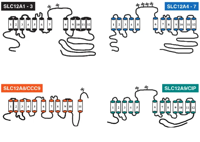
Almost all of the cotransporters share a similar 12 transmembrane domain with intracellular N and C-termini. The only exception is that one of the orphan cotransporters CCC9 has 11 transmembrane domains and a putative extra-cellular C-terminus. The other orphan co-transporter (CCC8 or CIP) is more closely related in structure to the K-Cl cotransporters. There are some differences between the Na+ dependent (NKCC1, NKCC2, and NCC) and Na+ independent (KCC1-4)transporters in structure and function, but overall, they appear highly homologous. For example, KCC1 shares about 25% sequence homology with NKCC/NCC whereas the four KCCs are approximately 65%-71% identical (Gillen et al., 1996; Mount et al., 1999). Na+ dependent transporters have an extracellular loop between transmembrane domains 7 & 8. Comparatively K-Cl co-transporters have an extracellular loop between transmembrane domains 5 & 6 (Figure 1-1).
4
Figure 1-1 Topology structures of the cation chloride cotransporters. All structures are based on hydropathy plot analysis. SLC12A1-3A (Black) are the Na+ dependent co-transporters. SLC12A4-7 (blue) are the Na+ independent co-transporters. SLC12A8/CCC9 (orange) and SLC12A8/CIP (teal) are the two orphan co- transporters. Figure adapted from Gamba 2005.
5
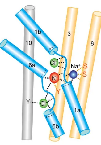
The recent cryo-electron microscopy (EM) structures of KCC1 and NKCC1 (Liu et al.
2019; Chew et al. 2019) confirmed the basic structure of the cotransporters which was based on hydropathy, i.e. cytosolic termini and 12 transmembrane domains. The cryo-EM structures will allow a more detailed understanding on how the cotransporters bind and transport ions across the plasma membrane. As the hydropathy plots provided valuable information on the number of transmembrane domains, and the position of the termini and putative glycosylation sites, the cryo-EM structures provide insights to the ion binding sites, and the path of ions through the protein which is embedded in the membrane.
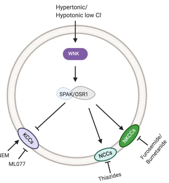
As for their function, the CCCs are phosphorylated by the WNK/SPAK-OSR1 kinases. WNK (With No Lysine-K)/SPAK (SPS1-related proline-arginine rich kinase)- OSR1(Oxidative Stress Response Kinase) exist in the same complex to reciprocally regulate the cation chloride cotransporters (Alessi et al., 2014; Gagnon et al., 2006; Piechotta et al., 2003; Piechotta et al., 2002). Specifically, the WNK/SPAK-OSR1 kinase cascade activates the Na+-dependent cotransporters while inactivating Na+-independent (K-Cl) cotransporters.
WNK kinases act upstream of SPAK-OSR1, which are the terminal kinases as they bind and phosphorylate the cotransporters (Gagnon et al., 2006; Kahle et al., 2003) (Figure 1-2).
6
Figure 1-2. WNK/SPAK-OSR1 regulation of CCCs. WNK becomes activated in hypertonic or hypotonic low Cl- which then phosphorylates SPAK/OSR1. SPAK or OSR1 phosphorylate all of the cation chloride cotransporters activating NKCC/NCC activity while inactivating KCC activity.
7 Na+ dependent Transporters
The Na+ dependent cotransporters, NKCC1 (SLC12A2), NKCC2 (SLC12A1), and NCC (SLC12A3) are critical in several cell types (epithelial cells, neurons, etc.) to help establish the chloride gradient and regulate cell volume through the import of Cl- (Gamba, 2005). This set of transporters become active during cell shrinkage and helps the cells undergo regulatory volume increase (RVI). NKCC1 and NKCC2 transport 1 Na+,1 K+, and 2 Cl – ions into the cell, whereas NCC transports 1 Na+ and 1 Cl – into the cell. Interestingly, it has been found that hypertonic conditions and hypotonic low Cl- conditions further stimulate WNK/SPAK- OSR1 activity (C. Richardson & Alessi, 2008). In these conditions, Na+ dependent transporter activity is ramped up as WNK/SPAK-OSR1 works to phosphorylate conserved Threonine residues in the cytoplasmic N-terminus of NCC and NKCC1/2. In NKCC2 these residues have been identified as Thr95, Thr100, and Thr105 (C. Richardson et al., 2011).
In terms of their structure, initial hydropathy plots had suggested a 12 TM topology that form tight hairpin-like structures within the membrane (Gerelsaikhan & James, 2000). Further information about ion binding sites was elucidated from the recent cryo-Electron Microscopy (EM) structure of the zebrafish NKCC1 (Chew et al., 2019). The structure confirmed the presence of an extracellular loop between TM7 and TM8 and showed that it was stabilized by a disulfide bond. Furthermore, a structured element with TM5 and TM6 formed a cap domain that is important for cell surface expression (Chew et al., 2019). Most importantly, the structure revealed new information on ion coupling and showed that the Na+ and K+ binding
8
sites were in proximity and bind between the core and scaffold domains (Figure 1-3).
Comparatively, the Cl- ion is transported adjacent to the cations with its movement assisted by backbone amides in TM1 (Chew et al., 2019).
9
Figure 1-3. Ion binding sites elucidated from cryo-EM structure. Ion binding sites in the core domain (blue cylinders) of the cryo-EM structure of NKCC1. Figure from Chew et al., 2019.
10
An additional characterization of the cation chloride cotransporters has been based on which pharmacological treatments inhibit or stimulate them. Specifically, the sodium dependent transporter, NCC, is characterized by its thiazide sensitivity (Figure 1-2). NCC was first purified from winter flounder urinary bladder (Gamba et al., 1993). Later Stokes and colleagues found Na+ and Cl- transport were inhibited in a dose dependent manner by hydrochlorothiazide and metolazone, two thiazide diuretics (Stokes et al., 1984). Structurally, it was found that transmembrane domain 11 in NCC defined the affinity for thiazides (Castañeda-Bueno et al., 2010). It should be noted that NCC is not inhibited by other diuretics like amiloride or bumetanide/furosemide. Currently, thiazide diuretics are used to treat high blood pressure and swelling (Gamba, 2009). In the clinic, they are sometimes also given in addition to loop diuretics, such as furosemide. Comparatively, the NKCCs (NKCC1 and NKCC2) have been shown to be inhibited by bumetanide or furosemide (Figure 1-2) (Payne et al., 1995). These two drugs are called loop diuretics because they inhibit NKCC2 in the loop of Henle, a major site of Na+ reabsorption in the kidney. NKCC1 plays a critical role in establishing a chloride concentration gradient that promotes GABA-mediated excitation in immature neurons. Bumetanide has been considered for the treatment of neonatal epilepsy and autism (Ben-Ari, 2017; Kahle & Staley, 2008). Interestingly, a furosemide variant, azosemide, was found to be a more potent inhibitor of NKCC1 than bumetanide. It also more readily crosses the blood brain barrier (Hampel et al., 2018).
11 Na+ independent cotransporters
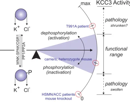
Like the Na+ dependent transporters, the K-Cl cotransporters are important transport mechanisms in several cell types (i.e. neurons, red blood cells, nephrons, etc.). They mediate the efflux of Cl- to maintain the concentration gradient and help regulate cell volume. All K- Cl cotransporters increase their activity in hypotonic conditions and thus are important regulators in cell swelling conditions (Rinehart et al., 2009). It should be noted that KCC2 is already active in isotonic conditions, and increases its activity in hypotonic conditions. In this context, the WNK-SPAK/OSR1 kinases phosphorylate K-Cl cotransporters in conserved threonine residues in the C-terminus to inactivate its activity. Specifically, in KCC3, T1048 and T991 are phosphorylated. I focus most of my dissertation on a KCC3-T991A mutant transporter which renders the transporter constitutively active. The two threonine are highly conserved among the K-Cl cotransporters (Kahle et al., 2016).
Like the NKCCs and NCC, initial hydropathy plots of K-Cl cotransporters were examined following their molecular cloning. A hydropathy plot of KCC2 also predicted the 12 TMD and the position of putative N-linked glycosylation sites on the extracellular loop between TM 5 & 6 (Payne et al., 1996). Furthermore, it was also found that a transmembrane domain residue at position 289 in mouse KCC3 (E289) was necessary for expression of the protein at the membrane (Ding et al., 2013). This site was conserved in SLC12A1-SLC12A7.
The recent cryo-EM structure of human KCC1 (Liu et al., 2019) confirmed the overall architecture of the transporter that was originally derived from hydropathy plots. What was surprising was how highly conserved the ion binding sites were between the KCC1 and
12
NKCC1 structures (Delpire & Guo, 2020; Liu et al., 2019). Specifically the K+ binding site is formed by the same five identical residues in KCC as it is in NKCC1 (Delpire & Guo, 2020), yet functional data had previously revealed NKCC1’s higher affinity for K+ (reviewed in Delpire & Guo, 2020). The cryo-EM structure of KCC1 revealed the possible path of ion permeation and provided important clues on the size of the hydrophobic cavities. Molecular docking studies suggested that ML077, a specific K-Cl inhibitor (Delpire et al., 2009), was binding deep in the ion permeation pathway and possibly interacting with a tyrosine residue important for the coordination of K+ (Delpire & Guo, 2020). This was consistent with previous kinetic studies which demonstrated that ML077 acted as a competitive inhibitor for K+ (Delpire et al., 2009). The cryo-EM structures of NKCC1 and KCC1 did not provide any insights into whether or not water molecules are co-transported alongside K+ and Cl- ions during a transport cycle, as suggested by functional studies (Hamann et al., 2010; Zeuthen, 1994). Additionally to ML077 and structural variants which are the most potent inhibitors of K-Cl cotransport, KCCs are also inhibited by furosemide, although at a much lower affinity (Mount et al., 1999) (Figure 1-2).
Localization of cation chloride cotransporters and their role in health and disease
Overall, because the CCCs are basic ion symporters that participate in ion homeostasis and cell volume maintenance and regulation, they are widely expressed in the body. Often, many of these cotransporters will overlap in expression in cells and tissues, as they have
13
reciprocal functions, such as transporting inward or outward and participating to regulatory volume increase or decrease. Because of their critical role in basic cellular physiology, the SLC12A family members have been implicated in various genetic disorders.
NCC (SLC12A3)
Initial characterization of NCC found that it was solely expressed in the kidney and the gene mapped to human chromosome 16q13 (Mastroianni et al., 1996). The cotransporter was localized to the distal convoluted tube where it mediates sodium and chloride reabsorption (Plotkin et al. 1996, Schmitt et al., 1999). Loss of NCC function results in a loss of NaCl reabsorption and Gitelman Syndrome (Gitelman et al., 1966). This syndrome is also characterized by an imbalance of K+ (hypokalemia), Ca2+ (hypocalciuria), and Mg2+
(hypomagnesemia). The disease manifests itself as extreme thirst, excessive urination, and metabolic deficits. Mice with NCC LOF displayed most of the phenotypes in patients with Gitelman (Schultheis et al., 1998). One major difference between the knockout mouse and Gitelman patients is the absence of clear hypokalemic alkalosis in the mouse (Schultheis et al., 1998). Currently, there is no cure for Gitelman syndrome, however, many patients manage their syndrome through increased intake of Na+-, K+-, and Mg2+-rich foods and supplements.
Comparatively, mutations in upstream kinases can result in increased NCC function.
Specifically, patients with mutations in WNK1 or WNK4 (Wilson et al., 2001) result in the increased NCC that develops into a disorder called pseudohypoaldosteronism type II or Gordon syndrome (Gordon, 1986). In these patients, the increased NaCl reabsorption results
14
in hyperkalemia and hypertension. This phenotype has also been reproduced in several mouse models: WNK4 knock-in mice (Chowdhury et al., 2013; Yang et al., 2010) and a constitutively active SPAK mouse (Grimm et al., 2017; Harris et al., 2018).
NKCC1 (SLC12A2)
Human SLC121A2 has been mapped to chromosome 5 (5q23.3). Unlike the Na-Cl cotransporter which is almost exclusively expressed in kidney, NKCC1 is expressed in almost all tissues throughout the body. This includes lung, heart, skeletal muscle, liver, stomach, pancreas, intestine, bladder, testis, uterus, endocrine glands, brain, spinal cord, etc. The transporter has been studied in many systems, mostly Cl- secreting epithelia (such as lung, gastrointestinal tract, and exocrine glands), but also the inner ear, and the nervous system (For comprehensive review, see (Delpire & Gagnon, 2018). As this dissertation is primarily focused in Neuroscience, cotransporters that are expressed in the central and peripheral nervous systems will be discussed in more detail. It should be noted that in neurons, NKCC1 and KCC2 functions are reciprocal, helping the neurons maintain or regulate their intracellular chloride concentration (Watanabe & Fukuda, 2015).
Several LOF mouse models of NKCC1 were generated and published in 1999 (Delpire et al., 1999; Dixon et al., 1999; Flagella et al., 1999). They displayed vestibular dysfunction and exhibited a shaker/waltzer behavior (Delpire et al., 1999). Despite being highly expressed in the central nervous system, there were no overt neurological phenotypes. However, the mice exhibited a sensory perception phenotype, due to the collapse of the high Cl-
15
concentration existing in dorsal root ganglion neurons (Laird et al., 2004; Sung et al., 2000).
Additional phenotypes include smaller size, intestinal obstruction (Flagella et al., 1999), alteration in airway ion transport (Grubb et al., 2001), male infertility (Pace et al., 2000), and deficit in saliva production (Evans et al., 2000).
Our laboratory documented the first human patient with a mutation in NKCC1 (Delpire et al., 2016). The patient carries a de novo 11-base pair deletion in exon 22 of the SLC12A2 gene, leading to premature termination and truncation of a large portion of the carboxyl- terminus of the protein. The patient suffers from multi-system dysfunction, including lung, gastrointestinal tract, bladder, exocrine glands, etc. (Delpire et al., 2016). Because NKCC1 regulates Cl- in sensory neurons and in smooth muscle cells, it was hypothesized that perhaps what the multiple organ involvement was due to defects in sensory feedback and/or smooth muscle cell function (Delpire et al., 2016). Further work by our laboratory demonstrated that while there was no dominant-negative effect of the mutant transporter on the wild-type in fibroblasts, there was a significant dominant-negative effect in epithelial cells. Using epithelial cells in culture, the lab showed that truncation of the carboxyl-terminus resulted in mis-trafficking of the mutant transporter to the apical membrane of epithelial cells (Koumangoye et al., 2018). Because of dimerization, the mutant transporter was also able to mistraffic wild-type cotransporter to the apical membrane. This phenotype was also observed in salivary gland and intestine of mice engineered to carry the patient mutation (Koumangoye et al., 2018). Expression of the mutant transporter in mice results in decreased intestinal water secretion, abnormal mucus release, and invasion of endemic bacteria in the epithelial layer of
16
the intestine. This phenotype is consistent with the intestinal dysfunction observed in the patient (Koumangoye et al., 2018).
Last year, the case of a 5-year old boy with a complete loss of NKCC1 expression was reported (Macnamara et al., 2019). Consistent with the knockout mouse model, the patient suffers from sensorineural deafness, gastrointestinal obstruction. He also presented with developmental delay and accumulation of mucus in the lung.
NKCC2 (SLC12A1)
Similarly to NCC, NKCC2 is a renal specific cation chloride cotransporter. NKCC2 was initially mapped to mouse chromosome 2 by Quaggin and colleagues (Quaggin, et al., 1995). Soon after, it was mapped to human chromosome 15q; consistent with mouse to human homology mapping (Simon et al., 1996). In the kidney, the cotransporter is found in the thick ascending limb in the Loop of Henle and in macula densa (Kaplan et al., 1996;
Payne & Forbush, 1994). Disruption of NKCC2 in human results is Bartter’s syndrome, another salt wasting disorder. Bartter’s syndrome is an autosomal recessive disorder that affects salt reabsorption in kidney. Patients present renal salt wasting, hypotension, and in extreme cases, young children can have stunted growth and seizures (Simon et al., 1996). In a knockout mouse model of NKCC2, mouse pups were born normally, but within the first week, they showed severe signs of dehydration, renal failure, and high plasma potassium (Table 1-1) (Takahashi et al., 2000). Despite NKCC1 also being expressed in the kidney, its expression is not sufficient to compensate for a loss of NKCC2. This is because the function
17
of the basolateral NKCC1 in alpha intercalated cells and inner medullary collecting duct is completely different than that of the apical NKCC2 in the tick ascending limb. Interestingly, Takahashi and colleagues were able to pharmacologically mimic the knockout NKCC2 phenotype in wild-type animals by administering furosemide. Wild-type mice treated with furosemide developed mild hydronephrosis and had comparable urine outputs than NKCC2 knockout mice (Takahashi et al., 2000). Importantly, this knockout mouse model recapitulated many of the phenotypes observed in patients with Bartter’s syndrome (hydronephrosis, renal insufficiency, hypokalemia), but to differing degrees.
18
Gene (protein) Expression *indicates ubiquitous Mouse model phenotype Human disease
SLC12A1 (NKCC2) Kidney (thick ascending limb in the loop of Henle (Quaggin et al., 1995)
Hypotension, dehydration, renal salt wasting
Bartter’s Syndrome (Simon et al., 1996) SLC12A2 (NKCC1) Kidney, PNS, CNS, Epithelial Cells,
Salivary glands basolateral
membrane, epithelial cells* (Delpire et al, 1999; Nejsum et al., 2005)
Deafness & Shaker/Waltzer behavior
One documented human case (Delpire et al., 2016) Killquist patient (Killquist
et al., 2019) SLC12A3 (NCC) Kidney (Distal convoluted tubule of
nephron)(Schmitt et al., 1999)
Hypokalemic alkalosis, hypomagnesemia, hypercalciuria
Gitelman syndrome (Simon et al., 1996) SLC12A4 (KCC1) *Brain, Colon, Heart, Kidney (Gillen
et al., 1996), red blood cells
No phenotype when disrupted by itself
N/A
SLC12A5 (KCC2)
Neuronal specific *Ubiquitous in the CNS (Williams et al., 1999)
Motor deficits/abolished respiration
Linked to Idiopathic epilepsy (Kahle et al., 2014)
SLC12A6 (KCC3)
Broadly expressed throughout PNS/CNS (Mount et al., 1999; Byun
and Delpire 2007)
HSMN phenotype and tissue vacuolization
HSMN/ACC (Howard et al., 2002); Gain of function
(Kahle et al., 2016)
SLC12A7(KCC4) Lung, heart, and kidney (Mount et al., 1999), abundant throughout CNS
and PNS
Deafness and renal tubular acidosis
N/A
SLC12A8(CCC9) Small intestine, stomach, testis, thyroid and colon (Hewett et al.,
2002)
N/A Researched as a psoriasis
susceptibility candidate gene (Hewett et al., 2002) SLC12A9
(CCC6/CIP1)
Muscle, brain, and kidney (Caron, et al., 2000)
N/A N/A
Table 1-1. SLC12A family tissue expression and mouse model phenotypes. A range of phenotypes results from the disruption of each co-transporter with the physiological significance of some co-transporters yet to be determined.
19 KCC1 (SLC12A4)
Similar to NKCC1, KCC1 is ubiquitously expressed. KCC1 has been detected in the brain, colon, heart and kidney (Gillen et al., 1996). Importantly, KCC1 has also been implicated in regulating cell volume in red blood cells (Quarmyne et al., 2011; Rust et al., 2007). However, because of its ubiquitous distribution, it is hypothesized to be a “house- keeping” cation chloride cotransporter (Garneau et al., 2019; Marco B. Rust et al., 2007).
Based on its expression pattern, it would seem KCC1 would play an integral role in cell volume maintenance/overall cell homeostasis. However, no human disease has been directly linked with a mutation in KCC1 (Table 1-1). In knockout mouse models of KCC1 observing specifically red blood cells, there was no phenotype in mice lacking KCC1 (Marco B. Rust et al., 2007). However, when this mutation was compounded with a LOF of KCC3, only then did red blood cells exhibit a defect in cell volume regulation, suggesting that RBC cell volume is mainly regulated by KCC3 or regulated by both KCC3 and KCC1. Interestingly, KCC1 has been implicated in the growth of cervical and endometrial cancers (Shang et al., 2011; Zhang et al., 2009). When KCC1 was knocked down via RNA interference, cancer cell invasion decreased (Shang et al., 2011). Though LOF of KCC1 has not been directly linked to diseased mouse models or human genetic disorders, a gain of function of KCC1 resulted in sickle cell disease pathology in a humanized mouse model (Brown et al., 2015).
Constitutively active KCC1 resulted in sickling, tissue damage, and ultimately premature death. Thus far, there has been no direct link with human sickle cell disease other than indication of increased K-Cl cotransport activity (Brown et al., 2015). It is clear that the
20
increased KCC activity in sickle cell disease accelerates the dehydration of red blood cells and the sickling process. Thus, KCCs could become potential therapeutic targets in the future.
KCC2 (SLC12A5)
KCC2 was first cloned in 1996 and labeled as a “neuronal-specific” isoform of the K- Cl cotransporters (Payne et al., 1996). It is found in most brain structures (Lu et al., 1999) including cerebellum (Notartomaso et al., 2017), hippocampus (Kelley et al., 2018), and even the retina (Vu et al., 2000). Interestingly, KCC2 is not expressed in the peripheral nervous system (Sedmak et al., 2015). Recent work has showed that KCC2 might be expressed in pancreatic beta cells (Kursan et al., 2017). Because of its importance in the CNS, the knockout of KCC2 in mice results in severe motor deficits that lead to the premature death of mouse pups due to respiratory failure (Table 1-1) (Hübner et al., 2001). Note that expression of KCC2 is driven by two separate promoters: KCC2a accounts for 5-10% of total KCC2 levels while KCC2b accounts for 90-95% of total KCC2 expression. Knockout of the KCC2b isoform results in pups surviving the perinatal period and exhibiting severe tonic/clonic seizures (Woo et al., 2002). KCC2 expression increases during development (Lu et al., 1999) leading to a shift in GABA responses from depolarizing to hyperpolarizing. Mutations in KCC2 have been also been associated with idiopathic epilepsy (Kahle, et al., 2014) and febrile seizures (Puskarjov et al, 2014). Additionally, deficient KCC2 expression has also been implicated in Rett syndrome, a neurodevelopmental disorder characterized by breathing abnormalities and severe seizures (Hinz et al., 2019). KCC2’s role in establishing the chloride
21
gradient in the CNS in conjunction with NKCC1 will be discussed further below.
KCC3 (SLC12A6)
KCC3 is expressed throughout the PNS, CNS (Byun & Delpire, 2007; Pearson et al., 2001), and had a broad range of expression in the lung, heart, esophagus and several other tissue systems (Mount et al., 1999; Shiozaki et al., 2014). It should be noted that KCC3 also exists in two different isoforms: KCC3a and KCC3b. The KCC3a isoform encodes 99 amino acids and contains several sites for phosphorylation for inactivation. Comparatively, KCC3b encodes 39 amino acids and has a unique amino-terminus (Hebert et al., 2004). Moreover, KCC3a is expressed in the nervous system as well as the heart and skeletal muscle, while KCC3b expression is restricted to the kidney (Pearson et al, 2001; Arroyo et al., 2013) Several studies have confirmed KCC3a as the only expressed isoform of KCC3 in the nervous system (Le Rouzic et al., 2006; Pearson et al., 2001; Shekarabi et al., 2011). The focus of this dissertation will remain on KCC3a and will simply be referred to as KCC3.
KCC3 is the first K-Cl cotransporter that was directly linked with human disease. LOF of KCC3 results in hereditary sensorimotor neuropathy with agenesis of the corpus callosum (HSMN/ACC). HSMN/ACC is also referred to as Agenesis of the corpus callous with Peripheral Neuropathy (or ACCPN) as well as Andermann syndrome, following the first description of the disease in the medical literature (Andermann et al., 1972). HSMN/ACC is a genetic disorder that affects a mainly French-Canadian population in the Charlevoix/Lac St Jean region. Interestingly, researchers had first mapped HSMN/ACC to human chromosome
22
15q before they knew the gene that was disrupted (Casaubon et al., 1996). Later, Mount and colleagues mapped KCC3 to chromosome to human chromosome 15 (Mount et al., 1999). It was until individuals were actually screened for mutations in KCC3 that it was confirmed that mutations in KCC3 caused HSMN/ACC (Howard, Mount, et al., 2002). Furthermore, the LOF mouse model recapitulates the peripheral neuropathy phenotype (Howard, Mount, et al., 2002).
Since KCC3 is expressed in the PNS and CNS, a disruption in this cotransporter is more commonly associated with peripheral neuropathy as opposed to epilepsy or defects in red blood cells like the other isoforms (Howard et al., 2002; Kahle et al., 2016; Shekarabi et al., 2011). Interestingly, a GOF of KCC3 results in nearly the same phenotype in mice and humans as a LOF (severe peripheral neuropathy). As my dissertation work focuses on this aspect, gain-of-function mutations will be discussed more extensively in chapters 2-4.
Currently, there appear to be no compensatory mechanisms involved when KCC3 is disrupted. KCC3 can be co-expressed with other K-Cl cotransporters in multiple cell types, yet a KCC3 LOF appears to be not compensated by other mechanisms involved in cell volume regulation. For example, KCC3 happens to be co-expressed with KCC2 in many central neurons, yet tissue vacuolization still occurs in mouse models and patients with a LOF of KCC3 in the CNS (Auer et al., 2016; Boettger et al., 2003).
23 KCC4 (SLC12A7)
KCC4 also has ubiquitous expression, but is more abundantly expressed in the nervous system, lung, heart, and kidney (Mount et al., 1999). Importantly, like several other cotransporters, KCC4 is localized to the kidney, with the distal convoluted tubule and connecting tubule exhibiting the most abundant expression of KCC3 (Velázquez & Silva, 2003). However, within the nervous system, KCC4 has been localized to the forebrain and in sensory neurons and cranial nerves (Karadsheh et al., 2004; Le Rouzic et al., 2006).
Interestingly, a LOF mouse model of KCC4 demonstrated both deafness and renal tubular acidosis (Boettger et al., 2002). The mice exhibited no motor or vestibular deficits, but severe hearing loss, suggesting an important and unanticipated role for KCC4 in maintaining K+ in the inner ear. In contrast to NKCC1 which is expressed on the basolateral membrane of stria vascularis epithelium, where it participates in the secretion of the K+-rich endolymph, KCC4 is expressed in hair cells. In addition to the hearing loss, LOF mice demonstrate urinary acidification resulting in renal tubular acidosis (Boettger et al., 2002). Currently, there are no documented human cases of a disruption in KCC4.
Orphan Cotransporters: CCC9 (SLC12A8) and CCC8 (SLC12A9)
CCC9 and CCC8 (sometimes referred to as CIP1) are orphan members of the SLC12A family. Their function as ion transporters has yet to be determined. While CCC9 expression seems broad, the highest levels of expression were observed in the small intestine, stomach, testis, thyroid and colon (Hewett et al., 2002). The gene was first identified as a potential gene
24
for psoriasis susceptibility (Hewett et al., 2002). Hewett an colleagues detected ubiquitous expression, but A splice variant of CCC9 was found to promote polyamine amino acid transport in HEK293 cells (Daigle et al., 2009). Interestingly CCC9 is the only member of the family that lacks the key residues coordinating the first Cl- binding site (Delpire & Guo, 2020). Comparatively, CCC8 has been detected in the muscle, brain, and kidney (Caron, et al., 2000). However, its physiological relevance is still unclear.
Roles for KCC2 and NKCC1 in neurodevelopment
In addition to volume regulation, cation-chloride cotransporters play a role in establishing the chloride concentration gradient (Delpire, 2000; Kahle et al., 2008). As mentioned above, the electroneutral cation-chloride cotransporters transport Cl- with Na+ and/or of K+ in a 1 cation:1 anion stoichiometry (Gamba, 2005). In the nervous system, the level of Cl- defines the direction and strength of GABAergic and glycinergic neurotransmission. Neurotransmitter signals are necessary for patterning synaptic connections in the brain’s intricate network of electrical synapses (Ben-Ari, 2002). These connections are part of a dynamic and delicate balance of excitation and inhibition necessary for the development of synapses and overall brain homeostasis. Unsurprisingly, disruption in the maintenance of intracellular chloride in the developing or in the adult nervous system can lead to an array of nervous system disorders.
The primary regulation of chloride in the nervous system is dependent on CCCs.
Immature neurons have a relatively high [Cl-]i with a depolarized GABA reversal potential
25
(EGABA-A), while mature neurons have low levels of [Cl-]i and hyperpolarized EGABA-A. The developmental change in that occurs in EGABA-A is correlated with expression changes in NKCC1 and KCC2 (Plotkin et al., 1997; Lu et al., 1999; Glykys et al., 2009). KCC3 expression is also low during development and increases in the late prenatal period in humans and postnatal period in rodents (Pearson et al., 1991; Lucas et al., 2012) thus it seems that KCC3 could also participate in the GABA developmental shift. This shift, in which GABA switches from depolarizing to hyperpolarizing, seems to mainly occur in central neurons where KCC2 expression is predominant.
NKCC1 and KCC2 are reciprocally regulated during development. Expression and function of NKCC1 is highest when the neurons are born in the subventricular zone and migrate to their final destination (Owens et al., 1996; Owens & Kriegstein, 2002). Then, during neuronal maturation, expression of NKCC1 decreases (Plotkin et al., 1997; Sedmak et al., 2015). In contrast, expression of KCC2 in rodent is low at birth and increases during postnatal development (Clayton et al., 1998; Lu et al., 1999). As NKCC1 is a Cl- loader and KCC2 a Cl- exporter, the differential expression of these two transporter accounts for the developmental decrease in the neuronal Cl- concentration and the well-established “chloride- switch” leading to the change in GABA effect from depolarizing in immature neurons to hyperpolarizing in mature neurons (Kahle et al., 2015, 2008; Lucas et al., 2012).
It should be noted that each brain region can have differing levels of NKCC1 and KCC2 expression during the developmental period; the developmental switch occurs during the first two weeks in rodents, and late in pregnancy (prior to birth) in humans (Kahle et al., 2016).
26
The change also seems to occur progressively from hindbrain to forebrain. While NKCC1 and KCC2 have major roles, other transport mechanisms might be part of the developmental switch (Kaila et al., 1999; Lucas et al., 2012; Ludwig et al., 2003).
In sensory neurons, KCC2 is not expressed and the GABA shift does not occur. While expression of NKCC1 in sensory neurons somewhat decreases during development, it remains high during adulthood (Lucas et al., 2012). A separate K-Cl cotransporter, KCC3, is expressed in sensory neurons. It is interesting that this transporter does not fulfill the same role as KCC2 in central neurons, that is, affect intracellular Cl- levels. Indeed, changes in [Cl]i
in sensory neurons appears to be solely under the influence of NKCC1, but not KCC3. This might be due to absence of KCC3 function under normal isotonic conditions.
Despite having the exact basic function, i.e. transporting K+ and Cl- out of the cell, KCC2 and KCC3, seems to have somewhat different physiological roles and lead to different types of diseases. Due to its basal activity under isosmotic conditions, KCC2’s main function is the regulation of intracellular Cl-, and consequently strengthening of GABA hyper- polarizing response and GABA inhibition. Thus, disruption in that process leads to brain hyperexcitability and development of epileptic seizures (Kahle et al, 2014; Puskarjov et al, 2014; Saitsu et al, 2016). In contrast, KCC3, which is also expressed in central neurons but is inactive under isosmotic conditions, plays only a minor role in the GABA switch (Boettger et al., 2003). Instead, as a transporter activated by hypotonicity, KCC3 seem to play a critical role in cell volume regulation and its disruption leads to cellular swelling, vacuolization, and nerve pathology.
27
In addition to KCC2 and KCC3, there are two other K-Cl cotransporters, KCC1 and KCC4 (Mount et al., 1999; Su et al., 1999). These transporters are also expressed in the nervous system and as KCC3 they are also silent under isotonic conditions and activated by hypotonicity. While KCC1 mRNA is detected in all cells, KCC4 expression is highest in cranial nerves (Karadsheh et al., 2004). The role of KCC4 in cranial nerve has still yet to be examined. A global knockout mouse model of KCC4 was generated. The mice displayed hearing loss, disruption with their vestibular system, and renal tubular acidosis (Boettger et al., 2002). There were no phenotypes indicative of disruption of KCC4 in cranial nerves.
Peripheral nerves and Peripheral neuropathy
Peripheral neuropathy is defined by the damage of nerves outside of the brain and spinal cord. It interrupts communication signals between the PNS and CNS. Peripheral nerves consist of sensory nerves (nerves that control nociception, thermoception, proprioception, etc.), motor nerves (movement of muscles), and autonomic nerves (nerves that regulate unconscious activity, i.e. breathing, digestion, gland function). The focus of this dissertation will remain on sensory and motor nerve fibers as that is what is primarily affected in by disruption of KCC3.
Peripheral nerves can be distinguished between two different roots: the dorsal root (sensory), and the ventral root (motor). The bundle of dorsal root sensory neurons is known as the dorsal root ganglion because the somas exist as a cluster (ganglion) of neurons that have
28
nerve endings that synapse onto the spinal cord. In contrast, the ventral root simply comprises of motor nerves in which the motor neuron soma exists on the spinal cord with nerve endings that synapse onto the neuromuscular junctions. When either of these nerve endings or bundles is damaged, it results is peripheral neuropathy. Peripheral neuropathy can be classified as either sensory neuropathy, motor neuropathy, or both (sensorimotor neuropathy).
Causes of peripheral neuropathy can range from blunt force trauma, alcoholism, mismanagement of diabetes, autoimmune disorders, or as a result of inherited mutations.
Overall, there are multiple causes of peripheral neuropathy, but the effect is the same:
disruption of sensory signals to and from the brain with significant sensory and motor symptoms. Although nerve pathologies may be similar from the aforementioned causes (i.e.
axonal degeneration/demyelination) (Ammendola, 2001; Katona & Weis, 2018), the focus of this dissertation will remain on inherited neuropathies as a LOF of KCC3 results in inherited neuropathy.
Hereditary peripheral neuropathies
Hereditary peripheral neuropathies occur at a prevalence of 1:2500 (Eggermann et al., 2018). There are four major types of inherited neuropathies: (1) Hereditary sensorimotor neuropathy (HSMN), (2) hereditary sensory neuropathy (HSN), (3) hereditary motor neuropathy (HMN), and (4) hereditary sensory and autonomic neuropathy (HSAN). Both loss and gain of function of KCC3 result in HSMN. It should be noted that a KCC3 LOF can also
29
results in Agenesis of the Corpus Callosum (Filteau et al., 1991; Howard, Mount, et al., 2002;
Uyanik et al., 2006), although this phenotype does not have 100% penetrance. Furthermore a GOF results in only HSMN and no ACC (Kahle et al., 2016)
Patients with HSMN can experience a variety of painful sensory and motor symptoms from sharp stabbing, numbness, tingling, and overall motor weakness and muscle atrophy. In general, symptoms of sensory neuropathy will precede motor (Watson & Dyck, 2015). One of the most well characterized inherited neuropathies is Charcot-Marie-Tooth Disease (CMT) which is categorized as a HSMN. Unlike HSMN associated with KCC3 (HSMN/ACC) which a one gene to one disease direct connection, CMT is actually associated with mutations in several genes that contribute to the sub-categories of HSMN (Dyck, 1975).
Stratification of CMT and comparisons to HSMN/ACC
Because there are a variety of different genetic causes of CMT, there is heterogeneity in representation of phenotypes. Moreover, CMT can be considered as an all-encompassing name for a range of peripheral neuropathies, and thus has been categorized into 5+ different sub-types (Table 1-2). It should be noted the Type V/VI/VII are mainly categorized by their associated phenotypes and categorization has evolved; therefore, the focus will remain on types I-IV (NINDS, 2019). CMT I-IV/X presents extreme heterogeneity in causes as well in phenotypes. Though in some types of CMT, symptoms can appear as early as infancy, whereas other types may not have symptoms until adulthood (Reilly, 2009). This somewhat in
30
contrast to HSMN/ACC as individuals affected show delay in motor milestones immediately in infancy, with the neuropathy aggressively progressing (Andermann & Andermann, 1994).
31
Type Inheritance Pattern Phenotypes Cause
Ia and Ib Autosomal dominant Myelin Sheath absnormalities
(Ia) duplication of PMP22 (Ib) Disruption in Myelin protein zero
II Autosomal dominant Axonal degeneration Mutations in
Mitofusin 2 (most common) III (Dejerine Sottas
Syndrome)
No specific inheritance pattern
Demyelinating neuropathy and muscle
atrophy that begins in infancy
Demyelinating neuropathy that begins in infancy
IV Autosomal recessive Demyelinating motor and sensory neuropathy
5+ genes (CMT4, GDAP1, EGR2) etc.
CMTX X-linked inheritance Demyelinating
neuropathy
Point mutation in Connexin-32 gene on X chromosome
Table 1-2. List of sub-categories of HSMN (CMT). Although somewhat related in phenotypes, CMTI-IV and CMTX result from a variety of different genetic mutations.
32
Several of the proteins (nearly 90% of cases) with CMT have some direct relation with myelin and axonal maintenance (Ramchandren, 2017), whereas HSMN associated with KCC3 is strictly associated with its ability to transport ions and regulate cell volume (Flores et al., 2019; Kahle et al., 2016, 2008); in fact HSMN/ACC was the first documented HSMN to be associated with a disruption of an ion transporter (Howard, Mount, et al., 2002). An additional important distinction between these two types of inherited neuropathies is that HSMN with KCC3 is inherited as an autosomal recessive neuropathy, whereas CMT can be inherited as autosomal dominant or recessive (Table 1-2).
HSMN associated with KCC3 and CMT-associated HSMN display similarities in that both neuropathies affect proteins that have been implicated in maintaining the integrity of peripheral node (Morelli et al., 2017; Sun et al., 2016). Furthermore, both types of inherited neuropathies have neuropathology displaying axonal and myelin disintegration (Hantke et al., 2014; Howard et al., 2002b; Kahle et al., 2016; Li, 2014). Currently, there are no pharmacological treatments for either inherited neuropathies other than medication for pain management.
Diagnoses and physical presentations: CMT vs HSMN/ACC
In order to diagnose inherited neuropathies, most medical examinations include detailing family and personal history. As for physical symptoms, medical doctors will look for muscle atrophy, signs of “hammer toes”, deformity that causes the foot to bend downwards,
33
other general foot/hand deformities, and changes in sensory perception (thermo- and nociception). As an additional confirmation of CMT specifically, patients will undergo nerve conduction studies and sometimes electromyogram tests (EMG). When possible, whole exome sequencing can be done for patients on a panel of genes associated with CMT.
However, because sequencing can be expensive and individuals may have not access or resources to this, many medical doctors can diagnose HSMN (CMT) with a high degree of certainty from family history and nerve conduction studies.
Sporadic HSMN associated with KCC3 is typically detected by whole exome sequencing. For individuals that live in the Quebec region of Charlevoix/Saguenay Lac-St Jean region (Deleu et al., 1997; Mathieu et al., 1990), the prevalence (1 carrier in 20) of a specific mutation (Howard, Mount, et al., 2002) makes it easier to diagnose, and a targeted genetic test in now available. As a result, many individuals receive genetic counseling as they plan their family.
Many individuals that have a LOF of KCC3 Charlevoix/Saguenay Lac-St Jean region also display dysmorphic features, such as square faces, large angled mandibles, and high arched palates (Andermann & Andermann, 1994). It is unclear if these features are directly related to the loss of KCC3 function or to a side effect of improper development of the corpus callosum. There has been only one documented human case of gain of function mutation in KCC3. This case is the main focus of my dissertation.
34 Summary and dissertation goals
Ultimately, KCC3 has been shown to play an important role in the development of peripheral neuropathy. Being a part of a family of secondary active transport mechanisms involved in the transport of cations, Cl-, and water KCC3 plays a significant role in cell volume regulation. The past two decades, has seen the development of mouse models that lack KCC3 expression, either globally, or in specific cell types (Ding and Delpire, 2014;
Shekarabi et al., 2012; Howard et al., 2002). This dissertation work focuses on the physiological impacts of a gain of function of KCC3 found in a patient with a unique mutation in the cotransporter. We assess the consequences of expressing this mutant transporter using a mouse model we created to reproduce the patient mutation and assess the effect of the mutation on cell volume regulation in sensory neurons. In addition, through the use of inducible systems, we seek to determine the importance of KCC3 expression in the adult mouse. The goal of this work is to contribute foundational knowledge to the physiology of KCC3 and determine if the SLC12A6 gene could be considered as a candidate for gene therapy.
35 Specific aims
Specific aim I: Characterize a gain-of-function mutation in KCC3 in mice.
Hypothesis: A mouse model recapitulating a gain-of-function of KCC3 found in a human patient will exhibit locomotor deficits.
Specific aim II: Assess sciatic nerve fibers of homozygous T991A mice
Hypothesis: Nerve fibers from T991A mice will appear shrunken compared to nerve fibers isolated from wild-type mice.
Specific aim III: Assess cell volume regulation in gain-of-function, loss-of-function, and wild-type sensory neurons.
Hypothesis: KCC3 is involved in the mechanism of regulatory volume decrease in sensory neurons.
Specific aim IV: Assess the importance of temporal expression of KCC3 in adult mice.
Hypothesis: Tamoxifen-induced disruption of KCC3 in adult mice will result in severe locomotor deficits. Re-introducing a functional KCC3 in adult diseased mice will result in a rescue of locomotor phenotypes.
36 CHAPTER 2
Peripheral Motor Neuropathy is Associated with Defective Kinase Regulation of the KCC3 Cotransporter
Introduction
Inherited peripheral neuropathies are heterogenous, involving at least 75 different loci (Klein et al., 2013; Rossor et al., 2015; Tazir et al., 2014), and are classified in part by whether the affected gene product involves the myelin sheath, axon, or both in sensory or motor neurons or both. Consideration of both the gene mutation and the resulting pathological and clinical phenotype is required to develop an appropriate diagnosis. This classification scheme is complicated, however, because different mutations in the same gene can yield distinct disease phenotypes. For example, dominant gain-of-function (GOF) duplications in PMP22, encoding peripheral myelin protein 22, cause Charcot-Marie-Tooth disease 1A (CMT1A; OMIM#
601097); in contrast, recessive loss-of-function (LOF) deletion in PMP22 cause Hereditary Neuropathy with liability of Pressure Palsies (HNPP; OMIM# 162500) (van Paassen et al., 2014). CMT1A is characterized by distal muscle weakness with atrophy, sensory loss, slowed nerve conduction velocity, and foot deformities. HNPP is a slowly progressing pressure- induced demyelinating neuropathy causing mild symptoms, such as numbness and pain, to more severe symptoms involving muscle atrophy and paralysis of affected areas.