Transcranial direct-current stimulation (tDCS) on temporal lobe selectively affects the encoding of visual long-term memory
Chong Zhao Vanderbilt University Mentor : Dr. Geoffrey Woodman
Acknowledgements
This honor thesis would have not have been possible without the support of many people throughout the last several years. First and foremost, I wish to express my deepest gratitude to Dr. Geoffrey Woodman for being a tremendous mentor. His
enthusiasm to experimental psychological research has inspired me to pursue my future career in the field of psychological research. Meanwhile, I would also want to thank my committee members, Dr. Sean Polyn and Rebecca Cutler in providing me helpful advice for my honor thesis. I am also grateful to my Woodman Lab friends, Drs. Jason Rajsic, Sisi Wang, Christopher Sundby, and David Sutterer, in providing me with invaluable advice and support during my years as an undergraduate research assistant. I would also like to thank Emma Megla and Grace Kim, my Woodman Lab friends, for their interesting discussions and generous help.
Above ground, I am indebted to all my family members and friends for their encouragement and unconditional support throughout my research. Finally, I would like to pay my special regards to Bohan Jiang for his tremendous supports throughout all my endeavors in the past two years.
Abstract
Classical views of human visual long-term memory propose that people first encode the visual stimuli into a long-term store, and then retrieve the visual information during task period. In examining the temporal dynamics of visual memory encoding, previous human electroencephalogram (EEG) recordings have shown that an increase in the amplitude of a positive frontal event-related potential (ERP) and the suppression of posterior alpha band (8-12 Hz) oscillations both occur during the successful encoding of a memory. However, we do not know whether these two different signals are
functionally independent as EEG neural signatures. In this study, we used transcranial direct current stimulation (tDCS) to dissociate these two neural signatures in human subjects during a recognition memory task. We found that the parietal-occipital alpha suppression, but not the time-domain frontal positivity, followed the improvement in memory under anodal stimulation relative to when the same subjects were given sham stimulation. Meanwhile, our experiments also show that the improvement of recognition memory was not due to the attentional arousal of participants or a better retrieval
quality. Collectively, our findings show that the time-voltage ERP measure of memory encoding and the alpha oscillations clearly index independent mechanisms that contribute to how well we later remember a stimulus.
Introduction
Imagine that a student was taking a midterm exam: all items being tested seemed more or less familiar to the student as he or she should have been encoded them into the long-term memory storage the night before the test. Specifically, if the student recognized that the figure on the test was similar to an example on a
PowerPoint slide, he or she must have retrieved the information from the recognition memory storage. Recognition memory is the storage of objects in the human declarative long-term memory (Potter & Levy, 1969; Squire & Zola-Morgan, 1988). The primary role of recognition memory is to maintain a large number of visual information in human memory system for a long period of time (Shepard, 1967). In my current study, the possible neural correlates of recognition memory system in healthy human subjects will be examined.
Temporal Lobe and recognition memory
In primate neuroscience research, recognition memory had its neural origins in temporal lobe cortical areas. One of the earliest studies in lesioned primate examined the function of hippocampus on recognition memory maintenance (Scoville & Milner, 1957). Hippocampus is a gray matter structure located underneath the temporal area of the primate cortex. Scoville and Milner (1957) revealed that the macaque monkeys were unable to retrieve recent memories after the bilateral hippocampal lesions.
Anatomically, hippocampal structures have intensive connections to the adjacent temporal lobe areas. Thus, Horel and Misantone (1976) questioned that if these temporal lobe areas were also functioning in consolidating long-term memory in macaque monkeys. Horel and Misantone (1976) selectively performed a lesion on the
hippocampal-temporal connections on macaques. If the temporal lobe areas received neural output from the hippocampus, the lesion would greatly affect the ability for macaques to form recognition memory. The macaques were trained to learn the discrimination between several visual stimuli pairs before the lesion. The post-lesion recognition memory experiment tested the learned discrimination, and also tried to encode some new pairs of discrimination into the monkeys’ memory. The result
revealed that the monkeys had little ability to either encode new recognition memory or retrieve from existing recognition memory after the lesion. The diminished long-term memory function supported the hypothesis that temporal lobe is a crucial cortical target of the hippocampus, and both of them were involved in forming and maintaining
recognition memory. As more temporal lobe areas were tested in macaque lesion studies, the temporal lobe areas were then identified as the processing center of the encoded long-term memory through learning (Squire & Zola-Morgan, 1991). Squire and Zola-Morgan (1991) showed that the visual features or relationships acquired in
perceptual learning were temporarily passed through the medial temporal areas before becoming useful in recognition tasks. However, the lesion to medial temporal lobe areas would not affect the learnt relationships in the far past. This selectivity of the lesion implied that medial temporal lobe only consolidated recent knowledge but had less function in retrieving long-known facts, which made it a perfect candidate in recognition memory studies under the lab experimental settings.
The earlier animal models probed the role of temporal lobe in the long-term memory system using causal lesion methods. In human participants, the causal relationship between brain function and behavior was set up by patients with brain
lesion. Specifically, human dementia patients usually have a worse performance on long-term memory tests, and evidence shows that temporal lobe deficits are one of the primary deficits within these dementia patients. Perry and Hodges (2000) recruited dementia patients with Alzheimer’s disease (DAT) and semantic dementia patients (SD). They tested the patients’ recognition memory with Warrington Recognition
Memory Test (1984), and most DAT patients showed a decreased performance in all of the recognition memory tests compared to healthy controls and SD patients. Chan et al.
(2001) further used volumetric magnetic resonance imaging (MRI) to identify the brain regions related to the recognition memory in DAT patients. The structural MRI map showed a symmetric atrophy of entorhinal cortex, hippocampus and amygdala in DAT patients with deceased recognition memory. Thus, the studies on dementia patients showed a causal relationship between temporal lobe atrophy and recognition memory loss.
A parallel line of researchers noticed that epilepsy patients with temporal lobe seizures also had impaired recognition memory. Milner (1968) identified that epilepsy patients with temporal lobe seizures had deficits in multiple visual recognition tasks.
Specifically, the patients had a severe difficulty in performing delayed recognition memory tasks. Milner (1968) thus claimed that right temporal lobe may have critical function in forming and maintaining visual long-term memory. The electrophysiological signature of temporal lobe deficits was then identified with combined scalp ictal
electroencephalogram (EEG)-MRI imaging techniques in Ebersole and Pacia (1996).
The ictal EEG is the electrophysiological signals measured when the patients were in their active seizure periods. In this study, the ictal EEG pattern were used to classify
different electrophysiological signatures of epilepsy patients with different lesion areas in the temporal areas. The researchers discovered that the origin of the seizure onset in epilepsy patients was either temporal neocortical areas or hippocampus. The MRI imaging further confirmed that different levels of atrophies in temporal areas would have different impact on the ictal EEG pattern. The study served as one of the first scalp EEG studies on probing the subcomponents of temporal area that contributed to memory deficits in epilepsy patients. Different from dementia patients, neurologists usually had to perform neurosurgeries on epilepsy patients to cease any active seizures. During the surgery, neurologists could acquire intracranial electroencephalogram (EEG) signals while performing studies on epilepsy patients. The intracranial EEG studies had
suggested that interactions between entorhinal and hippocampal regions produced the cognitive deficits in epilepsy patients (Spencer & Spencer, 1994), and the intracranial EEG patterns supported the scalp ictal EEG patterns in linking specific temporal lobe subcomponents to patient memory deficits (Pacia & Ebersole, 1997). These behavioral, EEG and MRI studies in epilepsy patients supported the hypothesis that temporal lobe areas, specifically the medial temporal lobe and the hippocampus, were responsible for the formation and maintenance of recognition memory.
Following the animal and human patient studies, researchers started to test whether the relationship between temporal lobe areas and recognition memory function in healthy human participants. In healthy human participants, functional Magnetic Resonance Imaging (fMRI) was widely used in studying medial temporal lobe
activations in recognition memory tasks. Researchers usually adopted an event-related fMRI design to separate brain activities in encoding and retrieval phase of object
recognition. When people encode visual objects into recognition memory, the activation of temporal lobe during the encoding phase is more proficient in the remembered items than the forgotten items (Brewer et al., 1998). Moreover, Cabeza and Nyberg (2000) found out that posterior medial temporal lobe was more activated in the retrieval phase of the signal items than noise items. The temporal lobe area was made up of multiple areas, and Davachi et al. (2002) revealed that different subregions within temporal lobe has distinct functions in maintaining recognition memory. The hippocampus activation was related to source recognition, the retrieval of contextual information of the items.
The perirhinal cortex activation, however, only signaled the successful retrieval of the item recognition and the details of how the items look like. The heterogeneity of temporal lobe suggests that it might serve as a relay station of multiple memory systems across healthy human brain.
Measurement of recognition memory performance
As neuroscientists uncovered the brain systems involved in coding human memory, cognitive psychologists were developing various methods to evaluate the quality of recognition memory. Historically, the performance in recognition memory task was measured by the proportion of correctly identifying old and new stimuli. However, the signal detection theory (Swets, 1961; Nevin, 1969) stated that the accuracy index itself failed to capture the individual bias in detecting the memory strength of a specific stimulus. For instance, if a participant tended to be lenient and always responded “yes”
to all items, he or she will have a high accuracy if more old items than new items were used in experimental design. Therefore, the participant failed to discriminate between signal and noise memory strength despite of having a high accuracy index. If an ideal
memory strength index was developed, participants should have gotton high scores if they could differentiate noise from signals. In signal detection models, every single participant adopted his or her unique decision criterion. If the strength of memory exceeded this decision threshold, participants would report the proposed item as a familiar item in the recognition memory. In correcting the decision bias of accuracy index, the signal detection theory categorized the accuracy index into two different components: hit rate, the proportion of the correct retrieval of old objects, and correct rejection, the proportion of successful identification of new objects. This dichotomy led to the invention of “d prime”, the sensitivity index in recognition memory experiment coded for the participants’ sensitivity to objects independent of decision criteria (Reed, 1973). The d prime index is calculated by 𝑍 − 𝑍 , assuming that the signal and noise distribution were both Gaussian distributed. The d prime index thus
represented participants’ different sensitivity levels towards signal and noise signals (Macmillan & Creelman, 1990; Haatveit et al., 2010). The d prime index was thus used to measure how discriminating the signal was in recognition memory task. As the hit rate became higher and the false alarm rate became lower, people discriminated the signal and noise better, and thus the d prime would be higher. Thus, in my current study, d prime, but not raw accuracy index, would be used as the measure of recognition memory performance.
Neural Signatures of recognition memory
In addition to the behavioral measurements developed by cognitive
psychologists, cognitive neuroscience researchers had discovered several neural signatures related to the encoding and retrieval of recognition memory. Specifically, the
event-related potential (ERP) studies discovered that different components of
electrocortical activities were manifest when participants were performing recognition tasks (Rugg & Curran, 2007). When human subjects are shown objects that they need to remember for a later memory test there are two neural signatures of encoding that appear to predict subsequent recognition memory performance. One is the power of posterior parietal-occipital alpha oscillations (Klimesch, 1997; Medendorp et al., 2007;
Hanslmayr et al., 2012). The other is the amplitude of a broad frontal positivity (Wiese &
Daum, 2006; Herzmann et al., 2011). Because these neural signatures of be studied largely in isolation, the relationship between these two measures of electrical brain activity is not clear. Are these two measures indexing independent cognitive processes that jointly determine how well we will later remember a visual stimulus, or are these two manifestations of the same encoding mechanism that is visible in both the frequency and the voltage domain? Here we distinguished between these competing accounts by causally manipulating brain activity and then recording these two neural signatures during memory encoding.
The present study sought to distinguish between competing accounts of how human electrical brain activity underlies memory encoding. One account proposes that the posterior alpha activity is intimately linked to the frontal structures that generate the positivity that is predictive of whether a subject will later remember seeing that object.
This account is supported by the fact that the timing of these two neural signatures of encoding overlap in time. That is, both the posterior alpha suppression and frontal positivity emerge at about 300 ms after a to-be-remembered visual stimulus appears in
the only experiments that have measured both simultaneously (Fukuda & Woodman, 2015).
The other account that could explain how electrical brain activity is related to memory encoding is that the posterior alpha and frontal positivity index independent processes that both ultimately contribute to the fidelity of our memories, but function separately. Under such an account, the alpha band suppression signal could index the attentional state of the subjects (e.g., Hakim et al., 2019), whereas the frontal positivity indexes the activation of semantic memory (Kutas & Federmeier, 2011). To our
knowledge, the only support for this account is a report that single-trial correlations between alpha power and frontal amplitude are not significant during a visual memory task (Fukuda & Woodman, 2015).
Built on the behavioral and neuroscience research of recognition memory,
neurologists had developed noninvasive electrical stimulation devices that could modify real time memory performance in human beings. Among several different stimulation paradigms, transcranial direct current stimulation (tDCS) could potentially change the cation-gated channel activities (Nitsche et al., 2008). In particular, the anodal stimulation condition had been effective in reducing GABA activities and promoting functional
connectivities (Bachtiar et al., 2015). Empirical work using transcranial direct-current stimulation (or tDCS) of the brain has shown that the anodal stimulation can increase local neuronal activity during a range of cognitive tasks (Nitsche et al., 2008; Reinhart et al., 2017). Because the long-lasting after effects of 2 mA of tDCS for 20 minutes can last 1.5-5 hours, this causal manipulation of brain activity is well suited for pairing with EEG and ERP experiments in which these electrophysiological recordings can take
several hours to get enough data for clean single-subject averages (Woodman, 2010;
Reinhart et al., 2017). In the present study we used tDCS applied to the temporal pole to modulate the performance of our human subjects on a recognition memory task.
In Experiment 1a, we showed that 20 minutes of anodal tDCS delivered to the temporal pole at 2 mA before the encoding phase could significantly improve the
sensitivity of subjects’ recognition memory judements. These findings mirror those from invasive stimulation experiments showing the importance of the temporal pole for
human memory storage (Ezzyat et al., 2017). Since some previous research suggested that tDCS may affect the attentional or aroual state of the participants rather than
improve specific cognitive functions (Nelson et al., 2014; Ironside et al., 2016), we performed a visual search task after tDCS in Expeirment 1b to examine this possibility.
In Experiment 2, we reversed the current direction of tDCS and tested if the cathodal stimulation could supress targeted brain areas, as suggested by previous
literature(Javadi & Walsh, 2012; Keshvari et al., 2013). To further investigate whether the improvement in recognition memory results from a higher encoding efficiency in Experiment 1a, we conducted a combined tDCS-EEG recognition memory Experiment 3 to study how anodal tDCS may affect the neural signatures indicing visual long-term memory encoding. Lastly, we shifted the 20-minute anodal tDCS session to after the encoding phase to test whether the memory improvement may indeed reflect higher retrieval quality in Experiment 4.
Materials and Methods
Experiment 1a
Participants. Using a desired statistical power of 0.8, we estimated that we needed to collect data from thirty-two participants in each experiment. Thirty-two Vanderbilt University undergraduate students participated in Experiment 1a. In exchange for their participation, subjects received either $15 per hour or partial course credit for an introductory psychology course. All participants self-reported having normal or corrected-to-normal vision, no colorblindness, and no history of neuropsychiatric
disorders. Four subjects’ data were excluded from Experiment 1a because they did not return for the second session.
Stimulation. Each session of the experiment began with 20 minutes of 2mA tDCS applied over the temporal pole of the brain or a single-blind sham procedure that was identical to the active stimulation session. In Experiment 1a the stimulation was anodal.
The tDCS was administered using a battery driven, constant current stimulator (Mind Alive Inc., Alberta, Canada) and pair of conductive rubber electrodes (active: 19.25 cm2 reference: 52 cm2). The electrodes were placed in saline-soaked sponges and held in place by a headband. The active electrode was either placed at T3 or T4 of the
International 10-20 system (Jasper, 1958). The anodal electrode on the head was paired with a cathodal electrode centered over the ipsilateral cheek to avoid confounding effects from other brain regions. We modeled the current flow using COMETS (Jung, Kim, &Im, 2013). Half of the participants were randomly assigned to left side (T3) anodal stimulation location, and the other half were assigned to right side (T4) anodal stimulation location. Each participant received either right or left side anodal stimulation in one of the sessions, and a sham stimulation session in the other session.
The side of stimulation and the order for sham and anodal stimulation within a subject were randomized.
Subjects were given a set of
questionnaires at the end of each
experimental session to see if they could detect whether the stimulator was on during the 20-minute period during which the stimulation electrodes were on, which direction of current flow was applied (we included a description of the meaning of bi-polarity), and to determine whether discomfort was felt, using our previous established methods (Reinhart et al., 2017). To evaluate the effectiveness of blinding by the sham procedure, we analyzed the data separately for subjects that correctly identified the sham and active stimulation sessions. In
Experiment 1a, one subject correctly identified the sham and active session. The findings do not differ when this subject is excluded, so the analyses report findings with all of the subjects included.
Task. The experiment consisted of a study phase and a recognition-memory test phase (see Fig. 1) following anodal stimulation or sham. The task was a recognition memory test in which pictures of 500 real-world objects were shown for 250ms each during a
Encoding Phase Recognition Test
+
+ 500ms
250ms
1000ms
+ 500ms
Until Response Electrical Field Model
0.1 0.2 0.3
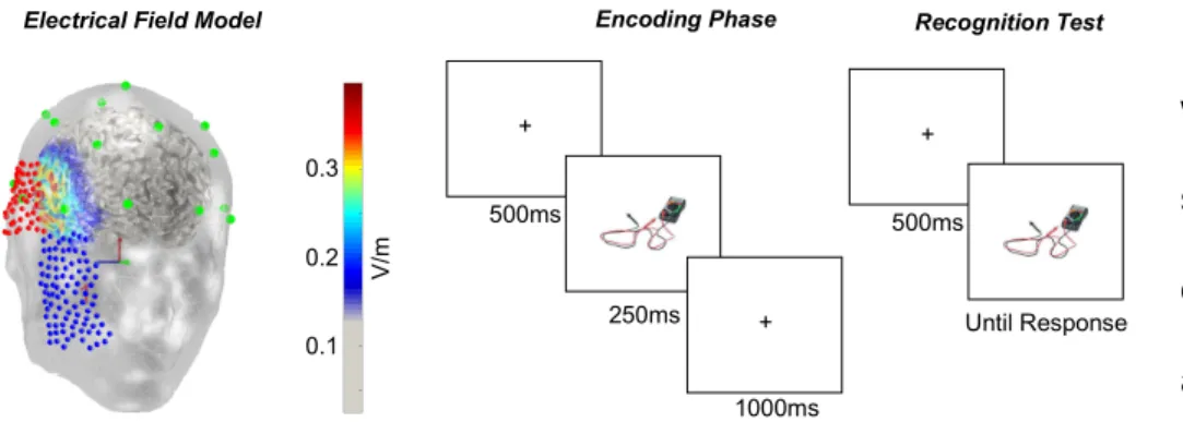
Fig. 1. The current distribution of the stimulation and examples of encoding and test phase trials. (Left) The current flow model illustrating right anodal stimulation. The anodal electrode was at T4 in this model and the cathodal electrode on the cheek. (Middle) An encoding trial starts with a fixation cross for 500 ms, followed by a 250-ms stimulus presentation and a 1000-ms post-stimulus break. This was repeated for all 500 stimuli. (Right) For the recognition test, we presented each picture on the screen until the participant pressed a keyboard button to indicate whether the picture was a new or an old stimulus and their confidence rating.
V/m
study phase (Fukuda & Woodman, 2015). The stimuli were adapted from a published set of photographs (Brady et al., 2008). Subjects were instructed to study each item while holding central fixation so that they could recognize them later. After a 500-ms pre-encoding period, in which the screen was blank except for a central fixation dot, each picture was presented for 250ms. Each picture was followed by a 1000-ms
encoding period, during which the computer screen remained blank. After the encoding task, the participants were presented with a 3-minute break between the encoding phase and the recognition test.
During the test phase subjects were shown 750 pictures of real-world objects (i.e., the 500 pictures shown during the encoding phase and 250 new pictures new), with the order randomized. During the test phase, each trial started with the onset of a central fixation dot for 500 ms. Participants were instructed to maintain central fixation until each trial was over. Following the 500-ms fixation period, a picture of a real-world object was presented at the center of the screen. The object was presented until the subject made a key press to indicate whether they had seen that object during the study phase, as well as their confidence. The number keys 1, 2, and 3 indicated that the item was old items, and the larger the number, the lower confidence level the subjects had for that response. The buttons 7, 8, and 9 indicated the item was new, and the larger the number, the higher confidence level the participants had for their response.
Design. The participant would finish both of the sessions at the other day under a different stimulation condition. The pictures used in the second session were
programmed such that no repeating pictures were reused in the second session of the same participant. Both of the sessions were designed to happen at the same time of a
day to minimize the interference of physiological conditions. Meanwhile, the time interval between two sessions for a single participant was designed to be at least forty- eight hours to avoid the remaining influence of stimulation on cortical activity.
The dependent variable in the experiment was the d prime sensitivity index, and the independent variable was the stimulation condition. The d prime sensitivity index was an interval variable calculated by 𝑍 − 𝑍 . The stimulation condition was a two-by-two nominal variable using mixed design. The first level was the condition of stimulation, and it was a within-subject variable containing anodal stimulation and sham stimulation. The second level was a between-subject variable coding for the side of stimulation the participant would get. A mixed-model ANOVA was performed on the d’
sensitivity index. The effect on d’ index was tested on the within-subject sham or anodal stimulation variable and the between-subject right or left hemispheric stimulation
variable.
Experiment 1b
Participants. Using a desired statistical power of 0.8, we estimated that we needed to collect data from thirty-two participants in each experiment. Thirty-two Vanderbilt University undergraduate students participated in Experiment 1b. In exchange for their participation, subjects received either $15 per hour or partial course credit for an introductory psychology course. All participants self-reported having normal or corrected-to-normal vision, no colorblindness, and no history of neuropsychiatric disorders. Two subjects’ data was excluded from Experiment 1b because they did not return for the second session.
Stimulation. The stimulation condition is the same as the model used in Experiment 1a.
In Experiment 1b, three subjects correctly identified both the sham and active sessions.
Across both experiments, the majority of subjects guessed that both sessions were sham.
Design. The dependent variable in the experiment was response time in the visual search task measured in second. The stimulation condition is a within-subject variable in that every participant received both sham and active stimulation in two separate sessions. Each participant would get either right or left side anodal stimulation in one of the sessions, and a sham stimulation session in the other session. The side of
stimulation and the order for sham and anodal stimulation within a subject were randomized. In each session, the participant would perform visual search under three different array sizes (2,4, or 8). This within-subject variable determines the difficulty of the visual search task and is a discrete variable.
Task. The visual search task is a modified version of the Landolt C task (Woodman, Vogel & Luck, 2001). Instead of searching for the direction of opening for Landolt C,
participants were always asked for the direction letter T is pointing to (see Fig. 2). The target T pointed to one of the canonical up, down, left or right directions. In the easy condition, one distractor stimulus L would appear on the screen with the target. The medium difficulty condition consisted of three L distractors, and the hard condition consisted of seven L distractors. The position of the target and the distractors were all randomized in each participant. In each trial, the fixation cross would first appear on the screen for 500 ms, followed by a stimuli pattern of either easy, medium or hard difficulty condition. The stimuli pattern would last until the participant pressed one of the four direction buttons on keyboard to indicate the direction letter T pointed to in each trial.
The long exposure time of the stimuli pattern is to ensure that participant acquire high accuracy in the visual search task so that response time could be our primary variable of interest in the present study. In each session, the participant would first have the
sham or active stimulation for twenty minutes, and complete nine-hundred trials of visual search task.
Experiment 2
Participants. Using a desired statistical power of 0.8, we estimated that we needed to collect data from thirty-two participants in each experiment. Thirty-two Vanderbilt University undergraduate students participated in Experiment 2. In exchange for their participation, subjects received either $15 per hour or partial course credit for an introductory psychology course. All participants self-reported having normal or corrected-to-normal vision, no colorblindness, and no history of neuropsychiatric disorders. All subjects completed both sessions for Experiment 2.
Stimulation. The stimulation condition is the same as the model used in Experiment 1a and 1b other than the direction of stimulation was reversed to cathodal stimulation. In Experiment 2, no subject correctly identified both the sham and active sessions. Across both experiments, the majority of subjects guessed that both sessions were sham.
Task. The task is the same recognition memory experimental paradigm as Experiment 1a.
Design. The design is the same as Experiment 1a.
Experiment 3
Participants. Twenty-six Vanderbilt University undergraduate students participated in the experiment. The participants received $15 per hour in exchange for their
participation. All subjects self-reported having normal or corrected-to-normal vision and no history of neuropsychiatric disorders. Two subjects’ data were excluded from
Experiment 3 because their EEG data had too many eye movement and other artifacts after our initial stages of data analysis (i.e., more than 20% of trials rejected due to artifacts).
Stimulation. The stimulation condition is the same as the model used in Experiment 1a.
In Experiment 3, one subject correctly identified both the sham and active sessions.
Across both experiments, the majority of subjects guessed that both sessions were sham or both active.
Task. The task is the same recognition memory experimental paradigm as Experiment 1a.
EEG acquisition and analysis. Subjects’ EEG data were recorded during both active and sham sessions, from the start of the encoding phase to the end of the recognition
memory test. The right mastoid was used as the reference site online and all signals were re-referenced offline to the average of the left and right mastoids. We used the international 10-20 electrode sites (Fz, Cz, Pz, F3, F4, C3, C4, P3, P4, PO3, PO4, O1, O2, T3, T4, T5, and T6) and a pair of custom sites, OL (halfway between O1 and OL) and OR (halfway between O2 and OR). Eye movements were detected using
electrodes placed 1-cm lateral to the external canthi for horizontal movement and an electrode placed beneath the right eye for blinks and vertical eye movements. All signals were amplified with a gain of 20,000, band-pass filtered from 0.01 to 100 Hz, and sampled at a frequency of 250 Hz. Trials accompanied by horizontal eye
movements (> 30 µV, mean threshold across observers) or eye blinks (> 100 µV, mean
threshold across observers) were rejected before further analyses. Two subjects who had more than 20 percent of trials rejected in either sham or active stimulation session and were eliminated from further data analysis.
To perform the time-domain analysis on the encoding data, we first time-locked the epochs to the onset of the stimulus in each trial. Each epoch started with 200 ms before the stimulus onset to 1250 ms after the stimulus onset. In the group-level time-domain analysis, the mean of the baseline activities (-200ms to 0ms) across all subjects was subtracted to correct for intrinsic fluctuations in individual data. The trials were further divided into different response conditions (high confidence hit, low confidence hit, and miss) and different stimulation conditions (active and sham). Each curve on the plot corresponded to the mean of time-domain epoch following a specific response and one of the stimulation conditions.
To perform the frequency-domain analysis on the data, we took the same epochs in the time-domain analysis and ran 400-ms sliding window over the epochs (with 380-ms overlapping window length). The band power in the frequency band of our interest was calculated and plotted as smooth curves. The trials were further divided into different response conditions (high confidence hit, low confidence hit, and miss) and different stimulation conditions (active and sham). Each curve on the plot corresponded to the mean band power within the epoch time length under a specific response condition and one of the stimulation conditions. To verify that our alpha band analyses were capturing the relevant memory effects in time-frequency space during encoding, we calculated power across time and frequencies for anodal relative to sham stimulation. These plots show that the difference between these trial conditions were confined to the alpha band.
Furthermore, we also plotted the scalp distribution of alpha-band suppression activities over all electrodes. The difference between anodal and sham conditions were plotted to examine the sites of significance manifesting in alpha suppression during anodal
condition compared to sham condition (see details in Fig. 8).
Design. The design is the same as Experiment 1a.
Experiment 4
Participants. Using a desired statistical power of 0.8, we estimated that we needed to collect data from thirty-two participants in each experiment. Thirty-two Vanderbilt University undergraduate students participated in Experiment 4. In exchange for their participation, subjects received either $15 per hour or partial course credit for an introductory psychology course. All participants self-reported having normal or corrected-to-normal vision, no colorblindness, and no history of neuropsychiatric
disorders. Three subjects’ data was excluded from Experiment 1b because they did not return for the second session.
Stimulation. The stimulation condition is the same as the model used in Experiment 1a.
In Experiment 4, four subjects correctly identified both the sham and active sessions.
Across both experiments, the majority of subjects guessed that both sessions were sham or both active.
Task. The task is the same recognition memory experimental paradigm as Experiment 1a other than that the anodal stimulation was performed after the encoding phase instead of before the encoding phase (for comparison across experiments, see Fig. 3).
Design. The design is the same as Experiment 1a.
Results
Experiment 1a
In Experiment 1a, anodal stimulation delivered to the temporal pole improved recognition
performance, regardless of which hemisphere was stimulated.
Specifically, we observed higher d’
sensitivity compared to the sham stimulation baseline (F(31)= 16.504, p<0.001, see Fig.
4). Anodal stimulation in Experiment 1a didn’t show a significant hemispheric preference (i.e., an interaction of stimulation condition x hemisphere, F(31)= 0.097, p=0.752).
We knew that visual attention would affect the quality of various types of visual memory, including categorized visual short-term memory (Olsson & Poom, 2005), visual scene memory (Hollingworth & Henderson, 2002) and recognition memory (Gardiner & Parkin, 1990). If the anodal stimulation improves the visual attention of participants, the
improvement of recognition memory could result from a downstream effect from increased attention. To test if tDCS could influence visual attentional state, we performed Experiment 1b using visual search task after the anodal tDCS to see if participants were better at visual attention paradigms as well.
Experiment 1b
In experiment 1b, the stimulation x difficulty ANOVA didn't reveal any significance on stimulation(F(31)=2.218, p=0.147), and no interaction as well(F(31)=0.660, p=0.524, see Fig. 5). In response time paradigm, though the response time may not be different across stimulation condition, the slope between set size and response time increase could be different (Yantis & Jonides, 1984). Therefore, we performed a paired t-test on the set size slope function, and the slope didn't change significantly(t(31)=0.5507, p=0.5858), nor did the intercept change reaches statistical significance(t(31)=1.6846, p=0.1021). Thus, we concluded that the anodal effect in improving recognition memory was not due to a better visual attentional state.
Experiment 2
To further examine the causal role of temporal lobe in coding recognition memories, we reversed the direction of current to cathodal, which supposed to inhibits local neuronal activities (Nitsche et al.,
2003). In Experiment 2
cathodal stimulation delivered to
the temporal pole deteriorated recognition performance (F(31)= 6.162, p=0.019, see Fig. 6). Furthermore, cathodal
stimulation in Experiment 2 didn’t show a significant hemispheric preference (i.e., an interaction of stimulation condition x hemisphere, F(31)= 1.034, p=0.317). To further investigate whether the changes in recognition memory was due to the encoding or retrieval-related activities, we collected the EEG data while participants were performing tasks in Experiment 3 to see if any of the encoding or the retrieval neural signature got changed under anodal stimulation.
Experiment 3
Behavioral Results. We found that anodal stimulation improved subjects’ visual recognition memory performance relative to when the same subjects received sham stimulation. Similar to our previous study of the effects of the temporal pole using tDCS, we did not find that which hemisphere was stimulated had an effect (i.e., an interaction of stimulation condition x hemisphere, F(23)= 0.209, p=0.652), so we collapsed across which hemisphere was stimulated. Thus, the paired t-test with the factor of stimulation
condition (sham versus anodal stimulation) showed that our anodal stimulation
delivered to the temporal pole improved the recognition memory performance (t(23)= - 2.988, p=0.007).
Encoding Phase ERP Results. The amplitude and scalp distribution of the frontal positivity following anodal stimulation and sham are shown, respectively, in Fig.
7 and Fig. 8. Recall that prior research has shown that when a stimulus elicits a more positive frontal potential that stimulus is remembered better than one that elicits a less positive potential (Fukuda & Woodman, 2015; Paller et al., 1988). Our findings replicate that basic effect, in that our subjects’ frontal positivities were larger amplitude for high confidence hits, smaller for low confidence hits, and smaller still for misses. However, we found that stimulation had a negligible influence on the amplitude of these potentials, indeed the waveforms were slightly more positive in the sham session than anodal, as confirmed with our analyses that we discuss next.
We calculated the mean voltage at electrode Fz from 400ms-1000ms following the to- be-remembered stimulus onset.
Next, we performed a
repeated ANOVA with the factors of subsequent response type (high confidence hit, low confidence hit or miss) and stimulation condition (sham or anodal stimulation). This yielded a significant effect of response type (F(2,24)=5.496, p=0.007) due to more positive potentials for high confidence hits than low confidence hits or misses, see Fig.
7. However, we did not observe a significant effect of stimulation (F(1,24)=1.028, p=0.321), or an interaction of response type X stimulation (F(2,24)=0.958, p=0.391).
Encoding Phase EEG Results. We next measured alpha band activity to determine whether stimulation changed these memory-related oscillations in the brain. The alpha- band activity measured during encoding is shown in Fig. 9 and Fig. 10. Recall that previous work showed that alpha power was reduced more following the onset of a stimulus that subjects would later report remembering (i.e., Hanslmayr, et al., 2009). We first observed that that we replicated this pattern in our data. However, most critically for the present study, we
found that stimulation had a strong effect on alpha in that is decreased alpha power across all to- be-remembered stimulation presentations
following anodal stimulation. We quantified this alpha activity by calculating alpha power within a 400-ms sliding window, with 380ms of overlap between windows, from 400ms- 1000ms following stimulus onset. These power values were entered into an ANOVA with the factors of subsequent response type (high confidence hit, low confidence hit, versus miss) and stimulation condition (anodal versus sham). This yielded a significant main effect of response type (F(2,24)=6.374, p=0.007), and a significant main effect of stimulation condition (F(1,24)=6.383, p=0.019). However, the interaction of these terms was not significant (F (2,24)=0.305, p=0.740).
To further investigate how alpha activities changed by anodal stimulation during the encoding phase, we also visualized the scalp distribution and time-frequency plot
difference between anodal and sham stimulation (see Fig. 10). By using paired t-test on every single electrode before and after stimulation, we discovered that other than PO4, the C3, C4, P3, P4, Fz, Cz electrodes also exhibit alpha band power suppression.
Moreover, the time-frequency plot on PO4 revealed that the suppression of band power mainly manifested within the alpha-band (8-12 Hz) range. These evidences together confirmed the potential role of parietal-occipital alpha suppression on recognition memory encoding.
Retrieval Phase EEG and ERP Results. The value of frontal positivity was calculated by the average activity level at Fz electrode site 400ms-1200ms following the stimulus onset.
In the frequency- domain, the alpha activity is
quantified by the 400-ms sliding window with 380ms overlapping window, and the points were calculated over 400ms-1000ms following stimulus onset. To examine the effect of anodal tDCS on ERP activities, we performed a repeated ANOVA on how response condition (high confidence hit, low confidence hit or miss) and stimulation condition (sham or anodal stimulation) could affect the frontal positivity and occipital alpha-band activities. In the time-domain frontal positivity analysis, we successfully replicated the prior work in that the higher confidence hit condition exhibited higher frontal positivity than the items later forgotton (F(2,24)=6.070, p=0.008, see Fig. 11). However, the Fz
site frontal positivity didn’t change with anodal stimulation (F(2,24)=0.091, p=0.766, see Fig. 11). In the frequency-domain occipital alpha-band power analysis, we successfully replicated the prior work in that the higher confidence hit condition exhibited more alpha-band suppression than the items later forgotton (F(2,24)=3.642, p=0.043, see Fig. 12). However, the PO4 site alpha-band power in retrieval phase didn’t change with anodal stimulation (F(1,24)=0.507, p=0.484, see Fig. 12).
Discussion. During the encoding phase, we saw that the anodal stimulation promoted the parietal-occipital alpha suppression and left the time-domain frontal positivity intact.
However, the anodal stimulation didn’t change either of the neural signatures during the retrieval phase. Given that these retrieval neural signatures still seemed to code for better memory states, one possibility was that the anodal stimulation only selectively affect the encoding state of the participants. Though previous research suggested that tDCS could last for more than 24 hours (Reinhart et al., 2017), a potential alternative hypothesis is that the stimulation before encoding phase didn’t last long enough till the retrieval state. To further test this hypothesis, we shifted the anodal stimulation to after the encoding phase of the experiment and saw if people could still benefit from the stimulation right before retrieval phase.
Experiment 4
In Experiment 4, anodal stimulation delivered to the temporal pole after the encoding phase didn’t improve recognition
performance. The d’ sensitivity in the anodal stimulation was not significantly different compared to the sham stimulation baseline (F(31)= 0.560, p=0.460, see Fig.
13). Meanwhile, anodal
stimulation in Experiment 4 didn’t
show a significant hemispheric preference (i.e., an interaction of stimulation condition x hemisphere, F(31)= 0.877, p=0.356).
Discussion
In our study, we first showed that anodal tDCS over temporal lobe area before the encoding phase could significantly improve the participant’s recognition memory performance. Additionally, this promotion is not a downstream effect from an enhanced visual attentional state. Further studies using scalp electrophysilgy reveals that the better recognition performance may result from a better encoding quality of the pictures, marked by a deeper parietal-occipital alpha supression during the anodal stimulation than the sham stimulation. On the contrary, the neural signatures of retrieval didn’t change after the stimulation. To further investigate whether the retrieval phase is beneifited from the anodal stimulation, our last experiment shows that stimulation after the encoding phase does not promote later recognition memory performance.
Dissociation of alpha suppression and frontal positivity. Here we showed that the improvement in human visual memory following anodal direct-brain stimulation of the temporal pole was due to deeper levels of alpha suppression over posterior cortex.
Most critically, the change in alpha activity was not accompanied by a change in the amplitude of the frontal positivity. These are theoretically important observations
because both of these encoding-related neural signals had been previously proposed to measure the quality of encoding a visual stimulus into long-term memory (Fukuda &
Woodman, 2015). Thus, the parsimonious conclusion would be that the alpha power and frontal positivity simply measure the same mechanism that encodes a visual representation into long-term memory. However, our findings provide causal evidence that these two electrophysiological signatures index independent mechanisms.
What are the mechanisms measured by posterior alpha power suppression and the amplitude of the frontal positivity when people are shown to-be-remembered
objects? One possibility is that the oscillatory neural activity in the alpha band measures for the level of coordination across brain areas, while the amplitude of the frontal ERPs directly reflects the activation level of the cortical structures that generate the potential (Hanslmayr & Staudigl, 2014). While this is a possible explanation, our findings would require such an explanation to propose the counterintuitive explanation that stimulation induced greater coordination or communication between brain areas, without changing the activation levels in the relevant areas. It is difficult to image how increasing the communication between regions of the brain could not result in each region having more information. However, additional tests of this idea need to be performed. If two
people talk on the telephone more frequently, we would assume that each who know more information about the other, but this assume each is listening.
An alternative theoretically impactful interpretation of the present findings is that they could suggest that posterior alpha band activity and the frontal positivity measure completely different cognitive mechanisms. One possibility is that perhaps the posterior alpha suppression measures the encoding of visual memory of the human subject immediately following the presentation of the stimulus, whereas the amplitude of the frontal positivity measures the integration of that stimulus into existing semantic memory structures of the human brain. Previous research proposed that the occipital-parietal alpha suppression could causally control the quality of perceptual input (Romei, Gross,
& Thut, 2010) and the encoding of visual short-term memory (Hsu et al., 2014).
Therefore, alpha suppression could be a potential biomarker for visual memory with no semantic component. Moreover, this visual memory signature has little relationship to the visual attention, since we demonstrated in Experiment 1b that visual search performance didn’t get improved under anodal stimulation. Different from the visually induced alpha suppression, frontal positivity has long been recognized as the FN400 component that coded for semantic memory (Paller, Voss, & Boehm, 2007; Rhodes &
Donaldson, 2008; Voss & Federmeier, 2011). In our recognition memory paradigm, an enhanced semantic memory that promotes the recognition of the name of the objects will hardly benefit people more given the short presence time of the encoding items.
Therefore, we could replicate the frontal positivity result for high confidence hit than items that later forgotton, but a higher encoding quality of the semantic information may not be the reason for a better recognition encoding performance.
Direct-brain stimulation and its practical use. The increase of parietal-occipital alpha suppression was observed after anodal stimulation in our study. This finding confirmed the recent empirical patient studies in that brain stimulation on hippocampal areas could modify the state of brain when encoding memories (Ezzyat et al., 2017; Ezzyat et al, 2018). Growing evidence reveals that anodal direct current stimulation could boost local neural activations and thus promote cognitive abilities (Nitsche et al., 2003; Reinhart &
Woodman, 2014; Reinhart et al., 2016). Our study proves that the anodal stimulation, even in healthy participants with intact memory functions, could activate the temporal lobe and improve recognition memory. In Alzheimer’s disease and epilepsy patients, functional imaging studies have shown that the memory loss resulted from decreased temporal lobe activations during memory tasks (Graham & Hodges,1997; Dupont et al., 2000). Therefore, the transcranial direct current stimulation could possibly be applied to the reduction of dementia symptoms in neural disorders in the future.
References
Bachtiar, V., Near, J., Johansen-Berg, H., & Stagg, C. J. (2015). Modulation of GABA and resting state functional connectivity by transcranial direct current stimulation.
Elife, 4, e08789.
Brady, T. F., Konkle, T., Alvarez, G. A., & Oliva, A. (2008). Visual long-term memory has a massive storage capacity for object details. Proceedings of the National Academy of Sciences, 105(38), 14325-14329.
Brewer, J. B., Zhao, Z., Desmond, J. E., Glover, G. H., & Gabrieli, J. D. (1998). Making memories: brain activity that predicts how well visual experience will be
remembered. Science, 281(5380), 1185-1187.
Cabeza, R., & Nyberg, L. (2000). Neural bases of learning and memory: functional neuroimaging evidence. Current opinion in neurology, 13(4), 415-421.
Chan, D., Fox, N. C., Scahill, R. I., Crum, W. R., Whitwell, J. L., Leschziner, G., ... &
Rossor, M. N. (2001). Patterns of temporal lobe atrophy in semantic dementia and Alzheimer's disease. Annals of neurology, 49(4), 433-442.
Davachi, L., & Wagner, A. D. (2002). Hippocampal contributions to episodic encoding:
insights from relational and item-based learning. Journal of neurophysiology, 88(2), 982-990.
Detre, J. A., Maccotta, L., King, D., Alsop, D. C., Glosser, G., D'Esposito, M., ... &
French, J. A. (1998). Functional MRI lateralization of memory in temporal lobe epilepsy. Neurology, 50(4), 926-932.
Dupont, S., Van de Moortele, P. F., Samson, S., Hasboun, D., Poline, J. B., Adam, C., ... & Baulac, M. (2000). Episodic memory in left temporal lobe epilepsy: a functional MRI study. Brain, 123(8), 1722-1732.
Düzel, E., Yonelinas, A. P., Mangun, G. R., Heinze, H. J., & Tulving, E. (1997). Event- related brain potential correlates of two states of conscious awareness in
memory. Proceedings of the National Academy of Sciences, 94(11), 5973-5978.
Ebersole, J. S., & Pacia, S. V. (1996). Localization of temporal lobe foci by ictal EEG patterns. Epilepsia, 37(4), 386-399.
Ezzyat, Y., Kragel, J. E., Burke, J. F., Levy, D. F., Lyalenko, A., Wanda, P., ... &
Sperling, M. R. (2017). Direct brain stimulation modulates encoding states and memory performance in humans. Current biology, 27(9), 1251-1258.
Ezzyat, Y., Wanda, P. A., Levy, D. F., Kadel, A., Aka, A., Pedisich, I., ... & Gross, R. E.
(2018). Closed-loop stimulation of temporal cortex rescues functional networks and improves memory. Nature communications, 9(1), 365.
Fukuda, K., & Woodman, G. F. (2015). Predicting and improving recognition memory using multiple electrophysiological signals in real time. Psychological science, 26(7), 1026-1037.
Gardiner, J. M., & Parkin, A. J. (1990). Attention and recollective experience in recognition memory. Memory & Cognition, 18(6), 579-583.
Graham, K. S., & Hodges, J. R. (1997). Differentiating the roles of the hippocampus complex and the neocortex in long-term memory storage: Evidence from the
study of semantic dementia and Alzheimer's disease. Neuropsychology, 11(1), 77.
Haatveit, B. C., Sundet, K., Hugdahl, K., Ueland, T., Melle, I., & Andreassen, O. A.
(2010). The validity of d prime as a working memory index: results from the
“Bergen n-back task”. Journal of clinical and experimental neuropsychology, 32(8), 871-880.
Hanslmayr, S., & Staudigl, T. (2014). How brain oscillations form memories—a processing based perspective on oscillatory subsequent memory effects.
Neuroimage, 85, 648-655.
Hollingworth, A., & Henderson, J. M. (2002). Accurate visual memory for previously attended objects in natural scenes. Journal of Experimental Psychology: Human Perception and Performance, 28(1), 113.
Horel, J. A., & Misantone, L. J. (1976). Visual discrimination impaired by cutting temporal lobe connections. Science, 193(4250), 336-338.
Hsu, T. Y., Tseng, P., Liang, W. K., Cheng, S. K., & Juan, C. H. (2014). Transcranial direct current stimulation over right posterior parietal cortex changes prestimulus alpha oscillation in visual short-term memory task. Neuroimage, 98, 306-313.
Jasper, H. H. (1958). The ten-twenty electrode system of the International Federation.
Electroenceph. clin. Neurophysiol., 10, 371-375.
Jung, Y. J., Kim, J. H., & Im, C. H. (2013). COMETS: A MATLAB toolbox for simulating local electric fields generated by transcranial direct current stimulation (tDCS).
Biomedical engineering letters, 3(1), 39-46.
Macmillan, N. A., & Creelman, C. D. (1990). Response bias: Characteristics of detection theory, threshold theory, and" nonparametric" indexes. Psychological Bulletin, 107(3), 401.
Milner, B. (1968). Visual recognition and recall after right temporal-lobe excision in man.
Neuropsychologia, 6(3), 191-209.
Nevin, J. A. (1969). Signal detection theory and operant behavior: A review of David M.
Green and John A. Swets' Signal detection theory and psychophysics1. Journal of the Experimental Analysis of Behavior, 12(3), 475.
Nitsche, M. A., Cohen, L. G., Wassermann, E. M., Priori, A., Lang, N., Antal, A., ... &
Pascual-Leone, A. (2008). Transcranial direct current stimulation: state of the art 2008. Brain stimulation, 1(3), 206-223.
Nitsche, M. A., Fricke, K., Henschke, U., Schlitterlau, A., Liebetanz, D., Lang, N., ... &
Paulus, W. (2003). Pharmacological modulation of cortical excitability shifts induced by transcranial direct current stimulation in humans. The Journal of physiology, 553(1), 293-301.
Olsson, H., & Poom, L. (2005). Visual memory needs categories. Proceedings of the National Academy of Sciences, 102(24), 8776-8780.
Pacia, S. V., & Ebersole, J. S. (1997). Intracranial EEG substrates of scalp ictal patterns from temporal lobe foci. Epilepsia, 38(6), 642-654.
Paller, K. A., McCarthy, G., & Wood, C. C. (1988). ERPs predictive of subsequent recall and recognition performance. Biological psychology, 26(1-3), 269-276.
Paller, K. A., Voss, J. L., & Boehm, S. G. (2007). Validating neural correlates of familiarity. Trends in cognitive sciences, 11(6), 243-250.
Perry, R. J., & Hodges, J. R. (2000). Differentiating frontal and temporal variant frontotemporal dementia from Alzheimer’s disease. Neurology, 54(12), 2277- 2284.
Potter, M. C., & Levy, E. I. (1969). Recognition memory for a rapid sequence of pictures. Journal of experimental psychology, 81(1), 10.
Reed, A. V. (1973). Speed-accuracy trade-off in recognition memory. Science, 181(4099), 574-576.
Reinhart, R. M., & Woodman, G. F. (2014). Causal control of medial–frontal cortex governs electrophysiological and behavioral indices of performance monitoring and learning. Journal of Neuroscience, 34(12), 4214-4227.
Reinhart, R. M., Cosman, J. D., Fukuda, K., & Woodman, G. F. (2017). Using
transcranial direct-current stimulation (tDCS) to understand cognitive processing.
Attention, Perception, & Psychophysics, 79(1), 3-23.
Reinhart, R. M., Xiao, W., McClenahan, L. J., & Woodman, G. F. (2016). Electrical stimulation of visual cortex can immediately improve spatial vision. Current Biology, 26(14), 1867-1872.
Rhodes, S. M., & Donaldson, D. I. (2008). Electrophysiological evidence for the effect of interactive imagery on episodic memory: Encouraging familiarity for non-unitized stimuli during associative recognition. NeuroImage, 39(2), 873-884.
Romei, V., Gross, J., & Thut, G. (2010). On the role of prestimulus alpha rhythms over occipito-parietal areas in visual input regulation: correlation or causation?.
Journal of Neuroscience, 30(25), 8692-8697.
Rugg, M. D., & Curran, T. (2007). Event-related potentials and recognition memory.
Trends in cognitive sciences, 11(6), 251-257.
Rugg, M. D., Mark, R. E., Walla, P., Schloerscheidt, A. M., Birch, C. S., & Allan, K.
(1998). Dissociation of the neural correlates of implicit and explicit memory.
Nature, 392(6676), 595.
Scoville, W. B., & Milner, B. (1957). Loss of recent memory after bilateral hippocampal lesions. Journal of neurology, neurosurgery, and psychiatry, 20(1), 11.
Shepard, R. N. (1967). Recognition memory for words, sentences, and pictures. Journal of verbal Learning and verbal Behavior, 6(1), 156-163.
Spencer, S. S., & Spencer, D. D. (1994). Entorhinal‐hippocampal interactions in medial temporal lobe epilepsy. Epilepsia, 35(4), 721-727.
Squire, L. R., & Zola-Morgan, S. (1988). Memory: brain systems and behavior. Trends in neurosciences, 11(4), 170-175.
Squire, L. R., & Zola-Morgan, S. (1991). The medial temporal lobe memory system.
Science, 253(5026), 1380-1386.
Swets, J. A. (1961). Is there a sensory threshold?. Science, 134(3473), 168-177.
Vogel, E. K., Woodman, G. F., & Luck, S. J. (2001). Storage of features, conjunctions, and objects in visual working memory. Journal of Experimental Psychology:
Human Perception and Performance, 27(1), 92.
Voss, J. L., & Federmeier, K. D. (2011). FN400 potentials are functionally identical to N400 potentials and reflect semantic processing during recognition testing.
Psychophysiology, 48(4), 532-546.
Yantis, S., & Jonides, J. (1984). Abrupt visual onsets and selective attention: evidence from visual search. Journal of Experimental Psychology: Human perception and performance, 10(5), 601.