A worldwide review of hermit crab species of the genus Sympagurus Smith, 1883
(Crustacea: Decapoda: Parapaguridae)
Rafael LEMAITRE
Department of Systematic Biology, National Museum of Natural History, P.O. Box 37012, Smithsonian Institution, Washington, D. C. 200713-7012, U. S. A.
ABSTRACT
A review of species of the genus Sympagurus Smith, 1883 (sensu Lemaitre) from the world oceans is presented. The study is based on the rich collections obtained during French campaigns in the Pacific and Indian Oceans, and on additional material in various museums and research institutions throughout the world. The 17 species recognised in this genus occur most frequently between 500 and 1000 m depth, and range from 80 to 2537 m. Some live in striking symbiosis with anthozoan or zoanthid coelenterates that can produce pseudo-shells. Three new species, S. aurantium, S. chani and S. symmetticus, are fully described and illustrated here. Sympagurus rectichela (Zarenkov 1990), a taxon originally described in Parapagurus Smith, 1879, has been found to be a junior synonym of S. dofleini (Balss, 1912); and S. papposus Lemaitre, 1996 is a junior synonym of S. hurkenroadi Thompson, 1943. All previously known Sympagurus species are diagnosed or redescribed and illustrated, and data on habitat, symbiotic associations, and coloration are provided. A key to aid in the identification of all Sympagurus species is presented, and their bathymetric and geographic distributions are summarised. The geographic distribution of 14 species (82.3%) includes the Pacific Ocean, 9 (52.9.%) the Indian Ocean, and 3 (1.8%) the Atlantic Ocean. New Caledonia and adjacent islands have the highest number of Sympaguras species in the world, with 12 species known to occur there.
RESUME
Revision mondiale des Bernards Termites du genre Sympagurus (Crustacea : Decapoda : Parapaguridae).
Une revision mondiale des especes du genre Sympagurus Smith, 1883 (sensu Lemaitre) est presentee. L'etude est basee sur les riches collections obtenues par les campagnes frangaises dans les oceans Pacifique et Indien et sur du materiel supplementaire provenant de differents musees et organismes de recherches. Les 17 especes reconnues dans ce genre se trouvent principalement entre 500 et 1000 m de profondeur, avec des extremes de 80 a 2537 m. Quelques especes vivent en etroite symbiose avec des anthozoaires ou des zoanthaires capable de fabriquer une pseudo-coquille.Trois nouvelles especes sont decrites et illustrees ici : S. aurantium, S. chani, et S. symmetticus.
Sympagurus rectichela (Zarenkov, 1990), decrit tout d'abord dans le genre Parapagurus Smith, 1879, s'avere etre un synonyme plus recent de S. dofleini (Balss, 1912) ; S. pappossus Lemaitre, 1996 devient un synonyme plus recent de S. hurkenroadi Thompson, 1943. Toutes les especes connues de Sympagurus sont redefinies et redecrites avec des illustrations. Les caracteristiques concernant les habitats, les
LEMAITRE R. 2004. — A worldwide review of hermit crab species of the genus Sympagurus Smith, 1883 (Crustacea: Decapoda: Parapaguridae), in MARSHALL B. & RICHER DE FORGES B. (eds). Tropical Deep-Sea Benthos, volume 23. Memoires du Museum national d'Histoire naturelle 191 : 85-149.
Paris ISBN : 2-85653-557-7.
associations symbiotiques et les couleurs sont foumies. Une clef d'identification pour toutes les especes de Sympagui-us est dorniee ainsi que leur repartitions geographiques et bathymetriques. La distribution geographique montre que 14 especes (82,3%) sont dans le Pacilique, 9 especes (52,9%) dans I'ocean Indien et 3 especes (1,8%) dans I'ocean Atlantique. C'est la Nouvelle-Caledonie et les lies proches qui ont le plus grand nombre d'especes de Sympagunis (12).
INTRODUCTION
The genus Sympagums Smith, 1883 was recently redefined to include 13 species, and is one of ten genera currently classified in the family Parapaguridae (Lemaitre 1996). Species of the genus inhabit the lower continental shelf and upper slope regions of most world oceans where they are frequently found in depths between 500 and 1000 m, although they range from 80 to 2537 m. Individuals of some species reach the largest sizes recorded for parapagurids (shield length up to 26 mm), and some species live in striking symbiosis with pseudoshell-producing anthozoans or zoanthids (Fautin Dunn et al. 1981; Fautin Dunn & Liberman 1983; Fautin 1987). Although various Sympagums species have been discussed in the last decade or so, details on their morphology and taxonomy are scattered in faunal studies of parapagurids from the western Atlantic (Lemaitre 1989), southeastern Pacific (Zarenkov 1990; Zhadan 1997), Antarctic and Subantarctic waters (Lemaitre Sr McLaughlin 1992), French Polynesia (Lemaitre 1994), Australia (Lemaitre 1996), Indonesia (Lemaitre 1997), New Zealand (Lemaitre 2000), and the western Indian Ocean (Zhadan in press).
While studying the rich and remarkable parapagurid collections obtained by French campaigns conducted in the Pacific and Indian Oceans, all but three of the previously known species of Sympagums were found to be represented.
Furthermore, three new species were discovered. Given that a good number of specimens of several poorly defined or rarely seen taxa were now available in these collections, a worldwide review of Symipagums species was undertaken, augmented by material in major museums and other institutions from throughout the world. As result of this review, a total of 17 species of Sympagums are recognised. Sympagwus rectichela (Zarenkov, 1990), a taxon originally described in Pampagums Smith, 1879 and subsequently transferred to Sympagums by Zhadan (1997), was found to be a junior synonym of S. dofleini (Balss, 1912). Moreover, S. papposus Lemaitre, 1996 was found to be a junior synonym of S.
hurkenroadi Thompson, 1943. The type and only known specimen of S. spinimamis (Balss, 1911), was determined as the juvenile stage of a species for which adults are yet unknown. Diagnoses and illustrations are provided for all species, and complete synonymies given. Information is supplied on habitat and symbiotic associations, and color photographs included for five species. A key to aid in the identification of the species is provided.
HISTORY OF GENERIC CLASSIFICATION
The genus Sympagums has been subjected to a number of revisions and familial classifications since it was originally proposed by Smith (1883) for a single species, S. pictus Smith, 1883. Smith also noted that Sympagums was very similar to a genus he had described earlier, Pampagums Smith, 1879, except that the former had phyllobranchiae instead of trichobranchiae, larger eyes, and shorter ocular peduncles and antennules. Smith (1883, 1884, 1886) did not indicate a familial placement for Sympagums, although he (Smith 1882) did propose the family Parapaguridae for Pampagums on the basis of the trichobranchiae alone. Henderson (1888: 52) placed Sympagums in the Paguridae sensu Dana, 1852, which along with the Coenobitidae made up his "Branch" Laminibranchiata, or forms with phyllobranchiae. Bouvier (1891) later criticized the use of gill type as a character for systematic grouping, and indicated that it was not natural to separate Sympagums from Pampagums based on differences of gill type alone. Milne-Edwards &r Bouvier (1893) then abandoned Smith's (1882) and Henderson's (1888) familial arrangement based on gill structure and placed Sympagums and Pampagums in the family Paguridae. However, at the generic level the use of branchial structure to separate Sympagum.s from Pampagums continued to be used (Milne-Edwards & Bouvier 1893, 1894, 1897, 1899, 1900; Alcock 1901, 1905;
Bouvier 1891, 1896, 1922, 1940; Przibram 1905; Fowler 1912; Melin 1939; Thompson 1943; Gordan 1956). To Balss (1912), the separation of these two genera was unsustainable, and he formally synonymized Sym.pagui'us with Parapagurus.
This arrangement was accepted by most subsequent carcinologists (e.g., Terao 1913; Forest 1955; de Saint Laurent 1972) until Lemaitre (1989) revised Parapagurus, and reinstated Sympagurus. The latter genus was at that time broadly defined by Lemaitre to include a morphologically diverse assemblage. More recently, Lemaitre (1996) restricted and redefined Sympagurus based on branchial characteristics and other morphological features, and proposed two other genera, Oncopagurus Lemaitre, 1996 and Paragiopagums Lemaitre, 1996, for a number of species previously placed in Sympagurus.
BRANCHIAL STRUCTURE AND TERMINOLOGY
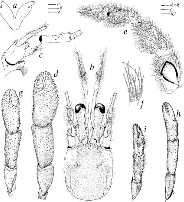
The parapagurid terminology used follows Lemaitre (1989, 1999), with some exceptions. The term quadriseiial, herein used for gill structure, was defined by McLaughlin &r de Saint Laurent (1998: 161, fig. 1) who pointed out that gills of parapagurids are not true trichobranchiae but rather a type of phyllobranchiae in which the lamellae inserted biserially on the rachis are divided. Although in Sympagurus species the division of the lamellae can vary from one end of the rachis to another, the maximum division of the lamellae (generally seen towards the midportion of the gill), can be of two kinds, herein described as distally divided (Figs Iflj, gi-!i), or deeply divided (Figs lbi-/i). In the former, each lamella is divided by a cleft that does not reach the midlength of the lamella, whereas in the latter the cleft reaches well beyond the midpoint of the lamella. The maximum division has been found to remain constant within each species.
Lemaitre's (1996) recent redefinition of Sympagurus was based primarily on a unique character among the Parapagu- ridae, i.e., the presence of a small subtriangular, flap-like structure on the wall of the last thoracic somite (Fig. 2). This structure is distally flexible, often delicate and transparent, and can be observed by gently raising the posterolateral portion of the posterior carapace just above the coxa of the fifth pereopod. This fiap-like structure, which lacks lamellae, was considered to be a rudimentary branchia by de Saint Laurent (1972), for which, the term vestigial pleurobranch proposed by Lemaitre (1989) is preferred here. Whether or not this vestigial structure actually serves a respiratory function is unknown.
Mouthpart morphology in Sympagurus species is very similar, except for the endopod of the maxillule. Depending on the species, the external lobe of the endopod of the maxillule varies from obsolete or weakly-developed (Figs laj, Cj, g^, ij) to moderately-developed (Figs Xh^, d^-j^, ^iJi'^- The internal lobe has at least one or more long distal setae, and the number of setae can vary intraspecifically. Although only a limited number of specimens have been studied for maxillule morphology, it appears that the degree of development of the external lobe is characteristic of each species.
The first and second ambulatory legs refer to the second and third pereopods respectively. The term semichelate is used as defined by McLaughlin (1997: 435).
MATERIALS AND METHODS
Treatment of the species and measurements follows the organisation and methods used by Lemaitre (1999) in his review of the genus Parapagurus Smith, 1879, although in the present review the species are discussed in the order that they appear in the key presented for their identification. The numbers or range in millimetres (mm) included in the "material examined" sections following the number and sex of specimens, are measurements for shield length (si), measured from the tip of the rostrum to the midpoint of the posterior margin of the shield.
The specimens from the French campaigns remain deposited in the Museum national d'Histoire naturelle, Paris (MNHN), with duplicates in the National Museum of Natural History, Smithsonian Institution, Washington D. C.
(USNM).
A significant number of specimens used came from other museums or institutions as follows.
AMS: The Australian Museum, Sydney;
BMNH: The Natural History Museum, London;
BPBM: Bernice P. Bishop Museum, Honolulu;
CBM-ZC: Natural History Museum and Institute, Chiba;
FRS: Fisheries Research Station, Hong Kong;
NTM: Northern Territories Museum, Darwin;
NTOU: National Taiwan Ocean University, Keelung;
QM: Queensland Museum, Brisbane;
SAM: South African Museum, Cape Town;
SAMA: South Australian Museum, Adelaide;
TAMU: Texas A &r M University, College Station, Galveston;
UMUTZ: University Museum, University of Tokyo, Zoology;
ZMA: Zoologisch Museum, Universiteit van Amsterdam;
ZMB: Museum ftir Naturkunde zu Berlin;
ZMK: Zoologisk Museum, Copenhagen;
ZMUM: Zoological Museum, Moscow State University;
ZSM: Zoologische Staatssammlung, Munich.
Figures 26 and 32 are digital images obtained with a Sony Mavica camera (model MVC-FD95). The images were processed in Photoshop©.
Station data for the French campaigns from which Sympagurus material was examined, can be found in the following publications or unpublished reports:
BATHUS 1-4: Richer de Forges & Chevillon (1996);
BFRYX 11: Lehodey et al. (1992);
BFNTHFDI: unpublished report (A. Crosnier, pers. comm.);
BIOCAL: Richer de Forges (1990);
BIOGEOCAL: Richer de Forges (1990);
BORDAU 2: unpublished report (A. Crosnier, pers. comm.);
CHALCAL 2: Richer de Forges (1990);
CORAIL 2: Richer de Forges (1991);
French Polynesia, Marara: Poupin et al. (1990), Poupin (1996a);
HALIPRO 1: Richer de Forges & Chevillon (1996);
Madagascar, NO Vauban: Crosnier (1978);
MUSORSTOM 4-6: Richer de Forges (1990);
MUSORSTOM 7: Richer de Forges <Sr Menou (1993);
MUSORSTOM 8: Richer de Forges et al. (1996);
SMIB 3, 4: Richer de Forges (1990);
SMIB 5: Richer de Forges (1993);
SMIB 8: Richer de Forges & Chevillon (1996);
SMIB 10: unpublished report (A. Crosnier, pers. comm.);
TAIWAN 2000: unpublished report (A. Crosnier, pers. comm.);
VOLSMAR: Laboute et al. (1989).
SPECIES LIST
(Listed alphabetically. Asterisk indicates species found in the New Caledonia region) Sympagurus acinops Lemaitre, 1989*
S. affinis (Henderson, 1888)*
S. andersoni (Henderson, 1896) S. aurantium n. sp.*
S. brevipes (de Saint Laurent, 1972)*
S. hurkenroadi Thompson, 1943 (= S. papposus Lemaitre, 1996)*
S. chani n. sp.
S. dimorphus (Studer, 1883)
S. dofleini (Balss, 1912) (= Parapagums rectkhela Zarenkov, 1990)*
S. pktus Smith, 1883
S. planimanus (de Saint Laurent, 1972)*
S. poupini Lemaitre, 1994 S. soela Lemaitre, 1996*
S. spinimanus (Balss, 1911) S. symmetncus n. sp.*
S. trispinosus (Balss, 1911)*
S. villosus Lemaitre, 1996*
SYSTEMATIC ACCOUNT
Family Parapaguridae Smith, 1882 Genus SYMPAGURUS Smith, 1883
Sympagurus Smitii, 1883: 37. Type species (monotypy): Sympagurus pktus Smitii, 1883 (gender masculine). — Henderson 1888: 52. — Bouvier 1891: 402; 1896: 127; 1922:21; 1940: 128.—Milne-Edwards SrBouvier 1893: 58; 1894:67; 1897: 131; 1899:55; 1900: 194.
— Alcock 1901: 223; 1905: 103. — Przibram 1905: 197. —Fowler 1912: 582. — Melm 1939: 20. —Thompson 1943: 418. — Gordan 1956: 341. — Lemaitre 1989: 36; 1996: 169; 2000: 210. — Ingle 1993: 19. — Zhadan m press.
Parapagurus - Balss 1912: 96 (in part). — de Saint Laurent 1972: 101 (in part).
DIAGNOSIS. — Twelve pairs of gills: 11 pairs of quadriserial (2 arthrobranchs on each third maxilliped, cheliped, and second through fourth pereopods, and 1 pleurobranch on seventh thoracic somite above fourth pereopod), with lamellae distally or deeply divided (Fig. 1); and 1 pair of vestigial pleurobranchiae on eighth thoracic somite above fifth pereopod (Fig. 2).
Shield about as broad as long, or broader than long; dorsal surface usually with irregularly-shaped, weakly calcified areas. Corneae weakly to moderately dilated.
Fourth segment of antennal peduncle unarmed, or with small dorsodistal spine.
Maxillule with external lobe of endopod (Figs la^-lj) obsolete or moderately developed, not recurved; internal lobe with 1 or more long setae distally. Epistomial spine short and straight, or absent.
Right chela with rounded dorsomesial and dorsolateral margins, or sometimes operculate with well delimited dorsomesial and dorsolateral margins.
Left cheliped usually well calcified.
Ambulatory legs with dactyls evenly curved.
Fourth pereopod with propodal rasp consisting of 1 or more rows of corneous scales or spines.
Second abdominal somite with left pleuron terminating ventrally in small subtriangular lobe.
Males with moderately to well developed, paired first and second pleopods modified as gonopods.
DISTRIBUTION. — Atlantic, Pacific and Indian Oceans, 80-2537 m (Fig. 34).
SPECIES INCLUDED. — (See "species list" above).
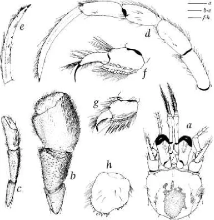
FIG. 1. Lamella (a,-/,) of posterior arthrobranch (midportion) of fourth pereopod, and distal end of endopod (52-/2) of l^ff maxillule (internal view) of Sympagurus species. a,_2. 5. poupini Lemaitre, 1994; b,_2, 5. acinops Lemaitre, 1989; c,_2, S. dimorphus (Studer, 1883); c(,_2. 5. soela Lemaitre, 1996; e,_2, 5. planimanus (de Saint Laurent, 1972); f,_2, S. aff/n/s (Henderson, 1888); g,_2, S. pictus Smith, 1883; /i,_2, S. brevipes{de Saint Laurent, 1972); /,_2, S. cfof/e/n/(Baiss, 1912);
y,_2. S. faurtenroacf/Thompson, 1943; k,_2, 5. villosus Lemaitre, 1996; /,_2, 5. trisp/nosus (BaIss, 1911). Scales = 0.5 mm (a,_2, c,, g^-l^.gj-h): ^nd 0.25 mm (fa,, fa2-f2. d^-i^).
FIG. 1. Lamelles (a,-I,) de I'arthrobranchie posteheure (partie centrale) du quatheme pereiopode et extremite distale de I'endopode (32-12) du maxillule gauche (vue interne) chezles especes de Sympagums. a,_2, S. poupini Lemaitre, 1994; b,_2, S. acinops Lemaitre, 1989; c,_2, S. dimorphus (Studer, 1883); d,_2, S. soela Lemaitre, 1996; e,_2, S. planimanus (de Saint Laurent, 1972); f,_2, S. affinis (Henderson, 1888); g,_2, S. pictus Sm/f/i, 1883; b,_2, S. brevipes (de Saint Laurent, 1972); i,_2, S. dofleini (BaIss, 1912); \,_2, S. burkenroadi Thompson, 1943; k,_2, S. villosus Lemaitre, 1996;
l,_2. 5. trispinosus (BaIss, 1911). Echelles = 0,5 mm (3,_2, c,, gj-lj, 92-^2), ^t 0,25 mm (b,, b2-f2, d,-f,).
FIG. 2. Left fifth pereopod and side wall of body with vestigial pleurobranch (arrow) of Sympagurus pictus Smith 1883, male, si 14.5 mm. Gulf of Mexico (USNM 265286). Scale = 2 mm.
FIG. 2. CInquleme pereiopode gauche et flanc du corps avec la pleurobranchie vestlgiale (fleche) de Sympagurus pictus Smith 1883, male, si 14,5 mm, Golfe de Mexico (USNM 265286). Echelle = 2 mm.
REMARKS. — The presence of a vesligial pleurobranch on ihe lasl ihoracic somile in Sympaguius species is unique in ihe Seclion Paguridea (cj. Forest 1987). Although the function of this vestigial structure has not yet been fully explored, it can be interpreted as the remnant of a former, fully functional pleurobranch. Fully developed pleuro- branchs on the last thoracic somite are known to occur in genera of other families of the Paguridea such as Coenobita Latreille, 1829 of the Coenobitidae; all genera of Pyloche- lidae; and at least Allodardanus Haig & Provenzano, 1965, Petwchirus Stimpson, 1858, Ciliopagums Forest, 1995, Strigopagufxis Forest, 1995, Tri^opagurus Forest, 1952, Ani- culus Dana, 1852, Cancellus H. Milne Edwards, 1836,
Dardanus Paul'son, 1875, and Tisea Morgan & Forest, 1991, of the Diogenidae. Thus, it would appear that the presence of a vestigial pleurobranch in Sympagums species represents a process of evolutionary reduction of this gill that is still under way.
KEY TO SPECIES OF SYMPAGURUS
1. Third to fifth pleopods paired, asymmetrical (left biramous, right consisting of small buds) juveniles (si < 5.0 mm) of Sympagurus species [juveniles have been documented for: S. spinimanus Balss, 1911 (this study. Fig. 3); S. pictus Smith, 1883 (see Lemaitre 1989); S. dimorphus (Studer, 1883) (see Lemaitre & McLaughlin 1992); S. brevipes (de Saint Laurent, 1972) (see Lemaitre 1996)].
- Third to fifth pleopods unpaired, present on left side 2 2. Uropods and telson symmetrical or nearly so 3 - Uropods and telson distinctly asymmetrical 4 3. Terminal margin of telson with corneous spines; shield distinctly longer than broad; antennal acicles reaching at most to mid-length of corneae S. symmetricus n. sp.
- Terminal margin of telson without spines; shield distinctly broader than long; antennal acicles exceeding distal margin of corneae S. poupini Lemaitre, 1994 4. Gills with lamellae deeply divided (Figs lbi-/i) 5 - Gills with lamellae at most distally divided (Figs laj^g-^^-l-i) 10 5. Corneae subconical, terminating bluntly or sharply (Figs 9a-c) S. acinops Lemaitre, 1989 - Corneae not subconical, rounded 6 6. Carpi of first and second ambulatory legs each with row of spines on dorsal margin; epistomial spine present S. dimorphus (Studer, 1883) - Carpi of first and second ambulatory legs each without spines on dorsal margin except for dorsodistal spine; epistomial spine absent 7 7. Palms of right and left chelae each armed dorsally with numerous spines. S. soda Lemaitre, 1996 - Palms of right and left chelae each unarmed dorsally or at most with scattered small spines or tubercles 8 8. Ocular acicles simple; dactylus of fourth pereopod terminating in long, slender claw considerably longer and slenderer in females (Fig. 13/) than in males (Fig. 13g). S. planimanus (de Saint Laurent, 1972)
- Ocular acicles bifid or multifid; dactylus of fourth pereopod terminating in short claw 9 9. Dactyls of first and second ambulatory legs each with 10 or more corneous spinules on ventromesial margin; right chela less than 2 x as long as broad S. afinis (Henderson, 1888) - Dactyls of first and second ambulatory legs each with less than 10, often minute corneous spinules on ventromesial margin; right chela 2-3 x as long as broad S. andersoni (Henderson, 1896) 10. Ventral surface of right chela armed with strong, corneous-tipped spines obscured by dense setae;
ventromesial margin of merus of right cheliped with dense fringe of long, bristle-like setae (usually yellow) S. aurantium n. sp.
- Ventral surface of right chela unarmed or at most with scattered small tubercles or spines, and moderately dense setae; ventromesial margin of merus of right cheliped without fringe of bristle-like setae 11 11. Shield distinctly broader than long 12 - Shield about as broad as long 14 12. Ventromesial margins of dactyls of first and second ambulatory legs each unarmed or at most with few microscopically small corneous spinules; dactylus of fourth pereopod straight, distinctly longer than propodal rasp, and with weak subterminal corneous claw (specimens si > 5.0 mm. Fig. 24g). . . S. pictus Smith, 1883 - Ventromesial margins of dactyls of first and second ambulatory legs each armed with 20 or more corneous spinules; dactylus of fourth pereopod curved, shorter than propodal rasp, and with strong terminal corneous claw 13
13. Telson strongly asymmetrical, terminal margin with strong corneous spines on left rounded projection (Fig. 25h); propodal rasp of fourth pereopod with ovate scales (4-6 rows) S. brevipes (de Saint Laurent, 1972) - Telson weakly asymmetrical, terminal margin with weak corneous spines on left rounded projection (Fig. 27g);propodalraspof fourth pereopod with conical scales (3 or 4 rows). S. dq/leini (Balss, 1912) 14. Propodal rasp of fourth pereopods with ovate scales (2 rows) S. chani n. sp.
- Propodal rasp of fourth pereopods with conical scales (2 or more rows) 15 15. Telson with left anterior ventrolateral margin with long, slender corneous spines and bristle-like setae (denser and stronger in females than in males. Fig. 30g) .... S. burkenroadi Thompson, 1943 - Telson with left anterior ventrolateral margin without corneous spines or bristles, at most with long setae 15 16. Ocular acicles simple; first and second ambulatory legs with numerous long bristle-like setae dorsally on 4 distal segments S. villosus Lemaitre, 1996 - Ocular acicles bifid or multifid; first and second ambulatory legs without numerous long bristle-like setae dorsally on 4 distal segments S. trispinosus (Balss, 1911)
Sympagurus spinimanus (Balss, 1911) Figs 3, 34
Parapagui-us spinimanus Balss, 1911: 1, fig. 1
Parapagums spinimanus - Balss 1912: 100, figs 10, 23, pi. 9, fig. 2. — Gordan 1956: 338. — de Saint Laurent 1972: 107.
Sympagunis spinimanus - Lemaitre 1989: 37; 1994: 412. — Zhadan 1999: 738, fig. 3a-j; in press.
Paragiopagurus spinimanus — Lemaitre 1996: 207.
TYPF MATERLVL. — Fastern Africa. Valdivia: stn DTF-254, 00°29'S, off Kenya, 42°47.6'F, 977 m, 25.03.1899: holotype (J si 4.3 mm (ZMB 16460).
MATERIAL EXAMINED. — The holotype (see above).
DFSCRIPTION (of holotype) (Fig. 3).— Gills with lamellae at most distally divided. Shield length 4.3 mm, distinctly longer than broad, dorsal surface weakly calcified posteromedially, anterior margins straight; lateral projections broadly subtriangular, terminating in small spine. Rostrum sub triangular, terminating in blunt apex, with low dorsal ridge.
Ocular peduncles more than 0.5 x shield length, with dorsal row of setae. Ocular acicles subtriangular, terminating in simple spine. Corneae slightly dilated.
Antennular peduncle exceeding distal margin of cornea by nearly full length of ultimate segment.
Antennal peduncle not exceeding distal margin of cornea. Fourth segment unarmed. Acicle reaching slightly beyond base of cornea; mesial margin setose, armed with 5 small spines. Flagellum with some articles having long setae about 4 flagellar articles in length, and some articles with short setae about 1 flagellar article in length.
Maxillule with external lobe moderately developed, not recurved; internal lobe with long distal seta. Sternite of third maxillipeds with strong spine on each side of midline. Fpistomial spine absent.
Chelipeds markedly dissimilar; with moderately dense long, simple setae on dorsal surfaces. Right cheliped with chela about 2.0 X as long as broad, and scattered tubercles on ventral surface. Palm with mesial and lateral surfaces rounded, dorsomesial and dorsolateral margins each with row of spines, dorsal surface with several longitudinal rows of spines.
Carpus with several longitudinal rows of spines on dorsal surface.
Left cheliped well calcified. Palm with 2 longitudinal rows of small spines on dorsal surface, several small spines on lateral margin proximally. Carpus with irregular row of spines on dorsal margin, and strong spine near dorsodistal margin mesially.
Ambulatory legs with long simple setae, right and left similar except for longer segments on right. Dactylus 1.7 x (first leg) or 1.9 X (second leg) as long as propodus; ventromesial margin with row of 5 long, slender corneous spines; with dorsal
and dorsomesial distal rows of long simple setae. Carpus with small dorsodistal spine. Merus 3.1 x (first leg) or 3.5 X (second leg) as long as high, unarmed. Anterior lobe of stemite of second legs setose, armed with 1 submarginal spine.
Fourth pereopod semichelate. Propodal rasp consisting of 2 rows of ovate scales.
Fifth pereopod semichelate. Propodal rasp extending to mid-length of segment.
Uropods asymmetrical, left exopod 2.6 x as long as broad. Telson (Fig. 3b) nearly symmetrical, longer than broad, with weak lateral indentations; posterior margin divided into 2 lobes by shallow, rounded (U-shaped) median cleft; lobes armed distally with 4 (left) or 3 (right) small corneous spines.
First pleopods paired, consisting of minute buds.
Second to fifth pleopods paired, asymmetrical; second pleopod biramous on left, uniramous on right; third to fifth pleopods biramous on left, and consisting of minute setose buds on right.
Colour in life unknown (preserved holotype white, with some iridescence).
HABITAT AND SYMBIOTIC ASSOCIATIONS.— Accor- ding to Balss (1911), the holotype was found inhabiting a Dentalium shell. Balss (1912) subsequently reported this shell as Fissidentalium chuni Plate.
DISTRIBUTION. Off Kenya, 977 m (Fig. 34).
REMARKS.— Based on information from the literature, Lemaitre (1989) assigned Balss' (1911) Pampagums spini- manus to Sympagurus. Subsequently, Lemaitre (1996) transferred this taxon to Pamgiopagurus. However, Zhadan (in press) examined the holotype and only known speci- men of S. spinimanus, and noticed the presence of vestigial pleurobranchs on the last thoracic somite. Based on this unique diagnostic feature, Zhadan correctly reassigned Balss' taxon to Sympagurus.
During this study the holotype of Parapagurus spinimanus was examined. As Zhadan (in press) suggested, the holotype is clearly a young, sexually immature individual. The holotype of S. spinimanus exhibits typical characters seen in juveniles of Sympagurus species: gonopores not yet open; rostrum broadly and obtusely subtriangular, well exceeding lateral projections of shield; antennal acicles short, just reaching the bases of the comeae; setation of chelipeds and ambulatory legs consisting of simple rather than plumose setae; first pleopods barely developed as buds, and vestigial right pleopods still apparent as buds on somites II-V; telson nearly symmetrical, with weakly armed terminal margin; and iridescent coloration. A similar juvenile morphology was documented by Lemaitre (1996) for S. hrevipes. However, the juveniles of
FIG. 3. Sympagurus spinimanus (Balss, 1911), holotype, male, si 4.3 mm, off Kenya (ZMB 15450): a, dorsal view (from Balss 1912: pi. 9, fig. 2); b, telson, dorsal view. Scales = 2 mm (a), and 0.5 mm (b).
FIG. 3. Sympagurus spinimanus (Balss, 1911), holotype, male, si4,3 mm, cotes du Kenya (ZMB 16460): a, vue dorsale (extrait de Balss 1912 : pi. 9, fig. 2) ;b, telson, vue dorsale. Echelles = 2 mm (a), et 0,5 mm (o).
S. brevipes differ from Balss' holotype in several important features: tfie rostrum is broader basally in S. brevipes than in Balss' holotype; the margin between rostrum and lateral projection forms a shallow, rounded (U-shaped) sinus in S. brevipes, whereas it is angled (V-shaped) in Balss' holotype; the lateral projections terminate bluntly in S. brevipes, whereas they terminate in a spine in Balss' holotype; and the right chela is armed with many more spines dorsally in S. brevipes than in Balss' holotype. The morphology of adult S. spinimanus remains unknown.
Balss (1912, pi. 9, fig. 2) depicted the right uropod slightly larger than the left, but in fact the left uropod is larger than the right.
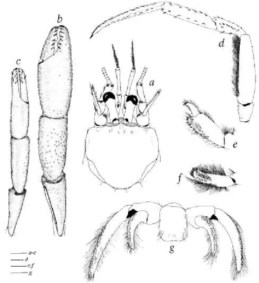
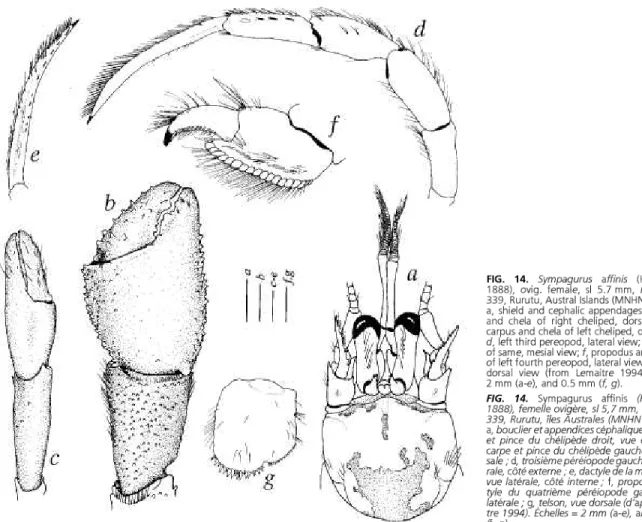
Sympagurus symmethcus n. sp.
Figs 4-7, 34
TYPE MATERIAL. — New Caledonia (holotype and paratypes). MUSORSTOM 4: stn CP241, 22°09'S, 167°12.20'E, 470-480 m, 03.10.1985: holotype 2 si 11.7 mm (MNHN-Pg 6050).— (no expedition name): stn Dr 3, 22°17'S, 167°12'E, 390 m, 23.05.1978: 1 S si 8.1 mm (USNM 1000005). — BIOCAL: stn CP 109, 22°10.03'S, 167°15.22'E, 495-515 m, 09.09.1985: 1 c? si 11.5 mm (USNM 1000004). — MUSORSTOM 5: stn DW 301, 22°06.9'S, 159°24.6'E, 487-610 m, 12.10.1986: 1 c? si 10.4 mm, 1 2 si 8.9 mm (MNHNPg 6051).— MUSORSTOM 6: stn CP 467,21=05.13'S, 167°32.11'E, 575 m, 21.02.1989: 1 3 si 9.4 mm (USNM 1000003); stn DW 471, 21°08'S, 167°54.10'E,460 m: 1 3 sl9.5mm(MNHN Pg 6052). — BERYX 11: stn CP 21, 24°44.35'-24°45.03'S, 168°06.72'-168°06.80'E, 430-450 m, 17.10.1992: 2 cj si 6.7 and 8.0 mm (MNHN Pg 6022). — SMIB 8: stn DW 178, 23°45'S, 168°17'E, 400 m, 30.01.1993: 1 2 si 10.4 mm (MNHN Pg 6023). — BATHUS 3: stn CP 811, 23°41'S, 168°15'E, 383-408 m, 28.11.1993: 1 3 si 10.5 mm (MNHN Pg 6024), 1 3 si 8.9 mm, 3 ovig. 2 si 9.7-10.6 mm (MNHN Pg 6025); stn CP 812, 23°43'S, 168°15'E, 391-440 m, 28.11.1993: 1 3 si 11.0 mm (MNHN Pg 6026); stn DW 817, 23°42'S, 168°15'E, 405-410 m, 28.11.1993: 2 cj si 8.0 and 10.2 mm, 2 2 si 8.9 and 9.0 mm (MNHN Pg 6027); stn DW 818, 23°43'S, 168°16'E, 394-401 m, 28.11.1993: 1 c? si 11.3 mm (MNHN Pg 6028); stn DW 838, 23°00'S, 166°55'E, 400-402 m, 30.11.1993: 1 3 si 7.7 mm, 1 2 si 8.4 mm (MNHN Pg 6029).—
BATHUS 4: stn CP 909, 18°57'S, 163°10'E, 516-558 m, 04.08.1994: 2 cj si 9.8 and 10.5 mm (MNHN Pg 6030);
stn CP910, 18°59'S, 163°08'E, 560-608 m, 05.08.1994: 2 c? si 10.4 and 11.1mm (MNHN Pg6031); stn CP911, 18°57'S, 163°08'E, 566-558 m, 05.08.1994: 1 3 si 10.9 mm (MNHN Pg 6032).
Vanuatu. MUSORSTOM 8: stn CP 963, 20°20'S,169°49'E, 400-440 m, 21.09.1994: 1 2 si 9.0 mm (MNHN Pg 6033);
stnDW 978, 19°23'S, 169°27'E, 413-408 m, 22.09.1994: 2 2 si 6.6 and 8.7 mm (MNHN Pg 6034); stn CP 1047, 16°53'S, 168°10'E, 486-494 m, 30.09.1994: 1 2 si 12.8 mm (MNHN Pg 6035); stn CP 1049, 16°39'S, 168°02'E, 469-525 m, 01.10.1994: 1 2 si 12.6 mm (MNHN Pg 6036); stn CP 1136, 15°40'S, 167°0TE, 398-400 m, 11.10.1994: 1 2 si 14.1 mm (MNHN Pg 6037).
MATERIAL EXAMINED. — The type material (see above).
DESCRIPTION. — Gills with lamellae at most distally divided (Tig. 4a). Shield length in males 6.6-11.5 mm, females 8.4-14.1 mm, ovigerous females 9.7-10.6 mm. Shield (Eig. 4b) distinctly longer than broad, dorsal surface weakly calcified medially, with pair of oblique rows of setae on anterior half and pair of longitudinal rows of setae on posterior half; linea d moderately marked; anterior margins weakly concave; lateral projections broadly rounded, or broadly subtriangular;
anterolateral margins slightly sloping; posterior margin broadly rounded. Rostrum rounded or subtriangular, overreaching lateral projections, with short mid-dorsal ridge. Anterodistal margin of branchiostegite broadly rounded, unarmed, setose.
Ocular peduncles long, about 0.7 x length of shield, constricted medially, weakly calcified laterally and mesially, with row of long setae dorsally. Corneae moderately dilated. Ocular acicles subtriangular, terminating in strong spine; separated basally by about basal width of 1 acicle.
i%y£&-^'l~k^.
e ^%:#fl
it'Iff'//
^ '/
^.K*!
FIG. 4. Sympagurus symmetricus n. sp.: a, ft, paratype, male, si 9.5 mm, MUSORSTOM 6 stn DW471, New Caledonia (MNHN Pg 6052); b-g, paratype, male, si 10.4 mm, MUSORSTOM 5 stn DW 301, New Caledonia (MNHN Pg 6051); /, paratype, female, si 8.9 mm, MUSORSTOM 5 stn DW 301 (MNHN Pg 6051): a.
Lamella of posterior arthrobranch (midportlon) of fourth pereopod; fa, shield and cephalic appendages; c, right antennal peduncle, lateral view; d, denuded right chellped, dorsal view; e, same lateral view; f, setae of same; g-i, left chellped, dorsal view. Scales = 0.5 mm (a), 2 mm (fa), 1 mm (c), 3 mm {d, e, g, h, i), and 0.25 mm (/).
FIG. 4. Sympagurus symmetricus n. sp. : A, H, paratype, male, si 9,5 mm, MUSORSTOM 6 stn DW471, Nouvelle-Caledonie (MNHN Pg 6052); B-G, paratype, male, si 10,4 mm, MUSORSTOM 5 stn DW301, Nouvelle-Caledonie (MNHN Pg 6051); I, paratype, femelle, si 8,9 mm, MUSORSTOM 5 stn DW301 (MNHN Pg 6051): A, lamelle de I'arthrobranchie posterieure (partle medlane) du quatrieme pereiopode; B, boudier et appendices cephaliques; C, pedoncule antennaire droit, vue laterale; D, chelipede droit denude, vue dorsale; E, le meme en vue laterale; F, soies du meme ciielipede; G-I, Chelipede gauche, vue dorsale. Echelles = 0,5 mm (A), 2 mm (B), 1 mm (C), 3 mm (D, E, G, H, I), et 0,25 mm (F).
Antennular peduncle exceeding distal margin of cornea by full length to 0.7 x length of ultimate segment. Ultimate seg- ment about 2.0 X as long as penultimate segment, with scattered setae. Basal seg- ment with strong ventromesial spine; late- ral surface with distal subrectangular lobe armed with 2 spines, and strong spine proximally. Ventral flagellum usually with about 8 articles.
Antennal peduncle (Fig. 4c) not excee- ding distal margin of cornea. Fifth segment with setae on lateral and mesial margins.
Fourth segment unarmed. Third segment with strong ventromesial distal spine.
Second segment with dorsolateral distal angle produced, terminating in strong, bifid or multifid spine; mesial margin with small setose tubercle or spine on dorsodis- tal angle. First segment with small spine on lateral surface (spine sometimes absent on 1 side); ventromesial angle produced, with row of spines laterally. Acicle short, at most reaching to about midlength of cornea, ter- minating in strong spine; mesial margin armed with 3-6 spines and with long setae.
Flagellum long, exceeding extended right cheliped and ambulatory legs; with setae > 1-3 flagellar articles in length.
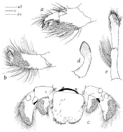
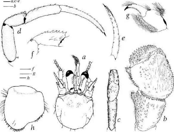
Mandible (Fig. 5a) with 3-segmented palp. Maxillule (Fig. 5b) with external lobe of endopod weakly developed, internal lobe with long terminal seta. Maxilla (Fig. 5c) with endopod at most reaching to distal margin of scaphognathite. First maxilliped (Fig. 5d) with endopod slightly exceeding exopod in distal extension.
Second maxilliped (Fig. 5e) without distinguishing characters. Third maxilliped (Fig. 5f) with crista dentata of about 14 corneous-tipped teeth; coxa and basis each with small mesial tooth. Sternite of third maxillipeds with spine on each side of midline. Epistomial spine absent.
Chelipeds markedly dissimilar, with dense, simple and plumose setae (Fig. 4/). Right cheliped (Figs 4d, e) spinose.
Fingers straight, each terminating in small corneous claw; cutting edges with irregularly-sized calcareous teeth, and rows of tufts of setae dorsally and ventrally near cutting edges; dorsal surfaces with strong spines (stronger proximally); ventral surfaces with small spines or tubercles. Dactylus nearly parallel to longitudinal axis of palm, slightly shorter than length of mesial margin of palm; mesial surface rounded, spinose distally, naked proximally. Fixed finger with dorsal and ventral surfaces similar to dactyl. Palm longer than broad; dorsolateral and dorsomesial margins with row of spines; mesial surface abruptly sloping ventrally, with small blunt or sharp spines; dorsal surface with numerous strong spines; ventral surface with small blunt spines distally and laterally, nearly smooth proximally. Carpus distinctly longer than broad, vidth numerous strong spines dorsally; dorsodistal margin with row of spines; mesial surface strongly sloping; ventromesial and ventrolateral distal margins each with row of spines; ventral surface with small tubercles. Merus with small tubercles on
FIG. 5. Sympagurussymmetricus n. sp., paratype, male, si 9.5 mm, MUSORSTOM 6 stn DW471, New Caledonia (MNHN Pg 6052). teft mouthparts, internal view: a, mandible; b, maxillule; c, maxilla; d, first maxilliped; e, second maxilliped; f, third maxilliped. Scales = 1 mm.
FIG. 5. Sympagurus symmetricus n. sp., paratype, male, si 9,5 mm, MUSORSTOM 6 stn DW471, Nouvelle-Caledonie (MNHN Pg 6052). Pieces buccales gauches, vue interne: a, mandibule; b, maxillule; c, maxllle; d, premier maxllllpede; e, second maxllllpede; f, trolsleme maxllllpede.
Echelles = 1 mm.
dorsal surface, and transverse dorsodistal row of setae; ventromesial margin with row of spines. Ischium with 1 or 2 small tubercles dorsally, and row of about 3 small tubercles ventromesially. Coxa with 1 ventromesial and 1 ventrolateral spine; ventral surface often with cluster of small spines proximally;
with ventromesial row of long setae.
Left cheliped (Figs 4g-i) well calcified, with dense simple and plumose setae; occa- sionally with abnormal left cheliped (Fig. 4g, see "remarks"). Fingers each termi- nating in sharp corneous claw; dorsal and ventral surfaces with tufts of setae. Dactylus about as long as or slightly longer than length of mesial margin of palm; cutting edge with row of closely-spaced small cor- neous teeth; dorsal surface usually with proximal spine. Fixed finger with 1 or 2 small lateroproximal spines; cutting edge consisting of irregularly-spaced, small, cal- careous teeth interspersed with small cor- neous teeth. Palm with row of often strong spines (Fig. 4i) on dorsomesial margin, and irregular rows of small spines on dorsolate- ral surface. Carpus with irregular rows of often strong spines on dorsal margin, and scattered small spines on dorsolateral sur- face. Merus unarmed except for setae dor- sally; ventromesial and ventrolateral mar- gins each with row of spines. Coxa and ischium each with row of setae on ventro- mesial margin.
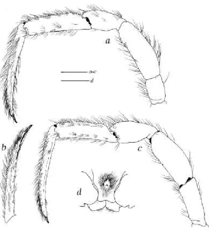
Ambulatory legs (Figs 6a-c) similar except for slightly longer segments on right; usually not exceeding extended right cheliped; meri, carpi, and propodi, with long simple setae on dorsal and ventral margins. Dactylus about 2.0 x as long as propodus, terminating in sharp corneous claw; with dorsal and dorsomesial rows of long simple setae, and ventromesial row of about 11-16 corneous spinules increasing in size distally (Fig. 6b). Propodus with transverse rows of short setae on dorsolateral and ventrolateral surfaces. Carpus with small dorsodistal spine; lateral surface sparsely setose, or with transverse row of short setae. Merus armed with spines on ventrodistal margin (first leg) or lacking spines (second leg).
Coxa and ischium with setose ventromesial margins. Anterior lobe of sternite of second legs (Fig. 6d) narrow, with bifid submarginal spine.
Fourth pereopod (Fig. 7a) semichelate. Dactylus subtriangular, terminating in sharp corneous claw; with ventrolateral row of small corneous spinules. Propodal rasp longer than propodus height; rasp consisting of 2 or 3 rows of ovate scales.
Carpus with long setae dorsally. Merus with long setae dorsally and ventrally.
Fifth pereopod (Fig. 7b) semichelate. Dactylus with row of ovate scales on lateral surface. Propodal rasp extending posteriorly to about mid-length or more of segment.
FIG. 6. Sympagurussymmefricusn.sp., paratype, male, si 10.4 mm, MUSORSTOM 5 stn DW301, New Caledonia (MNHN Pg 6051): a, left first ambulatory leg, lateral view; b, dactylus of same, mesial view; c, left second ambulatory leg, lateral view; d, sternite of second ambulatory legs.
Scales = 5 mm (a-c), and 2 mm (d).
FIG. 6. Sympagurus symmetricus n. sp., paratype, male, si 10,4 mm, MUSORSTOM 5 stn DW301, Nouvelle-Caledonie (MNHN Pg 6051): a, premiere patte marcheuse gauche, vue laterale, cote externe; b, dactyle de la meme patte, vue laterale, cote Interne; c, deuxieme patte marcheuse gauche, vue laterale; d, sternite des deuxlemes pattes marcheuses. Echelles = 5 mm (a-cj, and 2 mm (d).
Uropods and telson (Fig. 7c) symmetri- cal or nearly so. Exopods of uropod slightly more than 2.0 x as long as wide; anterior margin broadly rounded; with broad rasp.
Telson with sparse setae dorsally, and long setae on lateral margin of anterior half; with weak lateral indentations; dorsal surface with low, blister-like tubercles on posterior half; posterior margin divided into 2 lobes by angled (V-shaped) cleft; each lobe armed distally with about 12-20 corneous spines.
Males with paired first and second gono- pods moderately developed. First gonopod (Fig. 7d) with short distal lobe and weakly concave mesial surface. Second gonopod (Fig. 7e) with distal segment having roun- ded tip and weakly concave anterior sur- face; distal segment with row of short brist- les on distal half of lateral margin, and long setae on distal half of mesial margin and anterior surface; basal segment with long setae on posterior surface. Females with vestigial second right pleopod.
Color in life unknown.
FIG. 7. Sympagurussymmetricus n. sp., paratype, male, sM 0.4 mm, MUSORSTOM 5 stn DW 301, New Caledonia (MNHN Pg 6051): a, propodus and dactylus of left fourth pereopod, lateral view;
fa, propodus and dactylus of left fifth pereopod, lateral view; c, uropods and telson, dorsal view;
d, left first pleopod, mesial view; e, left second pleopod, anterior view. Scales = 1 mm.
FIG. 7. Sympagurus symmetricus fi. sp., paratype, male, si 10,4 mm, MUSORSTOM 5 stn DW301, Nouvelle-Caledonie (MNHN Pg 6051): a, propode et dactyle du quatrleme perelopode gauche, vue laterale; b, propode et dactyle du cinquieme perelopode gauche, vue laterale; c, uropodes et telson, vue dorsale; d, premier pleopode gauche, vue laterale, cote interne; e, second pleopode gauche, vue anterleure. Echelles = 1 mm.
HABITAT AND SYMBIOTIC ASSOCIA- TIONS. — In gastropod shells that someti- mes support an actinian.
DISTRIBUTION. — Off New Caledonia and Vanuatu, 383-610 m (Fig. 34).
REMARKS. — This new species is unique among Sympagurus species in the symmetry of uropods and telson, the short antennal acicles, relatively long eyes, and narrow anterior lobe of the sternite of the second ambulatory legs. Although S.
poupini also has symmetrical uropods and telson, that is where the similarity with S. symmetricus ends. The two species differ in numerous other characters, such as shape of the shield, relative length of the antennal acicles, ambulatory legs, fourth pereopods, and telson armature.
Some abnormalities were detected in specimens of S. symmetficus. A male (si 10.4 mm) has a left cheliped (Fig. 4g) similar in finger armature and shape to that of the right cheliped (Fig. 4d). A female from the same station (si 8.9 mm) has no right antenna, and the right antennule arises from where the antenna would normally be.
ETYMOLOGY. — From the Greek symmetros (symmetrical), in reference to the symmetrical condition of the uropods and telson.
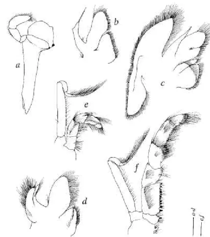
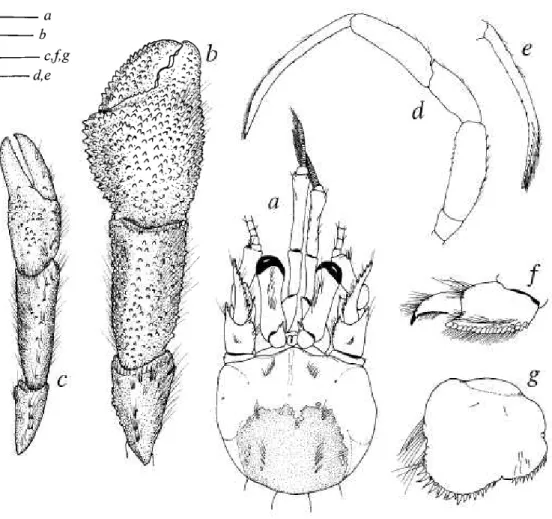
Sympagurus poupini Lemaitre, 1994 Figs Iflj, 2, 8, 34
Sympagurus poupini Lemaitre, 1994: 402, figs 20-23, 27k, 28h.
Parapagurus dofleini - Poupin et al. 1990: 94 (in part not Balss 1912), pi. 2f.
Sympagurus n. sp. - Poupin 1993: 51.
Sympagums poupini - Lemaitre 2000: 211. — Poupin 1996a: 20, pL 9d; 1996b: 96.
TYPE MATERIAL. — Tuamotu Archipelago. Makemo: stn 309, 16°34.2'S, 143°38.7'W, 580 m, 07.10.1990: holotype 3 si 18.5 mm (MNHN-Pg 5150). Paratypes recorded by Lemaitre (1994).
MATERIAL EXAMINED. — The type material (see above)
DESCRIPTION.— Gills with lamellae almost distally divided (Eig. la^). Shield length in males 8.6-21.1 mm, only known female 13.4 mm (ovigerous female unknown). Shield (Eig. 8a) distinctly broader than long, dorsal surface weakly and irregularly calcified, anterior margins weakly concave; lateral projections broadly subtriangular, with small terminal spine.
Rostrum triangular, with short mid-dorsal ridge. Anterodistal margin of branchiostegite rounded, unarmed.
Ocular peduncle less than half shield length, naked. Cornea weakly dilated. Ocular acicles subtriangular, terminating in strong spine.
Antennular peduncle slender, exceeding distal margin of cornea by length of ultimate segment.
Antennal peduncle exceeding distal margin of cornea by about 0.3 x length of fifth segment. Eourth segment unarmed.
Second segment with dorsolateral distal angle produced, terminating in strong, bifid or multifid spine (usually trifid);
mesial margin unarmed. Eirst segment unarmed. Acicle nearly straight, exceeding distal margin of cornea by about 0.5 x length of acicle, terminating in strong spine; mesial margin setose, armed with row of small blunt or sharp spines.
Elagellum naked or with scattered short setae < I article in length.
Maxillule with external lobe of endopod weakly developed, internal lobe with 4 long setae distally (Eig. Idj). Sternite of third maxillipeds with small spine on each side of midline. Epistome unarmed or with short, straight spine.
Chelipeds markedly dissimilar, with dense plumose setae on merus, carpus, palm, and proximal halves of fingers. Right cheliped (Eig. 8b) elongated. Lingers inwardly curved at tips. Dactylus straight. Palm about 1.6 x as long as broad; mesial and lateral surfaces rounded, armed with small spines; dorsal and ventral surfaces smooth except for few scattered small tubercles. Carpus with dorsal, lateral, and mesial surfaces armed with small, well-spaced spines. Merus with transverse row of setae near dorsodistal margin; surfaces unarmed except for few small tubercles on dorsolateral surface.
Left cheliped (Eig. 8c) well calcified. Palm smooth, except for few scattered small tubercles on lateral surface proximally.
Carpus with well-spaced, small spines on dorsal margin and dorsomesial surface.
Ambulatory legs (Eig. 8d) similar except for slightly longer segments on right. Dactylus about 1.7 x (first leg) or 2.2 x (second leg) as long as propodus; ventral margin armed with irregular row of about 25-30 small corneous spinules; dorsal margin with row of bristle-like setae. Carpus with small dorsodistal spine. Merus with distinct ventrolateral fringe of long setae, and row of blunt to sharp tubercles on ventral margin (tubercles more numerous on first leg). Anterior lobe of sternite of second legs, setose, armed with I or 2 strong submarginal spines.
Eourth pereopod (Eig. 8e) semichelate. Dactylus terminating in sharp, short corneous claw. Propodus elongate, more than 2.0 x as long as wide; rasp consisting of 7-12 well-spaced, corneous spines.
Eifth pereopod (Eig. 8J) semichelate. Propodus long, more than 3 x as long as wide; rasp consisting of 10-15 well-spaced, small corneous spines on distal third of segment.
Uropods and telson (Eig. 8g) symmetrical or nearly so. Uropods with endopods and exopods very elongated, exopods approximately 7.5 x as long as wide, endopods approximately 4.5 as long as wide; rasps of exopod and endopod consisting of 3 or 4 rows of small corneous spinules. Telson lacking lateral indentations; posterior margin weakly divided into 2 broadly rounded, unarmed lobes by broad shallow sinus.
FIG. 8. Sympaguruspoupini Lemaitre, 1994, Marara stn 309, Tuamotu Archipelago: a, d-g, Inolotype, male, si 18.5 mm (MNHN Pg 5150); b, c, paratype, male, si 15.9 mm (USNM 265395): a, shield and cephalic appendages; b, denuded right cheliped, dorsal view; c, denuded left cheliped, dorsal view; d, left first ambulatory leg, lateral view; e, propodus and dactylus of left fourth pereopod, lateral view; f, propodus and dactylus of left fifth pereopod, lateral view; g, uropods and telson, dorsal view (from Lemaitre 1994). Scales = 5 mm (a-d), and 3 mm (e-g).
FIG. 8. Sympagurus poupini Lemaitre, 1994, Marara stn 309, archipel des Tuamotu : a, d-g, holotype, male, si 18,5 mm (MNHN Pg 5150); b, c, paratype, male, si 15,9 mm (USNM265395): a, boudler etappendices cephaliques; b, chelipede droit denude, vue dorsale; c, chelipede gauche denude, vue dorsale; d, patte marcheuse gauche, vue laterale; e, propode et dactyle du quatrieme pereiopode gauche, vue laterale; f, propode et dactyle du cinquieme pereiopode gauche, vue laterale; g, uropodes et telson, vue dorsale (d'apres Lemaitre 1994). Echelles = 5mm ('a-dj, and3 mm (e-g).
Male first gonopod with moderately concave distal lobe. Second gonopod usually with small exopod on left side; distal segment setose on lateral and mesial margins.
Female often with paired rudimentary first pleopods.
Color (after Lemaitre 1994: 419, fig. 28h): overall cream yellow, dactyls and propodi of walking legs with faint white stripe.
HABITAT AND SYMBIOTIC ASSOCIATIONS. — All known specimens have been found living with an undetermined species of actinian that entirely covers the abdomen of the hermit crab (Lemaitre 1994).
DISTRIBUTION. — French Polynesia, Western Samoa and Wallis Island, 300-600 m (Fig. 34).
REMARKS. — As indicated by Lemaitre (1994), the specimens reported by Poupin et al. (1990) as Parapagurus dofleini are actually S. poupini. Individuals of this species can attain up to 170 mm in extended body length (from tip of dactyls of ambulatory legs to distal margin of telson).
Symmetry of the uropods and telson of Sympagurus poupini is also a character state of S. symmetricus n. sp., but, as mentioned under that species, the shape of the uropods and armature of the telson are markedly different.
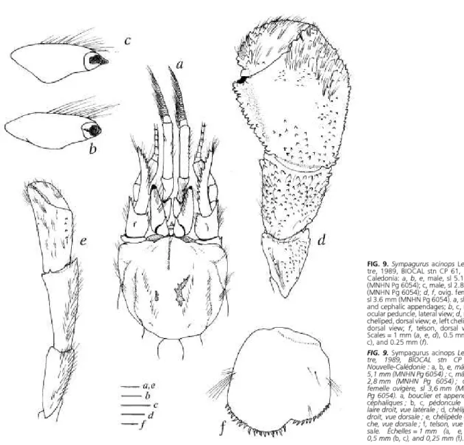
Sympagurus acinops Lemaitre, 1989
Figs Ibi, 2, 9, 10, 34
Sympagums acinops Lemaitre, 1989: 52, figs 24-27.
Sympagui-us acinops - Lemaitre 1990: 229; 1994:412; 1996: 169; 2000: 211. —Garcia Raso 1996: 739. — Udekem d'Acoz 1999: 177.
TYPE MATERIAL. — Bahamas. Columbus Iselin: stn 356, Tongue of the Ocean, 24°23.2'N, 77°25.5'W, 1561 m, 20.08.1975: holotype si 5.0 mm (USNM 228519). Paratypes recorded by Lemaitre (1989: 52).
OTHERMATERIALEXAMINED —New Caledonia. BIOCAL: stn 1000006). —MUSORSTOM 5: stn 323, 21°18.52'S, 157°57.62'E, CP 30, 23°09.65'S, 166°40.85'E, 1140 m, 29.08.1985: 1 3 si 970 m, 14.10.1986: 1 5 si 2.2 mm (MNHN Pg 6055).
1.7 mm (MNHN Pg 6053); stn CP 61, 24°11.67'S, 167°31.37'E, BIOGEOCAL: stn CP 238, 21°27.64'S, 166°23.4rE, 1300- 1070 m, 02.09.1985:2 6 si 2.8 and 5.1 mm, 1 ovig. 9 si 3.6 mm 1260 m, 13.04.1987: 1 3 si 2.9 mm (MNHN Pg 6056). — HALI- (MNHN Pg 6054); stn CP 62, 24°19.06'S, 167°48.65'E, 1395- PRO 1: stn CC 856, 21°44'S, 166°37'E, 311-365 m, 20.03.1994:
1410 m, 02.09.1985: 1 3 si 2.9 mm, 1 9 si 3.5 mm (USNM 1 S si 3.5 mm (MNHN Pg 5949).
DESCRIPTION. — Gills with lamellae deeply divided (Fig. Ibj). Shield length in males 1.7-6.5 mm, females 2.2-5.6 mm, ovigerous females 3.0-4.6 mm. Shield (Fig. 9a) about as broad as long, dorsal surface well calcified except for narrow median region extending posteriorly from tip of rostrum for about 0.3 x length of shield, and small irregular regions on posterior half; linea of dehiscence well marked; lateral projections broadly rounded. Rostrum rounded, slightly overrea- ching lateral projections; with short mid-dorsal ridge.
Ocular peduncles about half shield length, inflated ventrobasally, with row of long setae dorsally. Cornea (Figs 9b, c) reduced, subconical, terminating in blunt to sharp distal tip; more darkly pigmented distally than proximally. Ocular acicles subtriangular, terminating in strong spine; separated basally by less than basal width of 1 acicle.
Antennular peduncle exceeding distal margin of cornea by half length of penultimate segment.
Antennal peduncle exceeding distal margin of cornea by full length of fifth segment. Fourth segment unarmed. Second segment with dorsolateral distal angle produced, terminating in strong, usually simple spine; mesial margin with spine on dorsodistal angle. First segment with small spine on lateral surface. Acicles curved outwardly (dorsal view), exceeding distal margin of corneae by 0.3-0.5 length of acicle, terminating in strong spine; mesial margin densely setose, armed with 4-10 usually strong spines (sometimes unarmed or with fewer than 4 spines in small specimens si < 2.5 mm). Flagellum with numerous setae 1-4 flagellar articles in length.
Maxillule with external lobe of endopod weakly developed, internal lobe with 4 long setae distally (Fig. 1^2). Sternite of third maxillipeds with spine on each side of midline. Epistomial spine short and straight.
Chelipeds markedly dissimilar, with sparse to moderately dense simple and plumose setae. Right cheliped (Fig. 9d) massive. Fingers curved ventromesially. Dactylus set at strongly oblique angle to longitudinal axis of palm, dorsomesial margin well delimited by row of spines, ventromesial surface weakly concave. Fixed finger very broad basally. Palm about as long as broad (males) or broader than long (females), dorsolateral and dorsomesial margins well delimited by row of spines; mesial surface rounded, with numerous blunt to sharp spines or tubercles; dorsal surface with irregular rows of
FIG. 9. Sympagurus acinops Lemai- tre, 1989, BIOCAL stn CP 61, New Caledonia: a, b, e, male, si 5.1 mm (MNHN Pg 6054); c, male, si 2.8 mm (MNHN Pg 6054); d, f, ovig. female, si 3.6 mm (MNHN Pg 6054). a, shield and cephalic appendages; b, c, right ocular peduncle, lateral view; d, right cheliped, dorsal view; e, leftcheliped, dorsal view; f, telson, dorsal view.
Scales = 1 mm (a, e, d), 0.5 mm (fa, c), and 0.25 mm (/).
FIG. 9. Sympagurus acinops Lemai- tre, 1989, BIOCAL stn CP 61, Nouvelle-Caledonie : a, b, e, male, si 5,1 mm (MNHN Pg 6054); c, male, si 2,8 mm (MNHN Pg 6054); d, f, femelle ovlgere, si 3,6 mm (MNHN Pg 6054). a, boudier et appendices cephaliques; b, c, pedoncule ocu- iaire droit, vue laterale; d, chelipede droit, vue dorsale; e, chelipede gau- che, vue dorsale; f, telson, vue dor- sale. Echelles = 1 mm (a, e, d), 0,5 mm (b, c), and 0,25 mm (f).
small spines or tubercles on proximal half; ventral surface unarmed or with scattered small tubercles. Carpus with dense spines or tubercles dorsally, mesial margin strongly sloping.
Left cheliped (Fig. 9e) well calcified. Dactylus about as long as mesial margin of palm. Palm with dorsomesial row of few small spines or tubercles. Carpus with strong dorsodistal spine, and small lateral spine on dorsodistal margin.
Ambulatory legs (Figs 10a, h) similar except for slightly longer segments on right. Dactylus about 2.0 x as long as propodus, with dorsal and dorsomesial rows of long setae, and ventromesial row of about 5-7 small, well-spaced corneous spinules. Carpus with small dorsodistal spine. Anterior lobe of sternite of second legs setose, usually with small, blunt submarginal spine.
Fourth pereopod (Fig. 10c) semichelate. Dactylus terminating in short, usually sharp corneous claw. Propodal rasp consisting of single row of ovate scales distally, and 2 rows on proximal third.
Fifth pereopod semichelate. Dactylus considerably overreaching ventrodistal angle of propodus. Propodal rasp extending to about mid-length of segment.
Uropods and telson strongly asymmetrical. Left exopod of uropod about 1.8 x as long as broad;
anterior margin broadly rounded, with broad or narrow rasp. Telson (Fig. 9/) lacking or at most with weakly marked lateral indentations, posterior margin divided into 2 lobes by angled (V-shaped) cleft, lobes armed distally with corneous spines (often curved).
Males with paired first and second gonopods well developed. First gonopod with distal lobe elongate. Second gonopod with distal segment having row of short bristles on distal half of lateral margin, and long setae on distal half of mesial margin and anterior surface; basal segment with long setae on posterior surface.
Color in life unknown.
HABITAT AND SYMBIOTIC ASSOCIATIONS. — Gastropod shells, occasionally with one or more small actinian or zoanthid polyps, or completely overgrown by a zoanthid.
FIG. ^0. Sympagurus acinops lemaitre, 1989, male, si 5.1 mm, BIOCAtstn CP61, New Caledonia (MNHN Pg6054):a, left first ambulatory leg, lateral view; fa, dactylusof same, mesial view; c, propodus and dactylus of left fourth pereopod, lateral view.
Scales = 1 mm (a, fa), and 0.5 mm (c).
FIG. 10. Sympagurus acinops Lemaitre, 1989, male, si 5,1 mm, BIOCAL stn CP 61, Nouvelle-Caledonie (MNHN Pg 6054): a, patte marcheuse gauche, vue laterale, cote externe; b, dactyle de la meme patte, vue laterale, cote interne; c, propode et dactyle du quatrieme pereiopode gauche, vue laterale. Echelles = 1 mm (a, b), and 0,5 mm (c).
DISTRIBUTION. — Tongue of the Ocean, Bahamas; Canary Islands and New Caledonia, 311-2537 m (Fig. 34).
REMARKS. — This species was previously known only from the western and eastern Atlantic (Lemaitre 1989, 1990).
Morphologically it is a relatively stable species that exhibits minor, predictable variations related to size, such as the armature of the mesial margin of the antennal acicles (unarmed or with up to 10 spines), and shape of the tip of the cornea (blunt to sharp); or related to sex, such as the proportions of the carpus and palm of the right cheliped (as broad as long in males, and broader than long in females). The variations observed in the New Caledonia specimens fall well within the range documented for the Atlantic specimens (Lemaitre 1989).
Among parapagurids, subconical corneae similar to those of Sympagurus acinops are known only in Oncopaguius minutus (Henderson, 1896), a species distributed in the Indo-Pacific Lemaitre 1996). Such corneal condition can be considered a case of convergence.
Sympagurus dimorphus (Studer, 1883) Figs lci,2, 11, 34,35a
Eupagums dimorphus Studer, 1883: 24, figs 11, 12.
Parapagurus brevimanus Balss, 1911: 4, fig. 5.
lEupagurus modicellus Stebbing, 1914: 255, pi. 26, fig. D.
Sympagurus arcuatus johnstoni Hale, 1941: 279, fig. 13a-d.
Sympagurus arcuatus mawsoni Hale, 1941: 280, fig. 14a-c.
Parapagurus dimorphus-Henderson 1888: 86, pi. 10, fig. 1.—Murray 1895: 395; 1896: 434.—Alcock 1905: 172. — Stebbing 1910:
356. —Balss 1912: 97; 1924: 768. — Carlgren 1923: 265, pi. 1, figs 1, 13, 14, pi. 2, fig. 10. —Barnard 1950: 452, fig. 83c, d. — Haig 1955: 18.—Gordan 1956: 338. —Fuller 1958: 164.—Forest &rde Samt Laurent 1968: 115, pi. 1, figs 5, 6. —Coelho &Araujo-ramos 1972: 164. — de Saint Laurent 1972: 108. — Scelzo 1973: 166. — de Saint Laurent 1973: 791, fig. 6. — Hand 1975: 513. — Probert
etal. 1979:381. —Coelho&r Santos 1980: 143. — Kensley 1981: 33. — Schembri 1982: 860.—Macpherson 1983a: 12; 1983b: 472.
— Schembri & McLay 1983: 28, fig. 6a, b. — Zarenkov 1990: 238.
Parapagurus hrevimanus - Balss 1912: 100, fig. 9. — Forest &r de Saint Laurent 1968: 116. — de Saint Laurent 1973: 791.
Sympagums arcuatus johnstoni - Gordan 1956: 341. — Forest & de Saint Laurent 1968: 116.
Sympagunis arcuatus mawsoni - Gordan 1956: 341. — Forest &r de Saint Laurent 1968: 116.
?not Parapagui-us dimorphus - Milne-Edwards &r Bouvier 1893: 32.
IParapagurus sp. 2 - de Saint Laurent-Dechance 1964: 15, figs 2, 7, 11-19.
?Species S.A. 1 - Williamson & von Levetzow 1967: 181, figs 2a-m, 3a-g.
Parapagurus dimorphus - Williamson &r von Levetzow 1967: 184. — Bacardit 1987: 79.
Sympagums dimorphus - Lemaitre 1989: 71, figs 36-38, 40E-H; 1990: 229; 1994: 412; 1996: 176, fig. 7; 2000: 214, fig. 68, pL 7. — Branch et al. 1991: 6, 36 (key, unnumbered fig.). — Lemaitre & McLaughlin 1992: 747, figs 1-5. — Melo 1999: 154, figs 93, 94. — Zhadan 1999: 735, fig. lb, 2c-f; m press. — Boschi 2000: 128.
ISympagums dimorphus - Manning & Chace 1990: 40, fig. 22.
TYPE MATERIAL. — Eupaguius dimorphus. South Africa. Off Cape of Good Hope, 34°13.6'S, 15°00.7'W, 211 m: syntypes (ZMB-not seen); Pa mpagurusbrevimanus. VaMiviastn 167: New Amsterdam. 37°47'S, 77°33'E, 496 m, 4.1.1899: syntypes - 3 cj 2.0-4.7 mm, 4 ovig. 2 2.7-3.3 mm (ZMB 16459); Sympagums arcuatus johnstoni. Tasmania. BANZARE: stn 115, 41°03'S, 148°42'E, 128 m: syntypes (SAMA C4095 - not seen); Sympagurus arcuatus mawsoni. Macquarie Island. BAN- ZARE; stn 83, 54°42'S, 158°54'E, 69 m; syntypes (SAMA C4094 - not seen).
OTHER MATFRIAL EXAMINED. —Tasmania. Southern Surveyor: 54°42.7-41.9'S, 158°46.1-45.9'E, 100-300 m, 22.01.1999: 1 6 si stn SS03/99-62, NW side of St. Helens seamount, 41°12.7'S, 9.3 mm, 1 9 si 7.3 mm, 5 ovig. 9 si 4.8-7.3 mm (SAMA C5951).
148°45.LE, 850 m, 27.07.1999: 1 9 si 10.0 mm, 1 ovig. 9 si SouthAfrica. Gazelle: Agulhas Bank (no other data): 2 9sl9.4and 12.8 mm (SAMA C5952). 10.5 mm (ZMB 6377).
Macquarie Island. Southern Surveyor: stn SSO1/99-69, W coast,
DESCRIPTION. — Gills with lamellae (Tig. Icj) deeply divided. Shield length in males 2.2-29.5 mm, females 2.2- 22.0 mm, ovigerous females 3.9-29.5 mm. Shield (Fig. 1 la) usually as broad as long, dorsal surface often weakly calcified medially, anterior margins concave; lateral projections subtriangular, with small terminal spine. Rostrum rounded, with broad low dorsal ridge
Ocular peduncles more than half shield length. Ocular acicles subtriangular, terminating in strong simple or occasio- nally bifid spine. Corneae weakly dilated.
Antennular peduncle exceeding distal margin of cornea by nearly full length of ultimate segment.
Antennal peduncle at most slightly exceeding distal margin of cornea. Fourth segment with small dorsolateral distal spine. Second segment with dorsolateral distal angle produced, terminating in strong bifid or multifid spine. Acicle curved outwardly (dorsal view), usually not exceeding distal margin of cornea; mesial margin setose, armed with 13-19 strong spines. Flagellum with numerous short setae < 1 to 2 articles in length.
Maxillule with external lobe obsolete or weakly developed, internal lobe with usually 4 long setae distally (Fig. Icj).
Epistomial spine short and straight, sometimes absent. Sternite of third maxillipeds with strong spine on each side of midline.
Chelipeds markedly dissimilar, covered with moderately dense simple and plumose setae. Right cheliped massive.
Chela usually operculate (Fig. lib), proportions and armature strongly affected by size and sexual dimorphism; dorso- lateral margin in dorsal view evenly convex or semicircular in females and males of similar size, or straight in large males (si > 9.0 mm); armature of dorsal surface consisting of numerous sharp to blunt spines or small tubercles. Fingers strongly curved ventromesially. Dactylus with ventromesial surface concave. Palm with dorsomesial and dorsolateral margins each well delimited by row of spines.
Left cheliped (Fig. lie) well calcified. Palm with dorsomesial, dorsolateral, and often dorsomedian rows of small tubercles or spines. Carpus with dorsal row of spines.
Ambulatory legs (Figs 1 Id-j) similar except for longer segments on right, and for armature on meri, carpi and propodi frequently more developed on right. Dactylus (Fig. lie) shorter than propodus, with ventromesial row of 15-20 strong
FIG. 11. Sympagurus dimorphus (Studer, 1883): a, c-e, h, male, si 10.0 mm, Eltanin stn 740, Drake Passage (USNM 155045); fa, f, female, si 8.1 mm. New Zealand (NMNZ Cr 3204); g, male, si 9.2 mm. New Zealand (NMNZ Cr 8472): a, shield and cephalic appendages, dorsal view; fa, propodus and chela of right cheliped, dorsal view; c, propodus and chela of left cheliped, dorsal view; d, right first ambulatory leg, lateral view; e, dactylus of same, mesial view; f, carpus of right first ambulatory leg, lateral view; g, propodus and dactylus of left fourth pereopod, lateral view; h, telson, dorsal view (a, c-e, h, from Lemaitre 1989;
fa, f, g, from Lemaitre 1996). Scales = 2 mm (a-f), and 1 mm (g, h).
FIG. 11. Sympagurus dimorphus (Studer, 1883): a, c-e, h, male, si 10,0 mm, Eltanin stn 740, Passage de Drake (USNM 155045); b, f, femelle, si 8,1 mm, Nouvelle-Zelande (NMNZ Cr 3204); a, male, si 9,2 mm, Nouvelle-Zelande (NMNZ Cr 8472): a, boudler et appendices cephallques, vue dorsale; b, propode etpince du chelipede drolt, vue dorsale; c, propode etpince du chellpede gauche, vue dorsale; d, premiere patte marcheuse droite, vue laterale, cote externe;
e, dactyle de la meme patte, vuelaterale, cote interne; f, carpe de la premiere patte droite, vue laterale; g, propode et dactyle du quatrieme pereiopode gauche, vue laterale; h, telson, vue dorsale (a, c-e, h, d'apres Lemaitre 1989; b, f, g, d'apres Lemaitre 1996). Echelles = 2 mm (a-f), ancf 1 mm (g, h).
spinules, dorsal row of long setae, and 3 or 4 short, dorsomesial oblique rows of setae distally. Carpus (Fig. 1 If) with dorsal row of spines. Anterior lobe of sternite of second legs with 1-3 small submarginal spines, setose.
Fourth pereopod (Fig. 1 Ig) semichelate. Dactylus terminating in sharp corneous claw. Propodal rasp with 2-5 irregular rows of ovate scales.
Uropods and telson strongly asymmetrical. Telson (Fig. llh) with weak lateral indentations; terminal margin divided into 2 lobes by wide, shallow, rounded (U-shaped) cleft; lobes armed distally with short corneous spines.
Male first gonopod with moderately concave distal lobe. Second gonopod with distal segment spatulate, basal segment occasionally with short exopod.
Females lacking first pleopods, or occasionally with rudimentary paired or unpaired first pleopods; with vestigial right second pleopod.
Color (Fig. 35a). Lemaitre (2000: 217, pi. 7) indicated that the overall color of the body is cream. Based on additional photographs obtained during this study, and color patterns still present in formalin-preserved specimens, a more detailed account of coloration is now possible. Shield with light orange-red areas on calcified portions. Ocular peduncles with orange-red stripe on dorsal faces, ventral faces orange-red. Antennal peduncle with light orange-red spot on lateral face of