I also investigated the genetic interactions of the plc-1, cpe-1 and splA2 genes in the regulation of these cell functions in N. This chapter describes the cell functions of plc-1, cpe-1 and splA2 in the circadian clock , stress responses, and cellulose degradation in N. However, the Δplc-1, Δcpe-1 and ΔsplA2 single and double mutants were completely resistant to cell wall stress.
The plc-1, cpe-1, and splA2 genes act in coordination to maintain intracellular Ca2+ homeostasis.

The biology of Neurospora crassa, a model filamentous fungus
The life cycle of N. crassa
Nitrogen limitation, light and cold temperatures induce the sexual cycle, resulting in the formation of female reproductive structures known as protoperithecia (Raju 1992). After fusion of the trichogen with the conidium, the male nucleus is transported to the protoperithecium, where the formation of the multicellular sex apparatus known as the perithecium (mature protoperithecium) begins. During karyogamy, nuclei of the opposite mating type combine to produce a diploid zygote, which then undergoes two meiotic divisions and one postmeiotic mitosis, generating eight of eight ascospores, which are packed in a linear order in ascus.
The ascospores, after ripening, are ejected towards the blue light through a beak (ostiole) at the top of the perithecium, and the discharged ascospores eventually germinate and form their vegetative hypha in the presence of adequate nutrients (Fig. 1.1; Raju.
The versatility and universality of calcium
The Ca2+ binding loop between the two helices in the EF hand domain contains 12 amino acids rich in acidic residues that provide negatively charged oxygen atoms for Ca2+ coordination (Fig. 1.3A) in a pentagonal bipyramidal geometry (Fig. 1.3B) , which is the most preferred chemistry for Ca2+ coordination is (Gifford et al. 2007). Monocapped trigonal prism or split vertex octahedral coordination geometry of Ca2+ in the EF site of parvalbumin. Purple represents the conserved hydrophobic amino acid residue that forms a short β-sheet in the paired EF hand.
Ca2+ coordination in the canonical EF hand domain 1 (EF-1) of calmodulin (CaM) showing both pentagonal bipyramidal Ca2+ coordination (solid lines) and hydrogen bonding (dotted lines).

The calcium signaling machinery in N. crassa
- Ca 2+ -permeable channels
- Ca 2+ and cation-ATPases
- Ca 2+ /H + exchangers
- Ca 2+ /Na + exchangers
- Phospholipase C-δ subtype proteins
- Calmodulin
- Ca 2+ and/or CaM binding proteins
The NCU06703 gene encodes the mating-induced death-1 (MID-1) protein, a stretch-activated mechanosensitive Ca2+-permeable channel that plays a crucial role in the regulation of ion transport via Ca2+ homeostasis (Lew et al. 2008). Ca2+ is released from intracellular Ca2+ storage vacuoles when IP3 is present (Cornelius et al. 1989). Ca2+ and/or CaM binding proteins are involved in the control of [Ca2+]c and contain multiple conserved domains, including an N-terminal catalytic domain, a Ca2+ binding EF-hand domain, an autoinhibitory domain and an overlapping CaM binding domain ( Tamuli et al. 2013 ).
CaMK-1 mutants have a phase lag, a light-induced phase shift in the circadian conidiation rhythm, a short period lengthening, and a retarded growth phenotype (Yang et al. 2001).

The Ca 2+ signaling proteins are important for Ca 2+ homeostasis
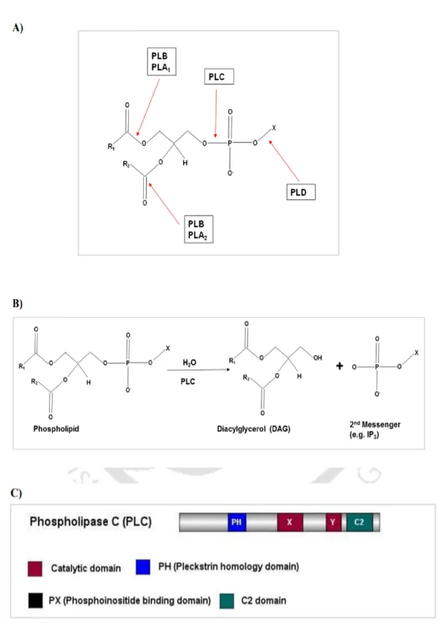
Phospholipase C
In the gray mold Botrytis cinerea, bcplc1 is an essential gene (Schumacher et al. 2008). The Candida albicans genome possesses three putative PI-PLCs, CaPLC1, CaPLC2, and CaPLC3 (Bennett et al. 1998; Andaluz et al. 2001). Furthermore, lac1 transcript did not accumulate when cplc1 was disrupted, indicating that cplc1 is important for lac1 gene expression (Chung et al. 2006).
In the filamentous fungus Trichoderma reesei, PLC functions as an essential link between cAMP and Ca2+ signaling in cellulase production (Chen et al. 2021).

Secretory Phospholipase A 2
The saprotrophic ascomycete Aspergillus oryzae has two unique putative sPLA2 proteins designated as sPlaA and sPlaB in its genome (Machida et al. Both sPlaA and sPlaB have different physiological characteristics; sPlaA has the maximum enzyme activity for Ca2+ under acidic conditions, whereas with external secretion conditions sPlaB has the highest enzyme activity for Ca2+ at a neutral to alkaline pH with intracellular localization (Nakahama et al. 2010) Only sPlaA expression was significantly increased in response to carbon deprivation, oxidative stress, heat shock and during or after conidiation, but sPlaB expression was barely detectable in either condition (Nakahama et al. 2010).

Ca 2+ exchangers
Materials and Methods
Materials
- Laboratory chemicals and reagents used in this study
- N. crassa strains used in this study
- Media for bacterial growth, antibiotics, and other commonly used reagents
- Primers used in this study
Methods
- Growth conditions
- Setting up crosses and harvesting ascospores
- Maintenance of stock
- Conidial cell count
- Scoring for antibiotic resistance in N. crassa
- Growth rate assay
- Aerial hyphae analysis
- Circadian regulated conidiation assay
- Thermotolerance assay
- pH tolerance assay
- Cell wall stress assay
- ER stress assay
- Cellulose degradation assay
- Isolation of N. crassa genomic DNA
- Isolation of RNA from N. crassa
- Quantification of nucleic acids
- Polymerase Chain Reaction (PCR)
- Reverse Transcription PCR for cDNA synthesis
- Real-time Quantitative PCR (qRT-PCR)
- Agarose gel electrophoresis
- Sequence analysis
Databases and Software Programs used in this study
Introduction
Results
- Circadian Regulated Conidiation Assay
- The plc-1 gene, but not cpe-1 and splA 2 , regulates the period length in N. crassa
- Loss of plc-1 influences temperature compensation in N. crassa
- Thermotolerance Assay
- The Δplc-1, Δcpe-1, ΔsplA 2 single and double mutants showed sensitivity to the heat shock
- pH Tolerance Assay
- The Δplc-1, Δcpe-1, and ΔsplA 2 single and double mutants are sensitive to alkaline pH
- Cell Wall Stress Assay
- The Δplc-1, Δcpe-1, and ΔsplA 2 single and double mutants are insensitive to cell wall stress
- Endoplasmic Reticulum (ER) Stress Assay
- The ΔsplA 2 mutant showed a growth defect in response to ER stress
- Cellulose Degradation Assay
- The splA 2 gene is involved in cellulose degradation in N. crassa
- The Δplc-1, Δcpe-1, ΔsplA 2 single and double mutants showed normal growth phenotypes
- Cellulose Degradation Assay during ER stress
- The ΔsplA 2 mutant is unable to utilize cellulose during ER stress
Discussion
Expression analysis of genes involved in the circadian clock, stress responses, and
Introduction
- Transcriptional analysis of the ∆plc-1, ∆cpe-1, and ∆splA 2 single and double mutants
- The period length change is correlated with frq and wc-1 transcript levels
- The plc-1, cpe-1, and splA 2 genes regulate the expression of heat shock proteins in N. crassa
- plc-1, cpe-1, and splA 2 positively interact to regulate pac-3 expression under alkaline
- ER stress induced growth defects in the ΔsplA 2 , Δplc-1; ∆cpe-1, and ∆plc-1; ∆splA 2 mutants
- The ΔsplA 2 and Δcpe-1; ΔsplA 2 mutants showed increased expression of cellulolytic genes,
- Promoter analysis
- Analysis of the promoter regions of the plc-1, cpe-1, and splA 2 genes in N. crassa
- Possible mechanism of the PLC-1, CPE-1, and sPLA 2 mediated pathways in circadian clock,
Therefore, further studies on the molecular players associated with PLC-1, CPE-1 and sPLA2 in the regulation of these cellular processes were critical to understand the detailed mechanism. I also tested the expression levels of plc-1, cpe-1 and splA2 under heat stress condition in WT. However, compared to the heat-shocked condition, the transcript levels of plc-1, cpe-1, and splA2 were decreased when cells were allowed to recover at 28 °C after heat shock for 30 min (Fig. 4.2B).
These results suggested that plc-1, cpe-1, and splA2 are induced by heat stress and possibly protect the cell by mediating the expression of heat shock proteins. Expression of plc-1 , cpe-1 , and splA2 was measured by qRT-PCR using RNA extracted from non-heat-shocked, heat-shocked, and heat-shock-recovered cultures of the WT strain. I also analyzed the expression of plc-1, cpe-1 and splA2 in the WT strain grown at ambient pH 5.8, and then subjected to acidic (pH 3.8) or alkaline (pH 7.8) stress conditions.
However, no significant changes in the expression of plc-1, cpe-1 and splA2 were observed in the acidic condition (Fig. 4.3B). To investigate the effect of ER stress, I also analyzed the transcription levels of plc-1, cpe-1 and. Since transcription factor binding elements regulate the expression of a gene, I used the MatInspector program (Quandt et al. 1995; Cartharius et al. 2005) to analyze the putative promoter regions of plc-1, cpe-1, and splA2 genes.
This analysis revealed putative binding sequences for different transcription factors in the promoter regions of plc-1, cpe-1 and splA2 genes (Table 4.1). The presence of these sequences further supports the findings that PLC-1, CPE-1 and sPLA2 are critical for circadian clock.

Discussion
The increased expression of plc-1, cpe-1 and splA2 after heat shock and gradually decreased when N. These results further supported that the effects of an alkaline pH on the growth sensitivity of these mutants (Fig. 3.3) were indeed due to the reduced level of PAC-3 transcription factor. The lumen of the ER serves as the reservoir for Ca2+ in the cell, and its oxidative environment is essential for disulfide bond formation and the correct folding of secreted proteins or membrane proteins.
Deviation from Ca2+ homeostasis in the ER causes protein unfolding due to the Ca2+-dependent nature of the molecular chaperones (Ma and Hendershot 2004). If the capacity of ER to handle adequate protein folding is exceeded due to an increase in protein load and/or a disruption in the conditions required for appropriate folding, the URP is activated (Bravo et al. 2012; Hetz 2012) . In addition, the expression level of splA2 was found to be upregulated during ER stress, while there was no change in the expression of plc-1 and cpe-1 (Fig. 4.4B).
The plc-1, cpe-1 and splA2 genes were not previously found to be involved in cellulose degradation; however, I observed a severe reduction in the expression of the cellulolytic genes, cbh-1, cbh-2 and endo-2 in the Δplc-1, Δcpe-1, Δplc-1; ∆cpe-1 and ∆plc-1; ∆splA2 mutants compared to WT when grown on Avicel (Fig. 4.5A). However, ∆splA2 and ∆cpe-1; ΔsplA2 mutants showed significantly increased expression of the cellulolytic genes, especially cbh-1 and cbh-2 (Fig. 4.5A). In addition, the expression levels of plc-1, cpe-1 and splA2 during growth on avicel (Fig. 4.5B) correlated with the phenotypes (Fig. 3.6) described in the previous chapter.
In addition, plc-1 exhibits epistatic interaction with cpe-1 and splA2 in regulating the cellulose degradation pathway in N. In addition, the presence of putative binding sites for several transcription factors (Table 4.1) further supported that plc-1, cpe-1 and splA2 are critical for circadian clock regulation, stress survival and cellulose utilization in N.
The splA 2 gene interacts with the transcription factor CRE-1 to regulate growth and
Introduction
In the previous chapter, I described the role of the splA2 gene in cellulose degradation and ER response pathways in N. The ΔsplA2 knockout mutant showed significantly higher expression of key cellulolytic genes, suggesting that splA2 plays a repressive role during growth on microcrystalline cellulose. Avicel). Moreover, mutants with enhanced lignocellulose degradation abilities showed an ER-sensitive phenotype, suggesting that lignocellulase secretion and ER stress response pathways are interconnected in N.
I found that the ∆splA2 mutant showed ER stress sensitivity and was unable to utilize microcrystalline cellulose during ER stress. A homokaryotic strain expressing the splA2 transgene complements the phenotype of the ΔsplA2 knockout mutant grown on microcrystalline cellulose and also results in the suppression of extracellular protein secretion, endoglucanase activity and ER stress sensitivity in N. In addition, the Δcre-1 knockout mutant consumed Avicel faster than the WT, secreted more extracellular protein and showed enhanced endoglucanase activity, when grown on 2% Avicel as sole carbon source.
Therefore, in this chapter I generated the double mutant of splA2 and cre-1 to understand the cell functions regulated by their genetic interactions in N. I also predicted the three-dimensional protein structures of sPLA2 and CRE-1 using the homology modeling approach. .
Results
- Generation and confirmation of the ∆cre-1; ∆splA 2 double mutant
- The ∆cre-1; ∆splA 2 double mutant showed distinct colony and hyphal morphology
- The ∆cre-1; ∆splA 2 double mutant showed severely reduced growth
- The ∆cre-1; ∆splA 2 double mutant showed reduced aerial hyphae and conidiation
- The ∆cre-1; ∆splA 2 double mutant showed irregular septation
- The ∆cre-1; ∆splA 2 double mutant was highly sensitive to cell wall stress drugs
- The ∆cre-1; ∆splA 2 double mutant was unable to grow in response to ER stress
- The ∆cre-1; ∆splA 2 double mutant exhibited enhanced cellulose degradation ability
- The splA 2 and cre-1 interacts to regulate the expression of UPR markers and the cellulolytic
- Validation of the modelled structures of sPLA 2 and CRE-1
- Secondary Structure Prediction in the simulated models of sPLA 2 and CRE-1
- Molecular Visualization of the MD simulated sPLA 2 and CRE-1 structure
A schematic representation of the crosses of ΔsplA2 and Δcre-1 single mutant strains of opposite mating type to generate the double mutant. Amplification of PCR products of size ~2.044 kb and 2.5 kb, respectively, indicates the presence of the ΔsplA2 and Δcre-1 knockout alleles in the double mutant. The ∆cre-1; ∆splA2 double mutant showed a colonial morphology different from that of the WT and the parent single mutants, with severely reduced mycelial growth and pigmentation (Fig. 5.2A, B).
The interaction between splA2 and cre-1 in growth and cellulose degradation CHAPTER 5. A) Colony morphology of the strains grown on VM agar medium in flasks. I also investigated whether the double deletions affected the cell wall integrity of ∆cre-1; ∆splA2 double mutant. I also assessed the ability of ∆cre-1; ∆splA2 double mutant to utilize different carbon sources.
I also measured the transcript levels of the UPR markers grp-78 and pdi-1 (as described in Chapter 4) to assess the impact of ER stress on Δcre-1; ΔsplA2 double mutant. The FASTA format of the obtained amino acid sequences was used to model the sPLA2 and CRE-1 proteins. To further validate the quality of the stereochemically reliable MD simulated protein structures of sPLA2 and CRE-1, ProSA analysis was performed.
I also analyzed the percentage of amino acids in the different secondary structures of the simulated models of sPLA2 and CRE-1. The three-dimensional structure of the MD simulated sPLA2 and CRE-1 structures was visualized using PyMOL (Fig. 5.14A, B).

Discussion
Conclusions and Future Perspectives
Although previous studies have shed light on the functions of PLC-1 homologues, CPE-1 and sPLA2. Although PLC-1, CPE-1 and sPLA2 are involved in the cytosolic Ca2+ sensing process in N. Therefore, I investigated the ability of WT, Δplc-1, Δcpe-1, ΔsplA2 single and their double mutants to use cellulose, a natural substrate.
I studied the ability of WT, Δplc-1, Δcpe-1, ΔsplA2 single and double mutants to use different carbon sources. In addition, growth defects of the double mutants under alkaline conditions indicated that plc-1, cpe-1, and splA2 positively affect survival under alkaline pH conditions.