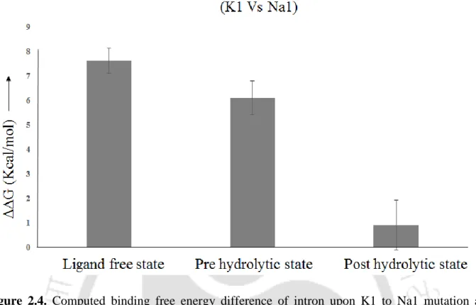
The Na+ in the active site of the post-hydrolytic state allows water input and fulfills the Na+ binding requirement, resulting in low discrimination (~ 1 kcal/mol). In Chapter 4 we showed divalent (Mg2+ vs. Ca2+) and monovalent (K+ vs. Na+) ion selectivity in the active site of lariat group II intron of a eukaryotic brown alga Pylaiella littoralis in the lariat-3' exon (pre-hydrolytic ) discuss condition) and post-hydrolytic condition, respectively (Figure 4).
Contents
Introduction, Methods, and Objectives
Introduction
Group II intron and splicing .1 Background
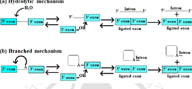
- Mechanism of group II intron splicing
- Structural studies of Group II intron 1.2.4 Hydrolytic splicing
- Pre-hydrolytic state
- Post-hydrolytic and ligand free state
- Branched splicing
- Pre-hydrolytic and Post-hydrolytic state
- Biochemical study of group II intron
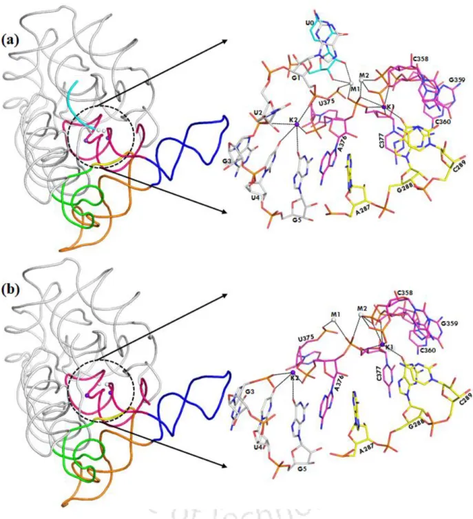
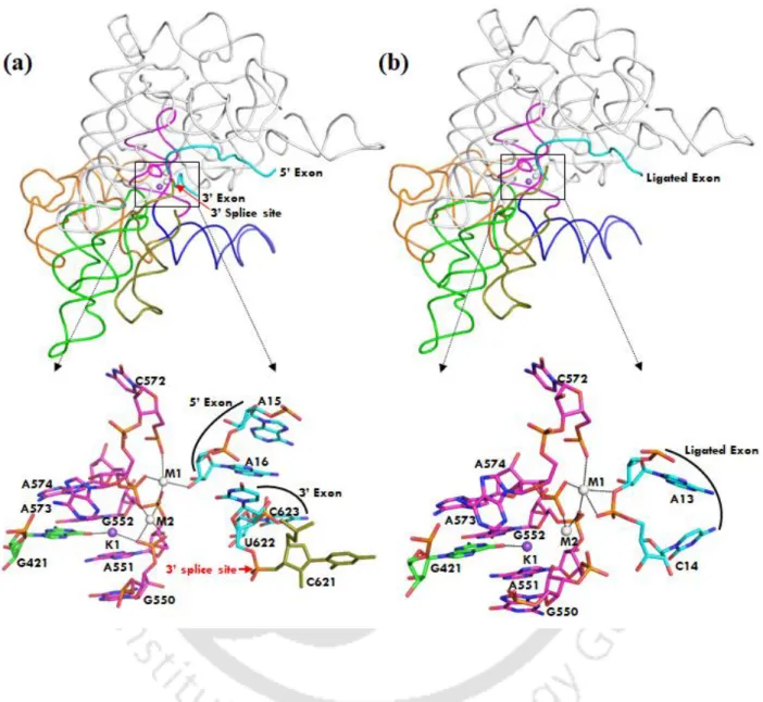
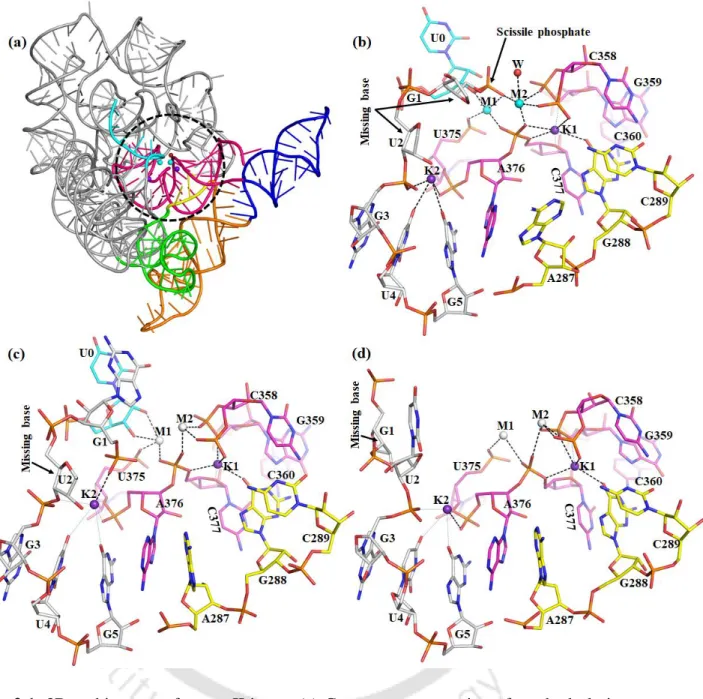
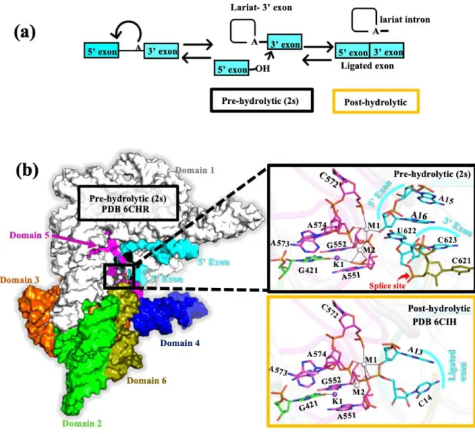
The folded structure of the group II intron in its prehydrolytic state is shown in Figure 1.3. The folded structure of the group II intron in the post-hydrolytic state is shown in Figure 1.4a.
Cas1 protein and its metal ion selectivity .1 Background
- Structural and Biochemical studies
The Cas1 protein is a specific metal-dependent DNA endonuclease that generates 80 base pair double-stranded DNA fragments ( Wiedenheft et al., 2009 ). Cas1-mediated DNA degradation activity is severely compromised by mutation of binding pocket residues as well as substitution of metal ions (Wiedenheft et al., 2009).
Methods
- Classical Molecular dynamics Simulations
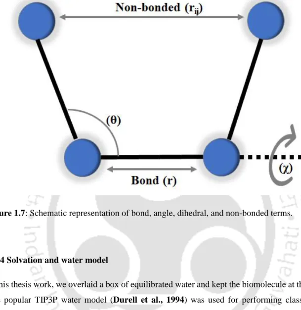
- Force Fields
- Solvation and water model
- Minimization and production dynamics
- Temperature and Pressure Control
- Truncation of short-range Van der Waals Interactions
- Periodic Boundary Condition
- Long-range Electrostatic Interaction
- MD setup adopted in this thesis
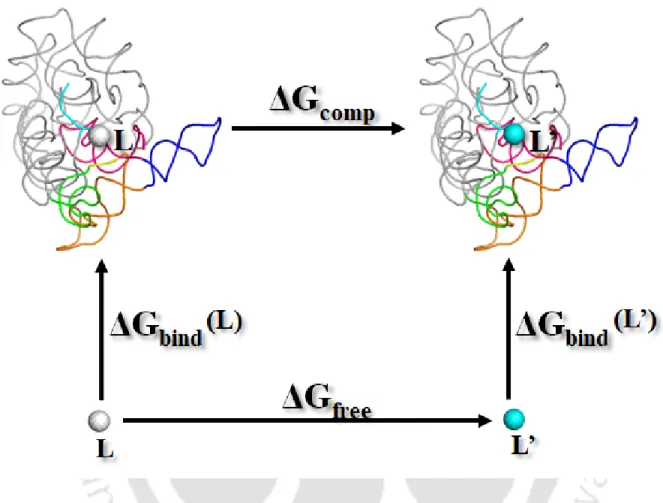
- Thermodynamic Cycle and Relative Binding Energy
But the disadvantage of the Verlet algorithm is that it does not include an explicit velocity term in the equation. A variation of the Verlet algorithm has been developed, known as the leap-frog algorithm (Hockney, 1970).
Objectives
Background
As discussed in Chapter 1, biochemical and structural studies of Oceanobacillus iheyensis group II introns (Pyle, 2006; Toor et al., 2008; and Marcia et al., 2012) have provided fundamental insights into the mechanism of RNA splicing. Monovalent ions are known to dictate the splicing pathway (Jarrell et al., 1998; . Erat et al., 2008) and modulate alternative conformations of the group II introns (Marcia et al., 2012).
Methods
- Molecular dynamics procedure
- Free Energy Calculations
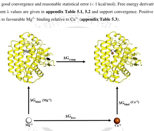
We calculated the change in free energy associated with the change in van der Waals radii (horizontal arms of Figure 2.3) and calculated the relative free energy of binding as ΔΔGbind = ΔGcomp – ΔGfree. Overall, a total of about 0.55 µs molecular dynamics free energy simulations were performed to obtain good convergence and a reasonable statistical error (1-2 kcal/mol), comparable to the previously reported force field uncertainty (Satpati et al., 2011). .
Results
- Structure-Based Energetics for K + vs Na + Recognition
- Robust structural features from MD and its comparison with X-ray Structures
- Ligand free group II intron with K + /Na + : MD vs X-ray
- Pre-catalytic group II intron with K + /Na + : MD vs X-ray
- Post-catalytic group II intron with K + /Na + : MD vs X-ray
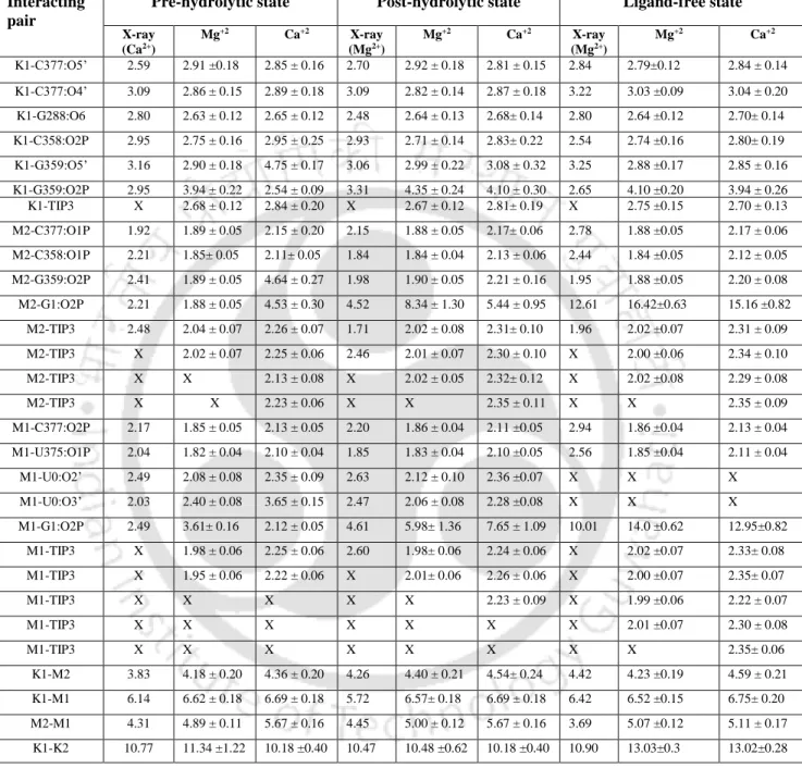
It should be noted that Na1 in the active site of the free intron is trapped in a dry desolvated pocket with. This leads to a very high discrimination of ΔΔG ~ 7 kcal/mol that does not favor Na1 in the active site of the intron. The pattern of K1 and M2 interactions in the pre-hydrolytic state is almost identical to the free state (Table 2.1 and Figure 2.7 a).
It should be noted that Na + bound in the active site of the intron has not been observed experimentally.
Discussion
Conclusion
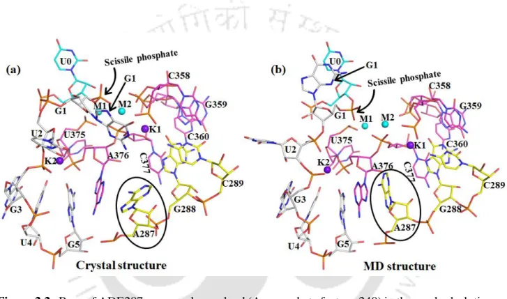
It is worth noting that ADE287 blocks the entry of water into the K1 binding site and the dryness of the binding pocket leads to discrimination. Thus, using the X-ray structures as a template, we performed molecular dynamics simulations to compare the structural and dynamic differences between the Mg2+ and Ca2+ bound active site of the group II intron at different stages of the splicing pathway (ie, prehydrolytic, posthydrolytic, and ligand-free state). In the post-hydrolytic state, the exonic part of the hydrolysis product is involved in direct interaction with M1, while the intronic part is highly flexible in our MD trajectories.
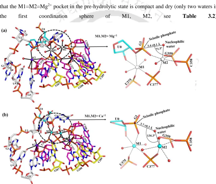
Solvent exposure of M1, M2 sites is least in the pre-hydrolytic state, highest in the ligand-free state, and intermediate in the post-hydrolytic state.
Background
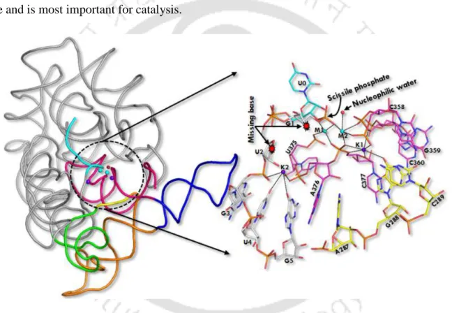
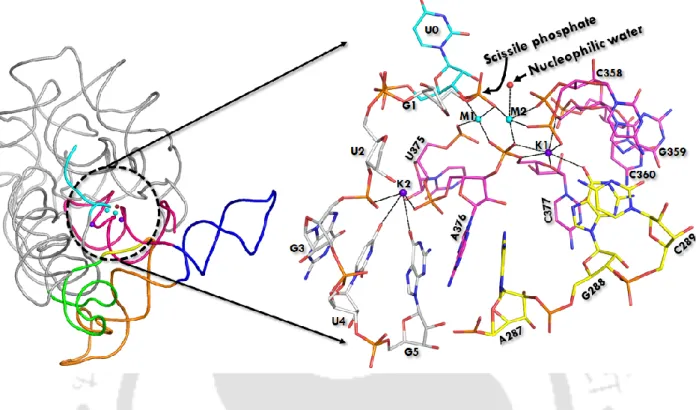
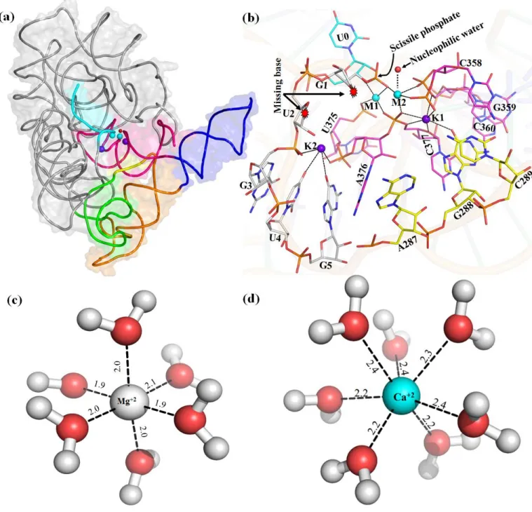
D5 (red) is highly conserved (~35 nucleotides in length) during evolution and forms the active site of group II intron. Heteronuclear metal ion cluster (M1, M2, K1 and K1) required for the structural integrity of the active site of group II intron. Previously, we quantitatively estimated the relative binding affinity (K+ versus Na+) at the K1 site of group II intron using computer simulations (Kumar et al., 2018).
However, the structure of the Mg2+-bound prehydrolytic state and the divalent metal ion selectivity (Mg2+ vs. Ca2+) with the group II intron at different levels are unknown.
Methods
- Molecular dynamics setup
- Free energy calculations
- Electronic structure calculation
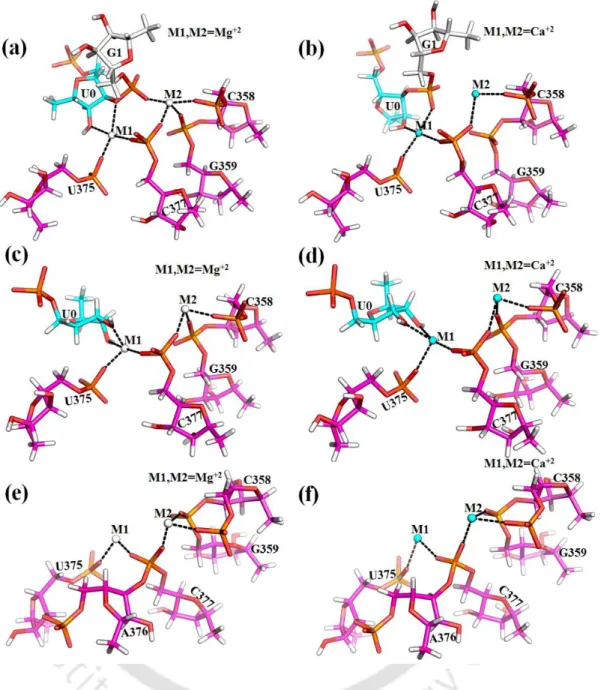
Thermodynamic cycle for binding of Mg2+ versus Ca2+ ions to the divalent ion binding sites M1, M2 of group II intron active sites. Representative MD structures of group II intron were selected and divalent metal ions M1, M2 and the nearby nucleotides were extracted and embedded in a dielectric continuum representing bulk water. Local environment around M1, M2 sites eligible for quantum chemical calculations. c, d) pos-hydrolytic state and (e, f) ligand-free state.
Single point energy differences evaluated between the right and left sides of eqn (3.1) approximate the relative divalent cation preference at the intron active site M1, M2.

Results
- MD vs X-ray structures of group II intron at various stage of splicing
- Active site of post-hydrolytic group II intron with bound Mg 2+ /Ca 2+
- Active site of ligand-free group II intron with bound Mg 2+ /Ca 2+
Replacement of Ca2+ in M1 and M2 sites of the ligand-free intron state leads to the entry of additional water molecules (Figure 3.6 b, Table 3.1), as seen in post-hydrolytic state (Figure 3.5 b). The same interaction network is stable and independent of the nature of divalent (M1, M2: Mg2+ or Ca2+) metal ions in the active site of group II intron (Table 3.1). These calculations involve the calculation of the change in binding affinity for divalent metal ions (M1, M2) in the active site of group II intron op.
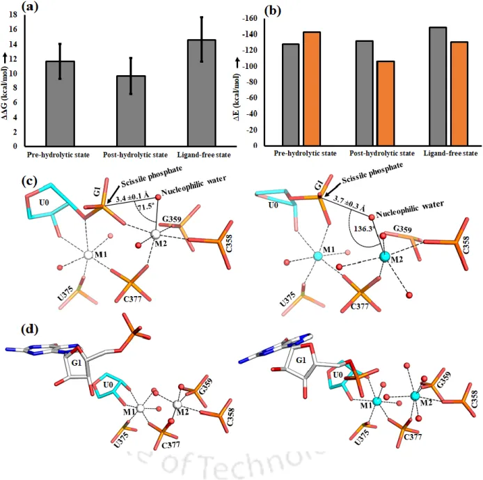
A large negative energy difference implies that Mg2+ is favored at M1 and M2 sites over Ca2+ in the intron active site at different stages of the cleavage pathway.
Discussion
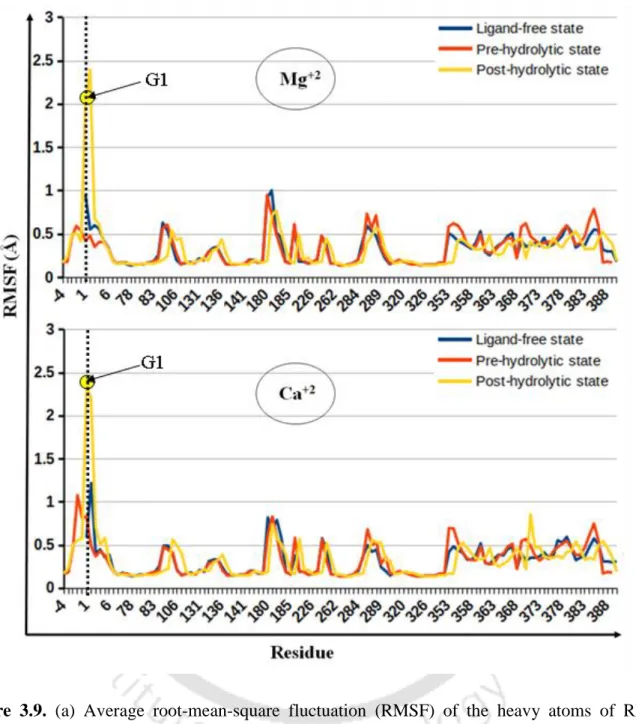
Large negative energy difference implies that Mg2+ is favored at M1, M2 sites relative to Ca2+ in the intron active site at various stages of the splicing pathway. pre-hydrolytic state, Figure 3.4 a) is lost, which is compensated by the recruitment of water molecule (Figure 3.5 a). Average RMSF (Figure 3.9) of nucleotide residues of group II intron indicates that the RNA flexibility of Mg2+ and Ca2+ bound group II intron at various conditions, e.g. After hydrolysis, i.e. in the post-hydrolytic state, the highest flexibility of the intron part (G1) was evident from the average RMSF (Figure 3.9).
Quantum chemical calculations also support Mg2+ preference over Ca2+ in the intron active site (Figure 3.8). a) Root-mean-square fluctuation (RMSF) of the heavy atoms of RNA nucleotides in group II introns.
Conclusion
- Background
- Computational methods
- Molecular Dynamics Simulations setup
- Relative Binding Free Energy Calculation
- Ab Initio Quantum Calculations
- Results
- Energetics of metal-ion selectivity by lariat group II intron
- Comparison MD and X-ray Structure
- Mg 2+ and Ca 2+ bound (M1, M2 site) pre-hydrolytic (2s) lariat group II intron
- Mg 2+ and Ca 2+ bound (M1, M2 site) post-hydrolytic lariat group II intron
- Na + /K + bound (K1 site) lariat group II intron
- Discussion
- Conclusion
The thermodynamics of metal ion selectivity in the active site of the lariat intron is unknown. The positive value of ΔΔG indicates that Mg2+ (K+) is favored over Ca2+ (Na+) binding in the active site of the lariat intron. The magnitude of metal ion preference in the lariat intron pocket (ΔΔG) was experimentally unknown.
The divalent metal ions (M1, M2 sites) were tightly bound in the dry catalytic pocket, while the monovalent ion (K1 site) was loosely associated in the solvent-accessible wet binding pocket of the lariat intron.
CRISPR-Cas system associated Cas1 protein
Background
The statistical-mechanical approach suggested a strong correlation between Cas9-mediated DNA splicing efficiency and the stability of the DNA-RNA loop in the protein complex (Xu et al., 2017). The first molecular dynamics (MD) simulations addressed the structure and dynamics of the Csy4-RNA complex (Csy4 is another protein component of the CRISPR system) and highlighted the limitations of the simulation techniques when studying protein-RNA systems (Estarellas et al., Estarellas et al., 2015). Classical MD simulations (Palermo et al., 2016; Palermo et al., 2017) of the CRISPR/Cas9 system revealed the importance of Cas9's HNH domain in the coordinated processing mechanism.
Biochemical studies showed that metal ion substitution, mutation of conserved metal ion binding residues, inhibits metal ion chelation Cas1-mediated DNA fragmentation (Wiedenheft et al., 2009).
Methods
- Molecular dynamics procedure
- Protocol for binding free energy calculation
- Ab initio quantum calculations
Free energy calculation for each replicate was based on 88–110 ns data collection averaged over 4–5 replicates with different initial velocities. A total of 704 ns of molecular dynamics free energy simulations were performed to obtain good convergence and reasonable statistical error (< 1 kcal/mol). Free energy associated with the horizontal legs of the thermodynamic cycle was calculated by MD simulations and thermodynamic integration.
Single point energy differences (∆E, see Appendix Table 5.4) evaluated between the right and left sides of eqn (5.1) and eqn (5.2) approximate the relative divalent cation preference (Mn2+ vs Mg2+) and (Mg2+ vs Ca2+ ) respectively in the Cas1 binding pocket.
Results
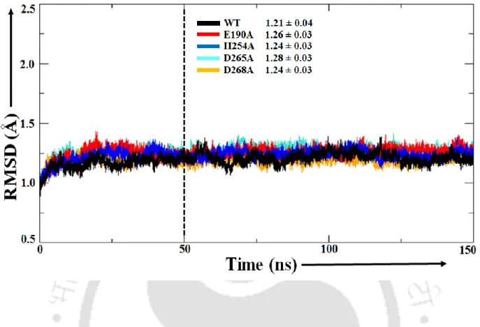
- MD insight into the structural convergence and overall flexibility of the protein
- Energetics of ion selectivity
- MD insight into the Mg 2+ and Ca 2+ bound binding pocket of WT Cas1
- MD insight into the Mg 2+ and Ca 2+ bound binding pocket of mutated Cas1
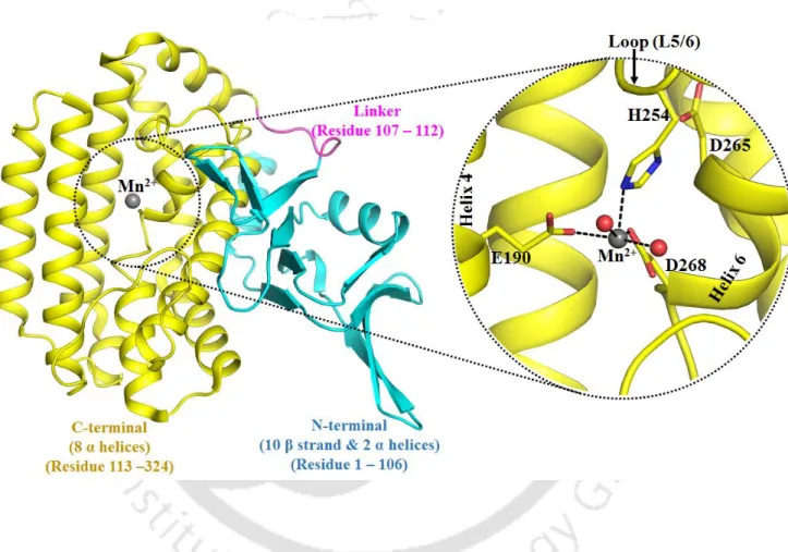
Cas1 protein active site flexibility by mutations. a) Root-mean-square fluctuation (RMSF) of the Cα atom. The calculations involve estimating the change in divalent metal ion binding affinity after Mg2+ → Ca2+ mutation in the Cas1 binding pocket and free in water (Figure 5.2). It appears that residues in the first coordination shell of the divalent metal ion were held in place by the residues from the second shell (R140, D265 and K271).
The second shell mutation (D265A) does not alter the first-shell interaction network of the divalent metal ion, resembling the WT binding pocket (Figure 5.9).
Discussion
It is known that a divalent metal ion with higher charge density binds strongly in the negatively charged protein binding pocket (Jing et al., 2018). Mutation of residues of the first coordination shell (E190A, H254A and D268A) of the divalent metal ion leads to water. D265A mutation does not disrupt the first shell interaction network of the divalent metal ion as observed in WT Cas1.
The coordination number of the divalent metal ion is the same as that observed in the free water simulation (6 for Mg2+ and 7 for Ca2+).
Conclusion
However, experimental verification can be challenging because our estimated selectivity for metal ions only pertains to a specific site of the Cas1 protein. It is believed that a single Mn2+ in the Cas1 binding pocket coordinates nuclease activity (Yang et, 2008). In addition, divalent metal ion substitution (Mg2+ by Ca2+) or mutations (E190A, H254A and D265A) alter the solvent accessibility of the divalent metal ion binding pocket, which may be related to its selectivity.
Overall conclusion and future perspective
Moreover, the direct interaction between Mg 2+ and the ribose sugar of the 5'-exon (observed in the X-ray and MD structures) was disrupted in response to replacement of divalent metal ions by Ca 2+ . Mutation of the conserved amino acid that coordinates to the divalent metal ion also results in water exposure of the metal ion binding pocket. The accessibility to water in the metal ion binding pocket appears to be related to the strength of recognition by biomolecules, eg, the group II intron and the Cas1 protein.
Dry binding pocket results in high selectivity, whereas water availability decreases the strength of recognition.
Appendices Appendix 2
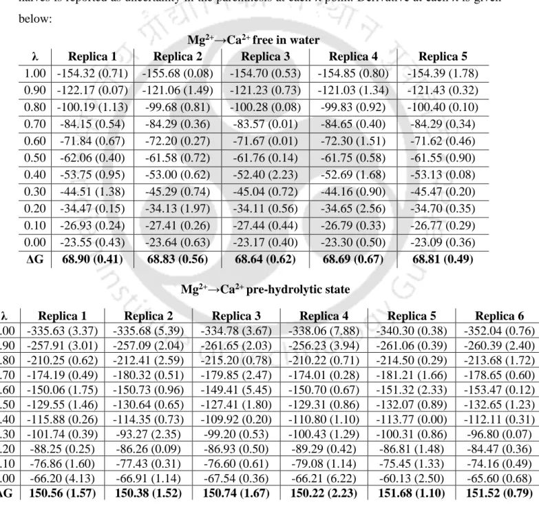
Average 68.77 ±0.11
Relative binding free energy of Na+ (ΔΔG in Kcal/mol) to the active site of the group II lariat intron. Determination of the intrinsic affinities of multiple site-specific Mg2+ ions coordinated to domain 6 of a group II intron ribozyme. Divalent metal ions modulate the self-splicing reaction of the mitochondrial group II intron of yeast Sc.
Divalent metal ions promote the formation of the 5' cleavage site recognition complex in a self-cleaving group II intron.