mutual interest in this fascinating bacterium
Edited by
C. Wray
Formerly of
Veterinary Laboratories Agency (Weybridge), Addlestone, Kent, UK and
A. Wray
Department of Oral Biology, College of Dentistry, University of Florida, Gainesville, Florida, USA
CABI Publishing
CABI Publishing CABI Publishing
CAB International 10 E 40th Street
Wallingford Suite 3203
Oxon OX10 8DE New York, NY 10016
UK USA
Tel: +44 (0)1491 832111 Tel: +1 212 481 7018
Fax: +44 (0)1491 833508 Fax: +1 212 686 7993
Email: [email protected] Email: [email protected]
© CAB International2000. All rights reserved. No part of this publication may be reproduced in any form or by any means, electronically, mechanically, by photocopying, recording or otherwise, without the prior permission of the copyright owners.
A catalogue record for this book is available from the British Library, London, UK.
Library of Congress Cataloging-in-Publication Data
Salmonella in domestic animals / edited by C. Wray and A. Wray.
p. cm.
Includes bibliographical references.
ISBN 0–85199–261–7 (alk. paper)
1. Salmonellosis in animals. I. Wray, C. (Clifford). II. Wray, A.
SF809.S24 S25 2000 636.089′6927--dc21
99–043803
ISBN 0 85199 261 7
Typeset by Columns Design Ltd, Reading.
Printed and bound in the UK by Biddles Ltd, Guildford and King’s Lynn.
Contributors vii
Preface ix
CHARACTERISTICS OFSALMONELLA
1 Taxonomy of the Genus Salmonella 1
P.A.D. Grimont, F. Grimont and P. Bouvet
2 Structure, Function and Synthesis of Surface Polysaccharides in
Salmonella 19
A.N. Rycroft
3 Fimbriae of Salmonella 35
C.J. Thorns and M.J. Woodward
VIRULENCE OFSALMONELLA
4 Virulence Mechanisms of Salmonellaand their Genetic Basis 57 A.J. Bäumler, R.M. Tsolis and F. Heffron
5 Host Susceptibility, Resistance and Immunity to Salmonella in Animals 73 P.S. Holt
6 Antibiotic Resistance in Salmonella 89
R. Helmuth
INFECTION INANIMALS
7 SalmonellaInfections in the Domestic Fowl 107
C. Poppe
8 Salmonella Infections in Turkeys 133
H.M. Hafez and S. Jodas
9 SalmonellaInfection in Ducks 157
R.R. Henry
v
10 SalmonellaInfections in Cattle 169 C. Wray and R.H. Davies
11 SalmonellaInfections in Pigs 191
P.J. Fedorka-Cray, J.T. Gray and C. Wray
12 SalmonellaInfections in Sheep 209
C. Wray and K.A. Linklater
13 Salmonellain Horses 219
J.K. House and B.P. Smith
14 SalmonellaInfections in Dogs and Cats 231
M.E. Carter and P.J. Quinn
15 Public-health Aspects of SalmonellaInfection 245
T. Humphrey
EPIDEMIOLOGY ANDPREVENTION
16 Environmental Aspects of Salmonella 265
C.J. Murray
17 Salmonellain Animal Feed 285
R.H. Davies and M.H. Hinton
18 Competitive Exclusion 301
C. Schneitz and G. Mead
19 Vaccination against SalmonellaInfections in Food Animals:
Rationale, Theoretical Basis and Practical Application 323 P.A. Barrow and T.S. Wallis
20 Epidemiology and Salmonellosis 341
K. Hollinger
LABORATORYMETHODS
21 Methods for the Cultural Isolation of Salmonella 355 W.D. Waltman
22 Methods for the Rapid Detection of Salmonella 373
H. van der Zee and J.H.J. Huis in’t Veld
23 Laboratory Aspects of Salmonella 393
Y.E. Jones, I.M. McLaren and C. Wray
24 Diagnosis of Salmonellaby ELISA and Other Tests 407 P.A. Barrow
25 Molecular Typing of Salmonella 429
J.E. Olsen
Index 447
P.A. Barrow, Institute for Animal Health, Compton Laboratory, Compton, Newbury, Berkshire RG20 7NN, UK
A.J. Bäumler, Department of Medical Microbiology and Immunology, Texas A&M University Health Science Center, 407 Reynolds Medical Building, College Station, TX 77843-1114, USA
P. Bouvet, Centre National de Référence des Salmonella et Shigella, Unité des Entérobactéries, INSERM Unit 389, Institut Pasteur, 75724 Paris Cedex 15, France M.E. Carter, Department of Veterinary Microbiology and Parasitology, Faculty of
Veterinary Medicine, University College Dublin, Ballsbridge, Dublin 4, Eire
R.H. Davies, Veterinary Laboratories Agency (Weybridge), New Haw, Addlestone, Surrey KT15 3NB, UK
P.J. Fedorka-Cray, Richard Russell Research Center, 950 College Station Road, Athens, GA 30605-2720, USA
J.T. Gray, University of Nebraska, Lincoln Veterinary Diagnostic Center, Lincoln, Nebraska, USA
F. Grimont, Centre National de Référence pour le Typage Moléculaire des Entérobactéries, Unité des Entérobactéries, INSERM Unit 389, Institut Pasteur, 75724 Paris Cedex 15, France
P.A.D. Grimont, Centre National de Référence des Salmonella et Shigella, Unité des Entérobactéries, INSERM Unit 389, Institut Pasteur, 75724 Paris Cedex 15, France H.M. Hafez, Free University of Berlin, Faculty of Veterinary Medicine, Institute of Poultry
Diseases, Koserstrasse 21, 14195 Berlin, Germany
F. Heffron, Department of Molecular Microbiology and Immunology, Oregon Health Sciences University, Portland, OR 97201-3098, USA
R. Helmuth, Federal Institute for Health Protection of Consumers and Veterinary Medicine BgVV, Laboratory for Molecular Biology and National Salmonella Reference Laboratory, Diedersdorfer Weg 1, 12277 Berlin, Germany
R.R. Henry, Cherry Valley Farms, North Kelsey Moor, Lincoln LN7 6HH, UK M.H. Hinton, Home Office, 50 Queen Anne’s Gate, London SW1H 9AT, UK
vii
K. Hollinger, FDA CVM, Division of Epidemiology and Surveillance, 7500 Standish Place, Rockville, MD 20855, USA
P.S. Holt, US Department of Agriculture, Agricultural Research Service, Southeast Poultry Laboratory, 934 College Station Road, Athens, GA 30605, USA
J.K. House, Department of Medicine and Epidemiology, School of Veterinary Medicine, University of California – Davis, Davis, CA 95616, USA
J.H.J. Huis in’t Veld, Department of Food of Animal Origin, Utrecht University, PO Box 80.175, 3508 TD Utrecht, The Netherlands
T. Humphrey, PHLS Food Microbiology Research Unit, Church Lane, Heavitree, Exeter EX2 5AD, UK
S. Jodas, Staatliches Tierarztliches Untersuchungasmt, Azenbergestrasse 16, 70174 Stuttgart, Germany
Y.E. Jones, Veterinary Laboratories Agency (Weybridge), New Haw, Addlestone, Surrey KT15 3NB, UK
K.A. Linklater, SAC, West Mains Road, Edinburgh EH9 3JG, UK
I.M. McLaren, Veterinary Laboratories Agency (Weybridge), New Haw, Addlestone, Surrey KT15 3NB, UK
G. Mead, Royal Veterinary College, University of London, Boltons Park, Hawkshead Road, Potters Bar, Hertfordshire EN6 1NB, UK
C.J. Murray, Institute of Medical and Veterinary Science, Box 14, Rundle Mall, Adelaide, SA 5000, Australia
J.E. Olsen, Department of Veterinary Microbiology, The Royal Veterinary and Agricultural University, Stigbøjlen 4, DK-1870 Frederiksberg C, Denmark
C. Poppe, Health Canada, 110 Stone Road West, Guelph, Ontario N1G 3W4, Canada P.J. Quinn, Department of Veterinary Microbiology and Parasitology, Faculty of Veterinary
Medicine, University College Dublin, Ballsbridge, Dublin 4, Eire
A.N. Rycroft, Royal Veterinary College, Hawkshead Lane, North Mymms, Hatfield, Hertfordshire AL9 7TA, UK
C. Schneitz, Orion Corporation, Animal Health, PO Box 425, FIN-20101 Turku, Finland B.P. Smith, Department of Medicine and Epidemiology, School of Veterinary Medicine,
University of California – Davis, Davis, CA 95616, USA
C.J. Thorns, Bacteriology Department, Veterinary Laboratories Agency (Weybridge), New Haw, Addlestone, Surrey KT15 3NB, UK
R.M. Tsolis, Department of Veterinary Pathobiology, Texas A&M University, College Station, TX 77843-4467, USA
H. van der Zee, Inspectorate for Health Protection, Regional Service East, De Stoven 22, 7206 AX Zutphen, The Netherlands
T.S. Wallis, Institute for Animal Health, Compton Laboratory, Compton, Newbury, Berkshire RG20 7NN, UK
W.D. Waltman, Georgia Poultry Laboratory, Oakwood, GA 30566, USA
M.J. Woodward, Bacteriology Department, Veterinary Laboratories Agency (Weybridge), New Haw, Addlestone, Surrey KT15 3NB, UK
A. Wray, Department of Oral Biology, College of Dentistry, University of Florida, PO Box 100424, Gainesville, FL 32610-0424, USA
C. Wray, Veterinary Laboratories Agency (Weybridge), New Haw, Addlestone, Surrey KT15 3NB, UK
ix It is now more than 40 years since the publica-
tion of ‘Salmonellosis in Animals’ by Professor Buxton and, although there have been books regarding diseases in domestic animals which contain a chapter on salmonellosis, there has been no English-language book that brings together all aspects of Salmonella infection in domestic animals.
Professor Buxton pointed out ‘salmonella are a large group of bacteria, which does not recognise international frontiers, shows little host specificity and from which there seems to be derived an ever increasing list of antigenic com- binations by which these organisms are classi- fied’. Since the publication of this book, the situation has gradually worsened and the preva- lence of Salmonella infection has increased markedly in both humans and domestic animals.
There are many factors for this increase, includ- ing the rapid changes in animal husbandry and production that have taken place. In the UK, the number of farms has decreased, as has the num- ber of farm-workers; at the same time, the num- ber of animals on the farm has increased through the adoption of intensive systems of animal pro- duction. To feed the increased number of farm animals, protein and vegetable by-products are imported on a large scale, which has resulted in widespread international outbreaks of salmonel- losis in animals and subsequently humans, e.g.
S. agona. These developments have been accom- panied by marked changes in food distribution and the eating habits of the human population;
chicken is now the cheapest source of animal protein in Western Europe and North America.
Over 2400 different Salmonella serovars have been described, but only a few predominate in an animal population or a country at any one time. There are currently global pandemics of S.
enteritidis and S. typhimuriumDT104 and we have little knowledge as to the factors that have resulted in these current pandemics and why the predominant phage types vary in different coun- tries. A consequence of the S. enteritidis outbreak was political concern and legislation has been enacted in many countries to control the preva- lence of Salmonella infections in farm animals in order to prevent food-borne infection. Likewise, the rise of the penta-resistant S. typhimurium DT104 has reopened the debate on the agricul- tural use of antibacterial drugs and the use of alternate methods of control.
Some Salmonella serovars are host-adapted;
thus S. choleraesuis is associated with pigs. The reasons for the host adaptation are largely unknown. In contrast,S. typhimuriummay infect most animal species. Infection is primarily by ingestion of the organism and large doses of Salmonella are usually required to cause experi- mental infections; yet epidemiological evidence suggests that the infective dose must be much smaller. Although Salmonella may multiply in the small intestine, disease is not an inevitable con- sequence, and most infections in pigs and poultry are asymptomatic.
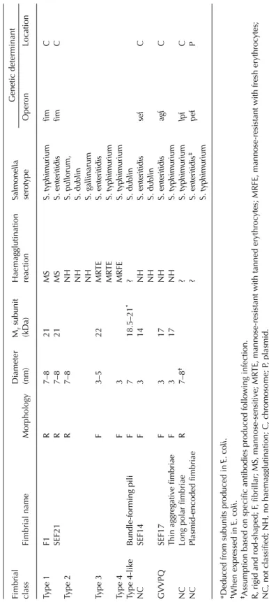
Fimbriae were first identified in Salmonella
more than 40 years ago, when it was suggested that they might assist Salmonella to attach to epithelial cells. However, although many studies claim to have demonstrated attachment, other studies have failed to detect any measurable effect. In recent years, other distinct fimbriae have been detected on some Salmonella serovars, but their role in the pathogenesis of disease requires clarification.
Although Salmonella may colonize the intes- tine without causing disease, close association and penetration of the intestinal mucosa are necessary for the induction of diarrhoea and systemic disease. Many possible virulence mechanisms have been identified in tissue culture systems and in experimental animals but there is still much to understand about the in vivo role of various Salmonella genes that may be involved in mucosal penetration. Likewise, evidence for the role of toxins is confusing and often contradictory.
Immunity to Salmonella infection has been studied predominantly in mice, but to what extent these studies are applicable to farm ani- mals is not known and the relative importance of humoral and cellular immunity in the resistance of farm animals to Salmonella infections has not yet been established. Despite our lack of knowl- edge on immunity and the pathogenesis of infec- tion, vaccines of varying degrees of efficacy have been used for many years. More recently, ratio- nally attenuated vaccines have been developed and some are undergoing field trials. The feeding of faeces from adult hens to young chickens has been shown to prevent Salmonella colonization and this competitive exclusion is being increas- ingly used in the poultry industry. However, despite many years of research, little is known about the ecology of Salmonella in the intestine and the bacteria that may prevent colonization.
The wider aspects of the ecology and epi- demiology of Salmonellaare, however, of impor-
tance to all those involved in Salmonella investi- gations. Salmonella are widespread in the envi- ronment and in recent years their prolonged persistence has been demonstrated on many calf and pig units. Indeed, some consider Salmonella to be primarily an environmental organism that is pathogenic for animals. Other survival mecha- nisms have been demonstrated and further stud- ies on the epidemiology and persistence of the organism are desirable, using the molecular tech- niques that have been used to study the patho- genesis.
The emphasis of the book is on the role of Salmonella in animal disease and it is written in five sections. In the first part, the characteristics of the microorganism are discussed. The follow- ing section considers its virulence, effect on the host and antibacterial resistance. The third part reviews current knowledge of Salmonella infec- tion in farm and companion animals. Each of the chapters in this section is intended to provide a comprehensive account, although more detailed information on some of the topics will be found in other sections of the book. Subsequent sec- tions discuss the epidemiology and prevention, and laboratory methods.
One problem that confronts all involved withSalmonella is the taxonomy and nomencla- ture of the bacteria. As discussed in Chapter 1 the full nomenclature of each serovar is compli- cated and, while taxonomically correct, it is cumbersome. As a consequence, the binomial nomenclature of the different serovars has been used.
It is hoped that the book will provide infor- mation for all those whose work involves Salmonella and that the book will assist in the control of Salmonella infections in animals to facilitate the economic production of food that is free from infection and safe for human consump- tion.
Taxonomy of the Genus Salmonella
Patrick A.D. Grimont,
1,2Francine Grimont
2and Philippe Bouvet
11Centre National de Référence des Salmonella et Shigella; 2Centre National de Référence pour le Typage Moléculaire des Entérobactéries, Unité des Entérobactéries,
INSERM Unit 389, Institut Pasteur, 75724 Paris Cedex 15, France
Introduction
The habitat of the genus Salmonellaseems to be limited to the digestive tract of humans and ani- mals. Thus, the presence of Salmonella in other habitats (water, food, natural environment) is explained by faecal contamination. Some serovars (serotypes) have a habitat limited to a host species, such as humans (serovars Typhi, Paratyphi A), sheep (serovar Abortusovis) or fowl (Gallinarum). Different infectious syn- dromes can be caused by Salmonellaserovars, e.g.
serovar Typhi causes typhoid in humans, serovar Typhimurium causes diarrhoea in humans and other animal species and a typhoid-like syndrome in mice, serovar Abortusovis is responsible for abortion in ewes and serovar Dublin has been associated with different extra-intestinal infec- tions in AIDS patients. The genetics of this pathogenic diversity is only beginning to be uncovered. Because no tools were available to identify virulence factors associated with the diverse salmonellosis syndromes, the genus Salmonella was subdivided into subspecific taxa (types), which could more or less be associated with a host species or syndrome. Furthermore, prevention of salmonellosis implies local (indus- try, hospital, district), national (national refer- ence centre) or international surveillance based on the systematic typing of strains.
History of Salmonella Taxonomy and Nomenclature
In 1884, Gaffky cultivated the typhoid bacillus (Kauffmann, 1978), which Eberth had observed in 1880 in spleen sections and mesenteric lymph nodes from a patient who died from typhoid (Le Minor, 1994). The organism now known as S.
choleraesuis was first isolated from pigs by Salmon and Smith (1886), when they considered the organism to be the cause of swine fever (hog cholera). Later, Pfeiffer and Kolle (1896) and Gruber and Durham (1896) discovered that the serum of an animal immunized with the typhoid bacillus agglutinated the typhoid bacillus. At the same time, Widal (1896) and Grunbaum (1896) found that the serum of a typhoid patient aggluti- nated the typhoid bacillus. This new test was called ‘serodiagnostic’ by Widal (1896). The same year, two isolates were recovered from patients with clinical symptoms of typhoid and negative Widal serodiagnosis (Achard and Bensaude, 1896). The organism was called
‘bacille paratyphique’.
This was only the beginning of an ongoing story and new serovars of what is now known as Salmonellaare described each year.
In an early stage, Salmonellastrains isolated from different clinical conditions or hosts were considered to be different species. This gave names such as ‘Eberthella typhosa’ (S. typhi), S. enteritidis,
‘S. abortusovis’, ‘S. gallinarum’, ‘S. bovismorbi- ficans’, S. choleraesuis or S. typhimurium. It was
© CAB International2000. Salmonella in Domestic Animals
(eds C. Wray and A. Wray) 1
soon realized that a number of these so-called species were ubiquitous.
Analysis of O and H antigens, initiated by White (1926) and extended by Kauffmann (1941), resulted in the description of a great number of serovars. The species was defined by Kauffmann (1961) as ‘a group of related sero-fer- mentative phage types’, with the result that each serovar was considered as a species. Names were given to more than 2000 serovars. These names were generally derived from the geographical location where the first strain was isolated (e.g.
‘S. london’).
This one serovar–one species concept was later found to be untenable since most serovars cannot be separated by biochemical tests.
Proposals were made to reduce the number of species. Borman et al.(1944) proposed that only three species (‘S. typhosa’, S. choleraesuisand ‘S.
kauffmannii’) should be recognized. Kauffmann and Edwards (1952) also proposed three species (‘S. typhosa’, S. choleraesuis and ‘S. enterica’).
Ewing (1963) proposed S. typhi, S. choleraesuis and S. enteritidis. All these proposals had in com- mon that, apart from S. typhiand S. choleraesuis, all serovars were placed into one species (‘S.
kauffmannii’, ‘S. enterica’ or S. enteritidis). The lat- ter was confusing as S. enteritidismeant either a precise serovar or a very large set of serovars.
Strains able to liquefy gelatin slowly and to ferment lactose were considered to form a sepa- rate genus, Arizona (Kauffmann and Edwards, 1952). After some nomenclature confusion, the name was validly published by Ewing with one species, Arizona hinshawii(Ewing, 1969).
Kauffmann (1966a,b) divided the genus Salmonella into four subgenera on the basis of biochemical reactions. These subgenera were designated by Roman numerals (I–IV) without formal nomenclature. The genus Arizonaconsti- tuted subgenus III. Later, Le Minor et al. (1970) considered Kauffmann’s subgenera to represent species named ‘S. kauffmannii’ (subgenus I), ‘S.
salamae’ (subgenus II), S. arizonae(subgenus III) and ‘S. houtenae’ (subgenus IV).
A landmark in bacterial nomenclature was the publication of the Approved Lists of Bacterial Names (Skerman et al., 1980). Names which did not appear in the Approved Lists lost standing in the nomenclature (when cited, these names should be printed with quotation marks).
All new names proposed after 1 January 1980 can
only be validated by publication or announce- ment in the International Journal of Systematic Bacteriology. The Approved Lists included five Salmonella species: S. arizonae, S. choleraesuis, S.
enteritidis, S. typhiand S. typhimurium.
DNA-relatedness studies showed that the so-called subgenera I–IV constituted a single DNA hybridization group with five subgroups delineated by studies of the thermal stability of hybridized DNA (Crosa et al., 1973; Stoleru et al., 1976; Le Minor et al., 1982, 1986). The sub- groups corresponded to the former subgenera except that subgenus III was split into DNA sub- groups IIIa and IIIb. Later, an additional sub- group (subgroup VI) was identified and a few rare serovars (Bongor group) were found to constitute a second DNA hybridization group (Le Minor et al., 1982, 1986).
However, in the absence of rules for delineat- ing bacterial species, Le Minor et al. (1982) con- sidered all Salmonella serovars to constitute a single species, which was named S. choleraesuis, since this is the name of the type species of the genus Salmonella. The species contained six sub- species: S. choleraesuissubsp. choleraesuis, S. choler- aesuissubsp. salamae, S. choleraesuissubsp. arizonae, S. choleraesuis subsp. diarizonae, S. choleraesuis subsp. houtenaeand S. choleraesuissubsp. bongori.
A new subspecies, S. choleraesuissubsp. indica,was added subsequently (Le Minor et al., 1986). This nomenclature, which strictly followed the rules of the International Code of Nomenclature of Bacteria (Rules Revision Committee, 1975) had a serious drawback, since the specific name (S.
choleraesuis) was also the name of a serovar. To overcome this, Le Minor and Popoff (1987) pro- posed the name S. entericafor the single Salmonella species, with the following subspecies, S. enterica subsp. enterica, S. entericasubsp. salamae, S. enter- icasubsp. arizonae, S. entericasubsp. diarizonae, S.
enterica subsp. houtenae, S. enterica subsp. bongori and S. enterica subsp. indica. This proposal requested an opinion from the Judicial Commission of the International Committee of Systematic Bacteriology. Unfortunately, the opin- ion has not yet been awarded, probably because the request was not limited to nomenclature (the only scope of the Judicial Commission) and included the recognition of a single species in the genus Salmonella(a taxonomic proposal).
From a taxonomic standpoint, a genomic species is now defined as a set of strains more
than 70% related by DNA–DNA hybridization with Tmvalues below 5°C (Wayne et al., 1987).
Application of these guidelines allowed the recognition of two species in the genus Salmonella– S. entericaand S. bongori(Reeves et al., 1989) – with six subspecies – S. enterica subsp. enterica, S. entericasubsp. salamae, S. enter- icasubsp. arizonae, S. entericasubsp. diarizonae, S.
enterica subsp. houtenae and S. enterica subsp.
indica. Although this nomenclature is not yet validated, it is widely used, since it is scientifi- cally based and less confusing than the S. choler- aesuisproposal.
Serovar names are no longer considered as species names and therefore should not be printed in italics. S. typhimurium becomes S.
enterica subsp. enterica serovar Typhimurium, or simply Salmonella serovar Typhimurium. Only serovars of S. enterica subsp. enterica are given names (usually geographical names). Serovars of other subspecies are designated by their O : H formula.
Phylogenetic Position of the Genus Salmonella
Bacterial classification is now based on phyloge- netic grounds. A phylogenetic tree can be derived from the comparison of 16S rRNA or other gene sequences. The two Salmonellaspecies (S. entericaand S. bongori) were separated by 16S rRNA sequence analysis. Within S. enterica, the diphasic subspecies entericaand indicawere sepa- rated from the monophasic subspecies arizonae and houtenae by 23S rRNA comparison. The genus Salmonella was found to be related to the Escherichia coli/Shigella genomic species and to Citrobacter freundiiby both 16S and 23S rRNA sequence comparison (Christensen et al., 1998).
Divergence within the genus Salmonella and proximity with E. coliand C. freundiimakes the choice of a Salmonella-specific oligonucleotide probe difficult (Lane and Collins,1991). It was discovered that 23S rRNA is fragmented in sev- eral Salmonella serovars (Winkler, 1979). This fragmentation is due to the presence of non-tran- scribed intervening sequences inserted in genes coding for 23S rRNA (Burgin et al., 1990).
More gene sequences are now used for phy- logenetic studies. A combined comparison of five gene sequences (proline permease, glyceralde-
hyde-3-phosphate dehydrogenase, malate dehy- drogenase, 6-phosphogluconate dehydrogenase and isocitrate dehydrogenase kinase/phos- phatase) yielded a phylogenetic tree consistent with DNA hybridization data (Barker et al., 1988; Beltran et al., 1988, 1991; Selander et al., 1990a,b). It is interesting that S. enterica sub- species enterica, salamae, indica and diarizonae, which are predominantly diphasic in flagellar expression, cluster apart from monophasic sub- species arizonaeand houtenae, whereas S. bongori branches apart.
From these sequence data, the following phylogenetic hypothesis has been drawn (Selander et al., 1996). The genera Salmonella and E. colimight have diverged from a common ancestor 120–160 million years ago, coincident with the origin of mammals. E. colievolved as a commensal and opportunistic pathogen of mam- mals and birds. The lineage of the Salmonella remained associated with reptiles (which are still the primary hosts of the monophasic subspecies of S. enterica) and evolved as intracellular pathogens through acquisition of genes that mediate invasion of host epithelial cells (inv/spa genes and others). Building a mechanism of fla- gellar antigen phase shifting (diphasic serovars) has permitted an extension of ecological range to mammals and birds as a pathogen (S. enterica subsp. enterica, salamae, diarizonae and indica).
S. entericasubsp. entericabecame highly special- ized for mammals and birds with some serovars adapting to single species.
S. enterica subsp. enterica serovar Typhi might have appeared when humans were avail- able as a host (3 million years ago). It has been hypothesized that serovar Typhi could have appeared first in Indonesia, where diphasic strains of this serovar (a supposedly ancestral form of the serovar) can still be found (Frankel et al., 1989a,b).
DNA Relatedness within the Genus Salmonella
A bacterial species can be defined as a DNA hybridization group. Strains within a species are generally more than 70% related and the thermal instability of reassociated DNA (Tm, diver- gence) does not exceed 5°C (Wayne et al., 1987). DNA hybridization studies (Crosa et al.,
1973; Stoleru et al., 1976; Le Minor et al., 1982, 1986), have shown the genus Salmonella to be composed of only two genomic species, S. enter- ica and S. bongori (Le Minor and Popoff, 1987;
Reeves et al., 1989). S. enterica (the most com- mon Salmonellaspecies) has been subdivided into six subspecies (Le Minor and Popoff,1987). The subspecific epithets are enterica, salamae, arizonae, diarizonae, houtenaeand indica. The habitat of S.
enterica subsp. enterica is the intestinal tract of humans and warm-blooded animals.
Population Genetics
The multilocus enzyme electrophoresis (MLEE) method has been used to assess allelic variation in multiple genes in a collection of isolates.
Electromorphs of an enzyme are equated with alle- les of the corresponding structural gene.
Distinctive allele profiles are designated as elec- trophoretic types (ETs). They represent multilocus enzyme genotypes. MLEE analysis indicates that S.
bongori is the most divergent group of Salmonella.
The other Salmonella show clusters corresponding to subspecies (Selander et al., 1990a,b).
Within S. enterica subsp. enterica, MLEE analysis shows serovar Typhi as a single clone, distinct from all other serovars studied. Serovars Paratyphi A and Sendai constitute a group, whereas serovars Typhimurium, Paratyphi B, Saintpaul, Heidelberg and Muenchen form a loose cluster.
MLEE analysis has identified serovar Enteritidis as a close relative of the non-motile serovar Gallinarum (Selander et al., 1996).
For Salmonella, a basic clonal population structure is evidenced by the presence of strong linkage disequilibrium among alleles at enzyme loci, the association of specific O and H serovars with only one or a small number of multilocus enzyme genotypes and the global distribution of certain genotypes (Selander et al., 1996).
Phenotypic Characteristics
Strains belonging to the genus Salmonellacomply with the definition of the family Entero- bacteriaceae: straight rods, generally motile with peritrichous flagella, grow on nutrient agar, aero- anaerobes, ferment glucose, often with produc-
tion of gas, reduce nitrate into nitrite and the oxidase test is negative. Some serovars have peculiarities: the avian serovar Gallinarum is reg- ularly non-motile and non-motile mutants of normally motile serovars are occasionally observed. Most Salmonella strains are pro- totrophic, i.e. they have no growth-factor requirement and can grow in a minimal medium with glucose as sole carbon and energy source and ammonium ion as nitrogen source. Some host-adapted serovars (e.g. Typhi, Paratyphi A, Sendai, Abortusovis, Gallinarum) are auxo- trophic and require one or more growth factors.
Some serovars (e.g. Typhi) never produce gas from glucose.
The following characteristics are used for Salmonella identification: urea not hydrolysed;
tryptophan and phenylalanine not deaminated;
acetoin not produced; lactose, adonitol, sucrose, salicin and 2-ketogluconate not fermented;
hydrogen sulphide (H2S) produced from thiosul- phate; lysine and ornithine decarboxylated;
growth on Simmons citrate agar; 4-methylumbel- liferyl caprylate (MUCAP) hydrolysed. Some serovars behave differently. Typhi never decar- boxylates ornithine and fails to grow on Simmons citrate agar. Paratyphi A fails to pro- duce H2S, to decarboxylate lysine and to grow on Simmons citrate agar. Subspecies other than S.
entericasubsp. entericamay ferment lactose. Tests allowing identification of these subspecies are shown in Table 1.1.
Some phenotypic properties of Salmonella are so specific that they have been used for enrichment, selective isolation or colony differ- entiation. Salmonella and other genera of Enterobacteriaceae are more resistant to novo- biocin, selenite, tergitol and bile salts, especially desoxycholate, than other bacteria. Salmonella are more resistant to brilliant green and mala- chite green than other genera of Entero- bacteriaceae. However, these properties are not sufficient for a true selective isolation and no medium is at present available with the ability to isolate only Salmonellaand no other bacteria. As
‘selective’ media are insufficiently selective, most of these media need to be more differential.
Lactose, sucrose, salicin, cellobiose or glycerol is often included with pH indicators in ‘selective’
media, since most Salmonellafail to produce acid from these substrates. Alternatively, a chro- mogenic substrate (e.g. 5-bromo-4-chloro-3-β-D-
galactopyranoside or X-gal) is used to detect β- galactosidase production. Thiosulphate and iron salts allow the production and detection of H2S unless the pH is acid. Thus, in Salmonella–Shigella (SS) agar, selective agents are bile salts and bril- liant green, substrates of interest are lactose and sodium thiosulphate and indicators are neutral red and ferric citrate. Typically, Salmonellastrains give colourless colonies with black centres.
Proteusstrains have a lower efficiency of plating than Salmonellastrains but their colonies are very similar. E. coli strains give red colonies and Citrobacter strains give red, pink or colourless colonies with or without black centres. Hektoen agar contains bile salts (selective agents), lactose, sucrose, salicin and sodium thiosulphate (sub- strates) and bromthymol blue, acid fuchsin and ferric ammonium citrate (indicators). Salmonella strains give green, black-centred colonies. XLT4 agar contains tergitol-4 (selective agent), xylose, lactose, sucrose, lysine and thiosulphate (sub- strates), a pH indicator and ferric ion. Salmonella strains produce acid from xylose. The low pH triggers lysine decarboxylation, which alkalinizes the medium around Salmonella colonies. Ferric sulphide accumulates, giving black colonies on a green background. Occasional sucrose- or lactose-fermenting Salmonella strains would give black colonies on a yellow background.
Chromagar (Rambach-agar) contains desoxy- cholate (selective agent), propylene glycol (sub- strate) and X-gal (chromogenic substrate). Most Salmonellacolonies are red or fuchsia. However,
serovars Typhi and Paratyphi A give colourless colonies. (The many different plating media are considered more fully in Chapter 21.)
Colonies that are suspected to be Salmonella can be submitted to the MUCAP test. Salmonella regularly produce caprylate esterase.
Strains of Citrobacter (when H2S-positive and lactose-negative) or Hafnia (lactose-negative at 37°C) are often misidentified as Salmonella.
However, Citrobacterstrains fail to decarboxylate lysine and often ornithine and Hafnia strains develop a characteristic reaction at 20–30°C [ortho-nitrophenyl-β-D-galactopyranoside (ONPG) hydrolysed, acetoin produced] and never produce H2S or grow on Simmons citrate agar at any temperature. Furthermore, most S.
enterica subsp. enterica strains are susceptible to phage O1 (Felix and Callow, 1943) and all are resistant to Hafnia phage (Guinée and Valkenburg, 1968), whereas Hafnia strains are often susceptible to Hafnia phage and all are resistant to phage O1.
Antigenic Diversity
Classically, three sorts of antigens are considered:
somatic (O), flagellar (H) and (mostly for serovar Typhi) surface (Vi) antigens. The antigenic structure of Salmonella has been revealed mostly by cross absorption of antisera, which subdivided antigens into different factors. The typing sys- tem, which was built up over more than 70 years Table 1.1.Phenotypic differentiation of Salmonellaspecies and subspecies.
Salmonella entericasubsp. Salmonella
Trait enterica salamae arizonae diarizonae houtenae indica bongori
ONPG test − − + + − d +
β-Glucuronidase d d − + − d −
α-Glutamyl transferase d + − + + + +
Acid from dulcitol + + − − − d +
Acid from sorbitol + + + + + + −
Acid from galacturonate − + − + + + +
Malonate alkalinized − + + + − − −
L(+) Tartrate utilized + − − − − − −
Gelatin hydrolysed − + + + + + −
Growth on KCN − − − − + − +
Phage O1 susceptible + + − + − + +
+, More than 90% strains positive; −, less than 10% strains positive; d, 10–90% strains positive; ONPG, ortho-nitrophenyl-β-D-galactopyranoside.
by White, Kauffmann and Le Minor, is a model of its kind.
O antigens
The chemical structure of diverse O factors (i.e.
the specific part of the bacterial lipopolysaccha- ride) has been determined and the genes involved in the production of essential enzymes for the assembly of some O factors have been located, cloned or sequenced. A new nomencla- ture has been proposed for the genes coding for those enzymes involved in polysaccharide syn- thesis (Reeves et al., 1996). A bacterial polysac- charide database is available on the Internet (http://www.microbio.usyd.edu.au/BPGD/default.
htm).
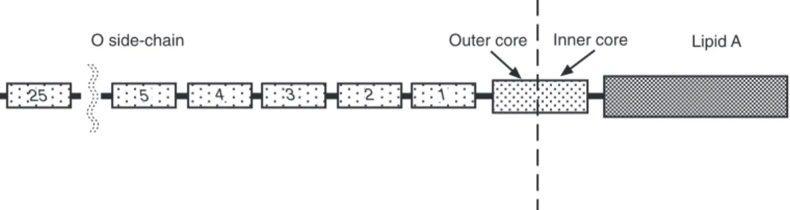
A core contains, in addition to 3-deoxy-D- manno-octulosonic acid and lipid A,
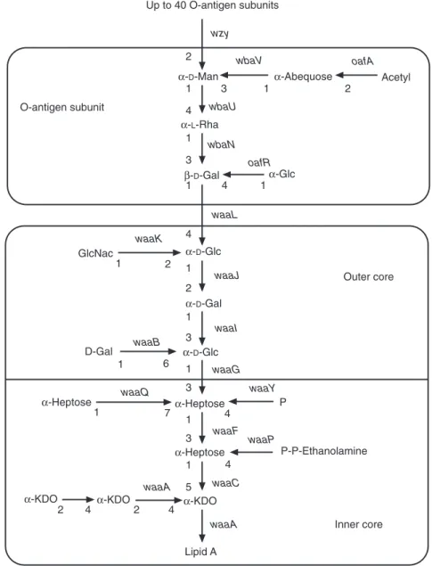
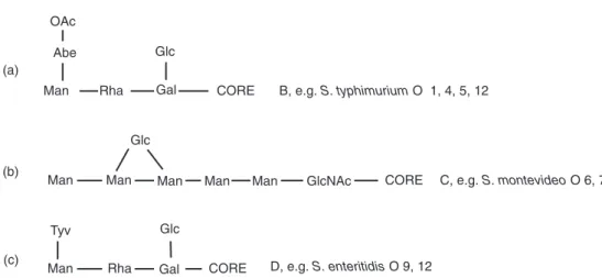
L-glycero-D-manno-heptose, D-glucose, D-galac- tose, N-acetylglucosamine and ethanolamine pyrophosphate. From this core, a poly-O side- chain extends to the bacterial surface. The poly- O side-chain is made of repeated monomers containing D-galactose, L-rhamnose, D-mannose and, for some serogroups, abequose (factor O4 in group B), paratose (group A) or tyvelose (factor 9 in group D) branched in position 1–3 on D- mannose (Rick, 1987; Jiang et al., 1991; Raetz, 1996; Fig. 1.1).
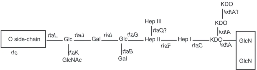
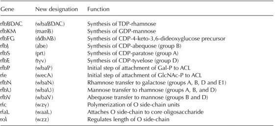
A genetic locus, rfa, located between genes cysEand pyrEat 79 minutes on the genetic map of strain LT2 (serovar Typhimurium), contains the structural genes coding for the glycosyltrans- ferases involved in core synthesis. Locus rfb, located in the vicinity of gene his (at 42 min- utes), contains the genes necessary for synthesis of an oligosaccharide monomer. Thus, from glu- cose-1-phosphate, CDP-4-keto-6-deoxyglucose is obtained by the action of glucose-1-phosphate cytidylyltransferase (coded by gene ddhA, for- merly rfbF) and CDP-glucose-4,6-dehydratase (coded by gene ddhB, formerly rfbG). Then, after action of enzymes coded by genes ddhC(formerly rfbH) and ddhD(formerly rfbI), CDP-4-keto-3,6- dideoxyglucose is obtained, and finally abequose synthase (gene abe, formerly rfbJ) produces CDP- abequose. In groups A and D, gene prt(formerly rfbS) codes for the final step yielding CDP- paratose and, in group D, the product of gene tyv
(formerly rfbE) turns CDP-paratose into CDP- tyvelose. A close examination (local G+C con- tent) of sequences in region rfb suggests that diverse parts could have been inserted or exchanged in the course of bacterial evolution.
Genes ddhC (rfbH), ddhD (rfbI) and abe (rfbJ), which control the last steps in abequose synthe- sis, could originate from genetic exchange with another species (Jiang et al., 1991). In region rfb are also located manB, formerly rfbK (phospho- mannomutase), and wbaN, formerly rfbN(rham- nosyl transferase). However, the genes, which are also involved in other syntheses (galEfor UDP- galactose-4-epimerase and pmifor phosphoman- nose isomerase), are located elsewhere on the chromosomal map (Rick, 1987; Jiang et al., 1991).
The oligosaccharide monomer is built by sequential transfer of galactose-1-phosphate, rhamnose, mannose and abequose moieties from UDP-galactose, dTDP-L-rhamnose, GDP-man- nose and CDP-abequose, on a lipid carrier, unde- caprenyl phosphate.
Oligosaccharide monomers are polymerized (action of gene wzy, formerly rfc, located in the vicinity of trp at 32 minutes) and then trans- ferred from undecaprenyl phosphate to the inde- pendently synthesized core. The lipopoly- saccharide is then translocated from the inner membrane to the surface of the outer membrane (Rick, 1987).
Mutations in regions rfaand rfbcause rough phenotypes, whereas a mutation in wzy(rfc) pre- vents the monomer polymerization (semi-rough mutants). When abequose is acetylated (effect of gene oafAlocated at 46 minutes on the genetic map), factor O4 becomes O5. Gene oafRcauses the α1–4 branching of a glycosyl residue on galactose, thus yielding factor 122.
Converting phages can modify O factor structure. Phage P22 changes the 1–4 link between glucose and galactose into a 1–6 link, thus yielding factor O1. Phage Φ27 changes the 1–2 link between monomers into a 1–6 link, thus yielding factor O27. Phages ε15 and ε34 alter several O factors in group E1 (Rick, 1987).
Plasmids can also change O factors.
A 7.5 kb plasmid has been found to deter- mine factor O54 (Popoff and Le Minor, 1985).
The lipopolyoside also carries receptors for phage binding (Ackermann and Dubow, 1987).
H antigens
H antigens are carried by flagella. These are com- posed of protein subunits called flagellin. H anti- gens are typically diphasic in Salmonella. The availability of two genetic systems (genes dis-
tantly located on the chromosome) expressing different flagellins could help the organism to survive the host’s defences (Macnab, 1987). The genes coding for the two sorts of flagellin are somewhat similar although not identical, thus suggesting that they may have resulted from the Up to 40 O-antigen subunits
wzy
wbaV oafA
wbaU
wbaN oafR
waaL waaK
waaJ
waaI
waaG waaQ waaY
waaF waaP
waaA waaC
waaA
1 3 1 2
4 1 3
1 4 1
O-antigen subunit
α-D-Man
α-L-Rha
β-D-Gal α-Glc
α-Abequose Acetyl
α-D-Glc α-D-Gal α-D-Glc
α-Heptose
α-Heptose
α-KDO
Lipid A
Inner core Outer core GlcNac
D-Gal
α-Heptose
P-P-Ethanolamine P
α-KDO α-KDO
1 2 1
2 1 3
1 6 1
3
1 7
1 4
3
1 4
4 4
2 2
5 waaB
4 2
Fig. 1.1.Chemical structure of Salmonella entericasubsp. entericaserovar Typhimurium lipopolysaccharide (adapted from Raetz, 1996, and the Bacterial Polysaccharide Gene Database). Gal, galactose; Glc, glucose;
GlcNac, N-acetyl-D-glucosamine; Heptose, L-glycero-D-manno-heptose; KDO, 3-deoxy-D-manno- octulosonic acid; Man, mannose; P, phosphate; P-P-Ethanolamine, ethanolamine pyrophosphate; Rha, rhamnose. Genes are indicated in italics following the new nomenclature (Reeves et al., 1996).
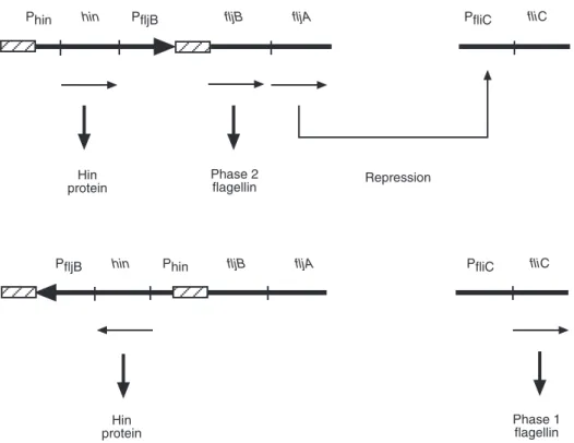
duplication of an ancestral gene. In a random fashion and after 1000–10,000 generations, the formerly silent gene is expressed and the expres- sion of the other gene is turned off (Jones and Aizawa, 1991). Gene fliC (formerly H1) is repressed by the product of gene fljA (formerly rh1) which is part of operon flj (Fig. 1.2). The expression of operon flj prevents expression of gene fliC. The alternating gene expression is due to the inversion of a 750 nucleotide-pair DNA fragment located upstream of gene fljBand which includes the promoter of fljB. In a given orienta- tion, the promoter can initiate transcription; in the other orientation, it cannot. The inversion region is flanked by inverted repeats, enabling homologous recombination. It also contains the promoter and the coding sequence of the gene hin(formerly vh2), whose product is necessary for the inversion process. It is noteworthy that the protein Hin (product of the gene hin) resembles protein TnpR of transposon Tn3. The phase inversion system could have evolved from a transposon occurring in Salmonella(Simon et al., 1980).
A non-motile Salmonellacan have structural genes fliCand fljB(and possibly have a defect in flagella assembly) and a monophasic Salmonella can be defective in phase inversion and still have the genes corresponding to both phases (Jones and Aizawa, 1991). This should be considered when nucleic acid probes are devised for the identification of major serovars. Confirming the results of an early transduction experiment (Lederberg and Edwards, 1953), Kilger and Grimont (1993) found that a fliCgene is present in the non-motile serovar Gallinarum.
There are more than 50 different alleles of gene fliC and more than 30 of gene fljB. To understand the molecular bases of such a diver- sity, some of these genes have been sequenced (Frankel et al., 1989a,b; Smith and Selander, 1990, 1991; Smith et al., 1990). Gene fliCcom- prises three parts: a 5 part containing 300 nucleotide pairs, a 3 part of 200 nucleotide pairs and a middle part of 350 nucleotide pairs.
It is remarkable that 5and 3distal parts have been largely conserved in their sequences across the diverse serovars. The sequence of the Hin
protein
fliC
Hin protein
Phase 2
flagellin Repression
PfliC
Phase 1 flagellin
Phin hin PfljB fljB fljA
PfliC fliC
PfljB hin Phin fljB fljA
Fig. 1.2.Flagellar phase inversion system in Salmonella. Promoters are indicated with capital P. Hatched zones indicate inverted repeats. Arrows indicate transcription directions.
middle part is hypervariable, with less than 32%
homology among serovars. The carboxy and amino distal parts of flagellin are essential for flagellin polymerization and secretion (hence the conserved sequences), whereas the middle part, which has no functional role, carries the major epitope of the H antigen. When compar- ing the global bacterial genotype (studied by MLEE), it appeared that strains with very simi- lar global genotypes could have very different fliC genes. Strains with very different global genotypes could have identical fliC genes (Smith and Selander, 1991). This strongly sug- gests that flagellin gene exchange has occurred among strains (possibly by transduction).
Interstrain exchange and recombination are major evolutionary mechanisms, generating both allelic variation in fliCand serovar diver- sity in natural populations of the Salmonella (Selander et al., 1990a,b).
Atypical strains of serovar Typhi have been isolated from Indonesia (Guinée et al., 1981).
Strains of serovar Typhi are normally monopha- sic with H:d. Some strains from Indonesia have a factor H:j in place of H:d and others are diphasic with H:z66. The following flagellar formulae are observed, H:d:–, or j:–, or d:z66, or j:z66. In fact, gene fliC,which codes for factor j, is identical to that which codes for factor d except for a dele- tion of 261 nucleotide pairs, which occurs in the middle part of the gene (Frankel et al., 1989a,b).
Factor z66 is coded by gene fljB. The current hypothesis is that serovar Typhi, originally dipha- sic, appeared first in Indonesia (when humans had appeared as a species), where mutations or deletions would have occurred, thus yielding monophasic variants or H:j variants. Serovar Typhi would have spread throughout the world from a monophasic H:d clone (Frankel et al., 1989a,b).
The Salmonella flagella can also carry a receptor for flagellotropic phage χ binding (Ackermann and Dubow, 1987).
Vi antigen
Vi antigen is a linear homopolymer of 2- acetamido-2-deoxy-D-galacturonic acid linked by α(1–4) bonds. This capsular polysaccharide is found in serovars Typhi, Paratyphi C and Dublin.
Three loci (viaA, viaBand ompB) are involved in
the genetic control of Vi production. Loci viaA and ompBare not limited to Vi producing species and genera and are involved in the regulation of Vi synthesis. Locus viaB is found only in strains able to produce Vi antigen. It contains genes coding for proteins involved in polysaccharide synthesis (wcdABCD), polysaccharide exporta- tion (wza, wzm, wzt, wzf) and anchoring to the bacterial surface (wcdE). A nucleic acid probe targeting viaB has been proposed for the detec- tion of serovar Typhi (Rubin et al., 1985). Such a probe also reacted with occasional other serovars which are able to produce Vi antigen. The Vi antigen is the receptor of phages ViI, ViII and ViIV (Craigie and Yen, 1938a).
Epidemiological Typing
Serotyping
The White–Kauffmann–Le Minor (WKL) (Popoff et al., 1998) scheme is a practical sum- mary of the antigenic structure of Salmonella serovars. O antigenic factors (numerals) which are easily modified by mutation are indicated in brackets and those that are determined by bacte- riophages or plasmids are underlined. In 1998, 2449 serovars were listed in the WKL scheme, 1443 in S. entericaand 20 in S. bongori. Within S. enterica, 1443 serovars belonged to subspecies entericaand were given names, 488 corresponded to subspecies salamae, 94 to subspecies arizonae, 323 to subspecies diarizonae, 70 to subspecies houtenae and 11 to subspecies indica (Popoff et al., 1998). However, only a limited number of serovars are encountered in practice. In the French National Reference Centre for Salmonella and Shigella, 19,174 strains isolated from humans were distributed into 194 serovars. Fifteen serovars represented 91% of the strains and serovars Typhimurium and Enteritidis repre- sented 69% of all Salmonellaisolates from human sources (P.A.D. Grimont and P. Bouvet, unpub- lished data).
Salmonella isolates should be sent to a national reference centre. In some countries, lab- oratories are requested to keep a minimum set of sera (anti-O4, 5 – O6, 7, 8 – O9), enabling the agglutination of 90% of isolates from human sources. Anti-Hi–Hb–Hd–HG (mixture) and Hr sera are also useful.
Phage typing
Phage typing evaluates the susceptibility (or resistance) of isolates to a set of selected bacte- riophages. Most bacteriophages were wild phages, which were isolated from sewage. Bacteriophage can also be produced by lysogenic bacteria.
International phage-typing schemes The most elaborate system had been described by Craigie and Yen (1938a,b) and Craigie and Felix (1947) for typing serovar Typhi. This sys- tem uses 87 variants derived from a single phage strain (ViII). These variants were ‘adapted’ by passage on different strains and thus submitted to different host restriction and modification systems. This set of phages allowed the subdivi- sion of serovar Typhi into 110 phage types.
Since the phage receptor is antigen Vi, isolates must produce Vi to be susceptible to any phage in the set. The specificity of typing phages is partly governed by the presence of prophages.
This system has been standardized and, to avoid host-range drift, laboratories should not multi- ply these phages for production but rather request phage suspensions from the Central Public Health Laboratory, Colindale, UK.
Unfortunately, 70% of isolates are distributed into only three phage types (A, E1 and C1). Vi- negative isolates (2.5%) and ‘degraded Vi strains’ (DVS) (9%) are untypable. A comple- mentary set composed of 13 unadapted Vi phages can subdivide phage type A into ten phage subtypes (Nicolle et al., 1954, 1958).
Type A subtype Tananarive, which is not lyso- genic, is considered as the precursor of all other phage types of Salmonella serovar Typhi.
The international scheme for serovar Paratyphi B uses a set of 12 adapted and unadapted phages and contains 48 phage types (Felix and Callow, 1943, 1951). Serovar Paratyphi B is split into biotype Paratyphi B (d-tartrate negative, associated with paratyphoid) and bio- type Java (d-tartrate positive, associated with diarrhoea). Unfortunately, phage typing cannot differentiate these biotypes (Vieu et al., 1988). In France, the most frequent phage types are 1var3 (35%) and 1010 (24%) among biotypes Paratyphi B and Java (F. Grimont, unpublished data).
The Paratyphi A scheme uses six phages and contains six phage types (Banker, 1955;
Anderson, 1964). Isolates encountered in France (imported cases) often correspond to phage types 1 (56% of isolates) and 2 (18% of isolates).
Other phage-typing systems
The Colindale scheme for serovar Typhimurium uses 37 phages and contains more than 210 phage types (Felix and Callow, 1943; Callow, 1959;
Anderson and Wilson, 1961; Anderson, 1964;
Anderson et al., 1977). Phage type DT104 shows multiple resistance to antibiotics in different European countries and is considered more fully in Chapter 6. Two other phage-typing systems have been published (Guinée and van Leeuwen, 1978).
Several phage-typing schemes have been published for serovar Enteritidis. The most widely used is that of Ward et al. (1987), which now differentiates 65 phage types by use of 16 phages. Phage type (PT) 4 has been associated with a pandemic in Europe and elsewhere. The scheme of Vieu et al. (1990) was first designed for serovar Dublin and 14 phages delineated 101 phage types. When applied to serovar Enteritidis, 85 phage types were differentiated. In this scheme, phage types correspond to either serovar Dublin or serovar Enteritidis, never to both.
Enteritidis phage type 33 corresponds to PT4 of Ward et al. (1987). There might be a relationship between phage type and insertion position of transposon IS200 (Stanley et al., 1991).
Other phage-typing schemes have been proposed for serovars Adelaide, Anatum, Bareilly, Blockley, Braenderup, Bovismorbificans, Gallinarum, Newport, Panama and Weltevreden.
These have been reviewed (Guinée and van Leeuwen, 1978). More recently described schemes were for serovars Bareilly (Jayasheela et al., 1987), Hadar (De Sa et al., 1980), Infantis (Kasatiya et al., 1978), Montevideo (Vieu et al., 1981) and Virchow (Chambers et al., 1987). Although Salmonellaphage-typing is cheap and requires no expensive equipment, the method should be in the hands of well-trained personnel. This means that it is generally limited to reference laboratories.
Bacteriocin typing
Bacteriocin typing of serovar Typhimurium has been described (Barker, 1980). However, it is sel- dom used.
Biotyping
Biotypes have been described in serovar Typhimurium (Cordano et al., 1971; Pohl et al., 1973; Duguid et al., 1975; Descamp et al., 1982).
However, the choice of tests for biotyping has often been empirical.
Utilization of d-tartrate (= L-tartrate) is used to separate two biotypes in serovar Paratyphi B. Biotype Paratyphi B cannot utilize d-tartrate whereas biotype Java can. Biotype Java is commonly associated with diarrhoea and iso- lated from stools, whereas biotype Paratyphi B is often associated with paratyphoid and isolated from blood or stools.
Plasmid profiling and plasmid restriction profiling
It is now easy to extract plasmid DNA and to separate plasmids of different sizes by agarose gel electrophoresis and ethidium bromide staining.
Isolates derived from the same epidemic strain will have plasmids with identical sizes. However, this test requires the presence of at least one plas- mid type. Some phage types in serovars Enteritidis (Threlfall et al., 1989) and Typhimurium (Threlfall et al., 1990) were subdi- vided by plasmid profiling.
More precise results are obtained when plas- mid DNA is extracted, digested by a restriction endonuclease and the fragments separated by agarose gel electrophoresis. Identical plasmids should have the same restriction pattern (Tacket, 1989).
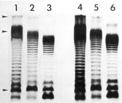
rRNA gene restriction patterns (ribotyping) When bacterial DNA is extracted, purified, digested by a restriction endonuclease and the fragments separated by agarose gel electrophore- sis, fragments in restriction patterns are too numerous for these patterns to be compared.
Instead, the fragments in the gel are transferred to a nylon membrane, to maintain their relative positions, and hybridized with a labelled mixture of 16+23S rRNA. Revealing the label (autoradi- ography or immunoenzymatic reaction) yields simpler patterns, often referred to as ribotypes (Grimont and Grimont, 1986).
Ribotyping has uncovered a wide hetero- geneity within serovar Typhi (Altweg et al., 1989). A diversity of ribotypes has been found in the most frequent phage types, A and E1. Such diversity is in contrast with the homogeneity of MLEE (Reeves et al., 1989) and could be explained by the presence of ‘intervening sequences’ inserted in 23S rRNA genes (Burgin et al., 1990).
Ribotyping of other serovars gives very dif- ferent results. Some serovars have almost a serovar-specific ribotype. However, for serovars of group B (e.g. Typhimurium, Paratyphi B), there is no relationship between ribotype and serovar (F. Grimont, unpublished data).
Pulse-field gel electrophoresis (PFGE)
PFGE uses restriction endonucleases, which have infrequently occurring restriction sites in a given bacterial DNA. A small number of fragments of a much larger size are produced. Conventional agarose gel electrophoresis cannot resolve DNA molecules larger than ~20 kb pairs. Thus, to sep- arate large DNA molecules above 50 kbp, PFGE which allows resolution of DNA molecules mil- lions of base pairs long is used. The most sophis- ticated configuration for this technique is called clamped homogeneous electric field (CHEF) electrophoresis, which uses an array of hexagon- ally arranged electrodes to generate uniform elec- tric fields at an angle of 120°C to each other, thus ensuring that large DNA fragments migrate through the gel in a straight line. Although PFGE is highly discriminative for Salmonellawith the endonucleases XbaI, BlnI or SpeI, it is expen- sive and time-consuming and standardization, analysis and comparison of restriction profiles require effort (Murase et al., 1995).
IS200 typing
DNA of most Salmonellaserovars contain several copies of a 708-base-pair insertion sequence, IS200. Stanley et al. (1991) found that isolates could be differentiated by comparing the restric- tion patterns of bacterial DNA after hybridiza- tion with an IS200 probe. Strains differed by the number of visualized fragments (IS200 number of copies) and the size of fragments.
Random amplification of polymorphic DNA (RAPD)
RAPD is a rapid genomic typing method of broad application. This technique uses the poly- merase chain reaction (PCR), in which a single arbitrarily chosen primer (usually 10-mer) can be annealed at multiple sites throughout the genome. The profiles of amplified products are characteristic of the template DNA. An RAPD method has been developed to differentiate S.
enteritidis isolates (Lin et al., 1996), providing more discrimination than any other subtyping method. This method proved to discriminate between isolates of different Salmonella serovars (Hilton et al., 1996). However, between-labora- tory reproducibility is a problem (Meunier and Grimont, 1993).
ERIC-PCR
A repetitive element, highly conserved, called the enterobacterial repetitive intergenic consen- sus (ERIC) sequence, about 126 bp in length, was described by Hulton et al. (1991). The chro- mosome locations of ERIC sequences can differ in different species and strains. This element has been identified in Salmonella and can be used for typing.
Restriction of amplified flagellin genes In an attempt to substitute molecular methods for serotyping, flagellin genes from 264 serovars were amplified by two phase-specific PCR sys- tems (Dauga et al., 1998). Amplification prod- ucts were cleaved by endonucleases HhaI or HphI. Restriction of 329 (195 phase 1 plus 134 phase 2) flagellin genes coding for 26 antigens yielded 64 HhaI profiles and 42 HphI profiles.
The phase 1 gene (fliC) showed 46 patterns with HhaI and 30 patterns with HphI. The phase 2 gene (fljB) showed 23 patterns with HhaI and 17 patterns with HphI. When the data from both enzymes were combined, 116 patterns were obtained: 74 for fliC, 47 for fljBand five shared by both genes. Of these combined patterns, 80%
were specifically associated with one flagellar antigen and 20% were associated with more than one antigen. Each flagellar antigen was divided
into two to 18 different combined patterns. The pattern corresponding to serovar Typhi H:d was specific and different from other H:d patterns.
The pattern corresponding to serovar Typhimurium H:i was also unique and differed from other H:i patterns. Overall, the diversity uncovered by restriction of flagellin genes did not precisely match that evidenced by flagellar agglu- tination.
New Approaches for the Detection of Salmonella
The procedure of the colorimetric Gene-Trak Salmonella assay used nucleic acid probes to detect polynucleotide sequences that are uniquely conserved among Salmonella bacteria.
This method was considered equivalent to the conventional Bacteriological Analytical Manual/AOAC culture method (Foster et al., 1992).
PCR will amplify DNA molecules 1000- fold, but the presence of a specifically amplified product must be identified. Several PCR primers for specific detection of Salmonella have been published utilizing specific gene sequences for targeting. Kwang et al.(1996) described the use- fulness of a primer set of oligonucleotides from the ompCgene of Salmonella. This primer set suc- cessfully amplified 40 Salmonellaserovars, but not 24 non-Salmonella bacteria. The sensitivity for boiled whole bacteria was 400 cells.
The magnetic immuno-PCR assay utilizes magnetic particles coated with monoclonal anti- bodies to extract bacteria from a sample. Fluit et al.(1993) applied this method to Salmonellaand detected the presence of 0.1 colony-forming units (cfu) of Salmonellag−1of chicken meat after enrichment.
An alternative method for analysing PCR products utilizes the oligonucleotide ligation assay (OLA). The OLA procedure used two adja- cent oligonucleotides. The first one (capture probe) is 5-biotinylated with the 3 end adja- cent to the second probe. The second (reporter) probe is 5-phosphorylated and 3-end-labelled with a reporter substance, such as digoxigenin. If the two oligonucleotides are hybridized to the target DNA, DNA ligase covalently joins the two oligonucleotides. The capture of the bio- tinylated probe is accomplished by binding the
biotin to immobilized streptavidin in a micro- titration plate. Stone et al. (1995) described a combined cultivation and PCR-hybridization procedure enzyme-linked immunosorbent assay (ELISA)-based oligonucleotide ligation assay, adapted to a 96-well microtitration PCR-OLA format for detection of the product from the invE and invAgenes of Salmonellaserovars. Sensitivity, specificity and predictive value were comparable to the conventional culture.
A digoxigenin-based ELISA (DIG-ELISA) following a PCR to detect the amplified lipopolysaccharide rfbSgene as a means for rapid screening of serogroup D Salmonellain stool spec- imens was described by Luk et al.(1997). In the presence of stool materials, the Salmonella were isolated by an immunomagnetic separation tech- nique with an O9-specific monoclonal antibody, followed by PCR and DIG-ELISA. The sensitiv- ity was 10–100 bacteria.
A recent technique for the separation/con- centration of bacteria from background food is the immunomagnetic separation (IMS) proce- dure, which uses a novel biosorbent consisting of a Salmonella-specific bacteriophage immobilized on to a solid phase (Bennett et al., 1997). This biosorbent could remove Salmonellafrom culture fluid and separate Salmonellafrom suspensions of other Enterobacteriaceae. The advantage is the ease of production of phage, the high affinity of phage–cell interaction and the ability of phage to infect host cells.
Chen et al.(1997) have developed a rapid, sensitive and automated fluorescence PCR method, the AG-9600 AmpliSensor assay, for the detection of Salmonella species in food
samples. The AmpliSensor assay comprises two steps: an initial asymmetric amplification with normal primers to overproduce one strand of the target and subsequent semi-nested amplification and signal detection, in which one of the outer primers and the AmpliSensor primer direct the amplification. The AmpliSensor primer is a double-stranded signal probe; one strand is labelled with fluorescein isothiocyanate and the other with Texas red.
During semi-nested amplification, one strand of the AmpliSensor duplex serves as a primer and the other as an ‘energy sink’. The semi-nested amplification results in strand dissociation of the duplex and disruption of the fluorescence signal. The extent of signal disruption is propor- tional to the amount of the primer incorpora- tion into the amplification product and can be measured cycle by cycle and used for quantifica- tion of the initial target.
Cocolin et al. (1998) described a primer set of oligonucleotides from the invA gene of Salmonellawhich amplified 33 Salmonellaserovars but did not amplify 16 non-Salmonella bacteria.
Moreover, after PCR amplification, it was possi- ble to identify serovar Typhimurium by HinfI restriction enzyme analysis.
A Salmonella-specific PCR system targeting a virulence gene with hybridization to a cova- lently immobilized oligonucleotide probe on a microplate (Chevrier et al., 1995) is now mar- keted under the name Probelia.
Another PCR system, involving elec- trophoresis, has been devised for Salmonella and is marketed under the name BAX (Bailey, 1998).
References
Achard, C. and Bensaude, R. (1896) Infections paratyphoïdiques. Bulletin et Mémoires de la Société de Médecine des Hôpitaux de Paris 13, 820–833.
Ackermann, H.-W. and Dubow, M.S. (1987) Viruses of Prokaryotes, Vol. 1. CRC Press, Boca Raton, Florida, 202 pp.
Altwegg, M., Hickman-Brenner, F.W. and Farmer, J.J., III (1989) Ribosomal RNA gene restriction patterns provide increased sensitivity for typing Salmonella typhistrains. Journal of Infectious Diseases160, 145–149.
Anderson, E.S. (1964) Phage typing of Salmonella other than Salmonella typhi. In: van Oye, W. (ed.) The World Problem of Salmonellosis. Junk Publishers, The Hague, pp. 89–110.
Anderson, E.S. and Wilson, E.M.J. (1961) Die Bedeutung der Salmonella typhimurium-Phagen-typisierung in der Human- und Veterinarmedizin. Zentralblatt für Bakteriologie, Mikrobiologie und Hygiene Series I. Abteilung Orig.
224, 368–373.
Anderson, E.S., Ward, L.R., De Saxe, M.J. and De Sa, J.D.H. (1977) Bacteriophage-typing designations of Salmonella typhimurium. Journal of Hygiene, Cambridge 78, 297–300.
Bailey, J.S. (1998) Detection of Salmonella cells within 24 to 26 hours in poultry samples with the polymerase chain reaction BAXtm. Journal of Food Protection 61, 792–795.
Banker, D.D. (1955) Paratyphoid A phage typing. Nature175, 309–310.
Barker, R.M. (1980) Colicinogeny in Salmonella typhimurium.Journal of General Microbiology120, 21–26.
Barker, R.M., Kearney, G.M., Nicholson, P., Blair A.L., Porter, R.C. and Crichton, P.B. (1988) Types of Salmonella paratyphi Band their phylogenetic significance. Journal of Medical Microbiology 26, 285–293.
Beltran, P., Musser, J.M, Helmuth, R., Farmer, J.J., III, Frerichs, W.M., Wachsmuth, I.K., Ferris, K., McWhorter, A.C., Wells, J.G., Cravioto, A. and Selander, R.K. (1988) Toward a population genetic analysis of Salmonella:
genetic diversity and relationships among strains of serovars S. choleraesuis, S. derby, S. dublin, S. enteritidis, S.
heidelberg, S. infantis, S. newport, and S. typhimurium. Proceedings of the National Academy of Sciences USA 85, 7753–7757.
Beltran, P., Plock, S.A., Smith, N.H., Whittam, T.S., Old, D.C. and Selander, R.K. (1991) Reference collection of strains of the Salmonella typhimurium complex from natural populations.Journal of General Microbiology137, 601–606.
Bennett, A.R., Davids, F.G.C, Vlahodimou, S., Banks, J.G. and Betts, R.P. (1997) The use of bacteriophage-based systems for the separation and concentration of Salmonella. Journal of Applied Microbiology83, 259–265.
Borman, E.K., Stuart, C.A. and Wheeler, K. (1944) Taxonomy of the family Enterobacteriaceae. Journal of Bacteriology 48, 351–367.
Burgin, A.B., Parodos, K., Lane, D.J., Pace, N.R. (1990) The excision of intervening sequences from Salmonella 23S ribosomal RNA. Cell60, 405–414.
Callow, B.R. (1959) A new phage typing scheme for Salmonella typhimurium. Journal of Hygiene, Cambridge 57, 346–559.
Chambers, R.M., McAdam, P., De Sa, J.D.H., Ward, L.R. and Rowe, B. (1987) A phage typing scheme for Salmonella virchow. FEMS Microbiology Letters 40, 155–157.
Chen, S., Yee, A., Griffiths, M., Wu, K.Y., Wang, C.-N., Rahn, K. and De Grandis, S.A. (1997) A rapid, sensitive and automated method for the detection Salmonellaspecies in foods using AG-9600 AmpliSensor Analyser.
Journal of Applied Microbiology83, 314–321.
Chevrier, D., Popoff, M.Y., Dion, M.Y., Hermant, D. and Guesdon, J.L. (1995) Rapid detection of Salmonellasub- species I by PCR combined with non-radioactive