Represents a graph showing the mean cell size in µm2 of both EOM-MSC and BM-MSC. These cells were termed central orbital adipose tissue mesenchymal stem cells (C-OAT-MSC) and medial orbital adipose tissue mesenchymal stem cells (M-OAT-MSC).
REVIEW OF LITERATURE
Stem cells
The bone marrow-derived mesenchymal stem cells and its properties The bone marrow is a soft spongy tissue localized within the hollow interior of.
The bone marrow-derived mesenchymal stem cells and its properties 11
MSCs from various fetal tissues
Both UCB-MSC and BM-MSC differentiated into hepatocyte cells with no significant difference in potency (K.-D. Lee et al., 2004). As a result, spindle-shaped UCB-MSC readily differentiated into adipogenic cells with downregulation of CD73, CD90, and CD105 markers (Sibov et al., 2012).
MSCs exist in all the tissues of the human adult
AT-MSC and BM-MSC efficiently differentiated into the three mesodermal lineages, while UCB-MSC did not form the adipocytes (Kern et al., 2006). Adipose tissue expanded in culture for a long time, but longer compared to UCB-MSC (Franco Lambert et al., 2009).
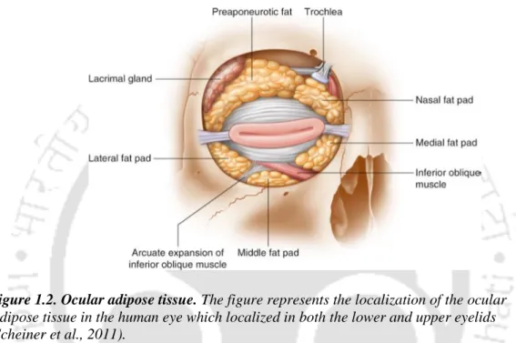
Stem cells of the eye
Similarly, MSC were detected in the bone marrow and adipose tissue of the horse (Iacono et al., 2015). Remarkably, nestin was only expressed by the central adipose tissue-derived adult stem cells (Korn et al., 2009).
Mesenchymal stem cells in neural repair
Neurological disorders
The impaired generation of new hippocampal neurons from endogenous NSCs in the subgranular zone of the dentate gyrus characterizes Alzheimer's disease (Vishwakarma et al., 2014). First, the stem cells were converted into NSCs or neural precursor cells via neurosphere generation or grown as monolayer culture (CN Svendsen et al., 1998).
Neurosphere formation
Differentiation of BM-MSCs into the neuronal lineage… 27
On the other hand, BM-MSC expressed β-ш-tubulin and MAP-2 when they were induced with bFGF followed by the addition of steroid hormones (β-estradiol, progesterone and testosterone) (Parivar et al., 2015). First, embryoid bodies were generated and secondly differentiated into neurons which expressed Nkx6.1 and Olig2 ( Karumbayaram et al., 2010 ).
Clinical trials of MSCs from BM and various tissues towards treatment
Overall, there was no increase in neurological recovery, although differentiated cells, undifferentiated and immature BM-MSC remained in the grafts (Zhao et al., 2002). These bone marrows entered the brain and differentiated into cells that displayed neuronal features and expressed neuronal markers NeuN and Kv2.1, although the survival rate was very low (Mezey, et al., 2003). Astrogliosis was significantly reduced and there was a marked decrease in the extent of demyelination and axonal loss in transplanted mice (Pluchino et al., 2003).
However, MSCs were more prone to form glial cells instead of neurons (K. N. Kim et al., 2006). Coordination between forelimb and hindlimb movements improved in the host (S. Wu et al., 2003). RPCs are preferred for retinal transplantation (Tomita et al., 2006), although BM-MSCs are the best option for the treatment of neurological disorders.
Neuroprotection
Whether or not true differentiation occurs after transplantation is of concern, as engraftment of a non-neural cell into an injured neural region can occur through spontaneous cell fusion (Terada et al., 2002). One advantage that occurred is that cell fusion did not occur because the cells did not reach a polyploid state after chromosomal analysis of BM-derived lung, kidney, and muscle (Herzog et al., 2003). BDNF-transfected BM-MSCs proliferated more, showed fibroblastic morphology, but secreted a higher concentration of BDNF in vitro, expressed nestin, NSE, and GFAP, which later differentiated into neural cells (Q. Liu et al., 2015) .
In vitro differentiation prior to transplantation is considered safer than direct in vivo implantation, although both the naive MSCs and MSCs differentiated into neural lineages could be implanted into a damaged tissue (Yaghoobi & Mahani, 2008) and showed a higher survival rate than naive MSCs within the damaged tissue (Deng et al., 2006). This was further supported by the study conducted by (Hayashi et al., 2013) when MSC isolated from Parkinson's macaque was converted into dopaminergic neurons and implanted in the macaque brain. That is, BM-MSC co-cultured with cortical neurons secretes BDNF that prevents neuronal cell death compared to cortical neurons cultured in chemically defined medium.
Factors involved in neuronal differentiation in vitro
Under serum and nutrient cell-free condition, in addition to EGF, bFGF and PDGF, BM-MSC differentiated into neurons. As a result of the differentiation, neurotransmitters were also expressed with 30% of the BM-MSC expressing NF-M and GABA, approximately 40-60%. Therefore, when BM-MSC was treated with cytokines, GABAergic, dopaminergic and serotonergic neurons were generated in vitro, respectively.
For example, BM-MSC viability was reduced when exposed to media containing DMSO/BHA followed by BME, but viability remained unaffected when exposed to ATRA and indomethacin/IBMX. Again, BM-MSC treated with indomethacin/IBMX expressed the highest level of MAP2 and GFAP while NSE expression was not affected by any inducer. It was therefore necessary to determine the efficacy of natural growth factors such as IGF, EGF, bFGF and NT-3 to induce neuronal differentiation in BM-MSCs.
OBJECTIVES
MATERIALS AND METHODS
MATERIALS
- Chemicals
- Cell culture media and reagents
- Media
- A. Transportation media for sample collection
- B. Media for maintenance of mesenchymal stem cells.44
- Cell Culture reagents
- Antibodies
- Antibodies used for flow cytometric analysis
- Antibodies used for immunofluorescence
- Growth factors
- Magnetic labelled beads for cell separation
- Primers used for quantitative real time PCR
- Preparation of working solutions
- Materials used
The vials were stored at 4°C until extraocular muscle tissue and ocular adipose tissue were collected. 250 µl of sodium nitrate solution was mixed with 250 µl of FBB alkaline solution and 11.25 ml of deionized water was added. Finally, 250 µl of Napthol AS-Bl-Alkaline solution was added and the substrate was ready for use.
Microcentrifuge tubes, microtips, and microtip boxes were treated overnight with DEPC against RNAses at a concentration of 0.02% in distilled water. Dissolve the solution by continuously adding NaOH and stir over a hot plate or water bath set at 55 °C - 65 °C until the milky appearance of the solution disappears. The remaining volume of the solution was supplemented and the solution was filtered and aliquoted into sample vials (10 ml) and stored at -20 °C.
METHODS
- Isolation of mesenchymal stem cells from human ocular tissue
- Fibronectin coating
- Isolation of mesenchymal stem cells from human ocular
- Isolation of mesenchymal stem cells from human extra ocular
- Clonal cell derivation
- Maintenance of mesenchymal stem cells in culture
- Cell harvesting
- Cell count
- Cell cryopreservation
- Cell freezing for RNA
- Growth properties of mesenchymal stem cells
- Population doubling time
- Colony forming unit-fibroblast
- Study of the morphology of MSCs using FESEM
- Karyotype Analysis
- Differentiation of MSCs into the mesodermal lineage
- Adipogenic differentiation
- Osteogenic differentiation
- Chondrogenic differentiation
- Neuronal differentiation of mesenchymal stem cells
- Formation of neurosphere
- Neuronal differentiation by direct conversion
- CD49B separation by magnetic-activated cell sorting
- Detection of GFAP using immunofluorescence
- Expression of surface protein markers expressed in MSCs
- Immunofluorescence staining
- Mitochondria staining using TMRE
- Stage specific embryonic antigen staining
- Phenotypic studies by flow cytometry
- Gene expression studies
- RNA Isolation
- cDNA synthesis by reverse transcription
- Real time quantitative PCR
- Statistical Analysis
The cells were diluted with trypan blue by a dilution of (1:1) and counted in the hemocytometer (Neubeau, Germany). Dilution factor – the number of times the cell mixture was diluted to obtain a count. Number of squares – the number of squares taken into account for the count 104 = the area of the hemocytometer. The cells were gold coated with a sputter coater and viewed under a Field Emission Scanning Electron Microscope (Zeiss, Sigma).
Cells were washed with PBS and fixed with neutral buffered 10% formalin for 20 min at room temperature. The cells were washed with PBS and incubated with anti-SSEA4 antibody for one hour. The cells were incubated at 4°C for 30 minutes, washed and analyzed by FACS caliper (Becton Dickinson).
Isolation and characterization of mesenchymal stem cells from ocular
Isolation of MSC from ocular adipose tissue
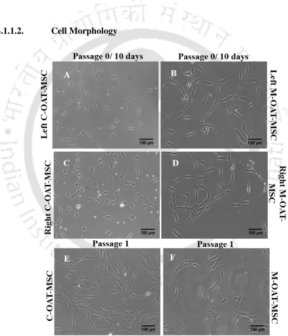
Cell morphology
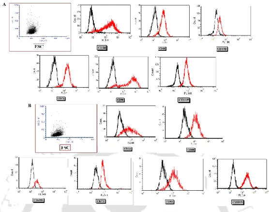
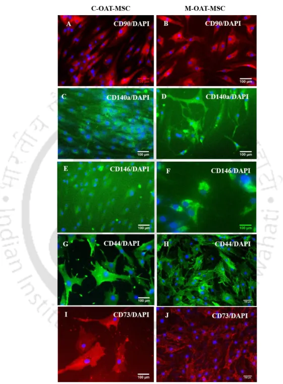
The surface marker expression of OAT-MSC
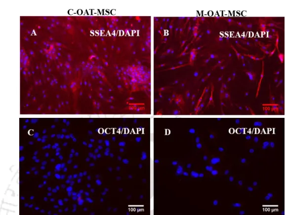
The expression of embryonic markers in OAT-MSC
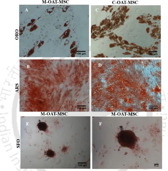
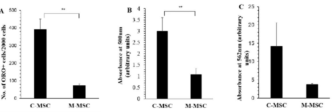
Differentiation of OAT-MSC into mesodermal lineage
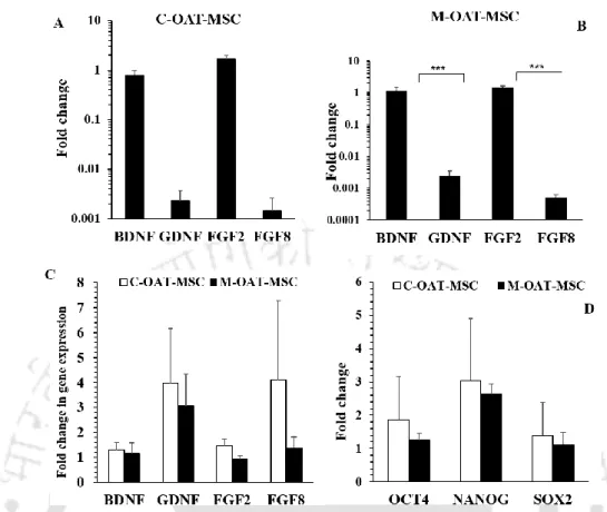
Gene expression studies
Neuronal differentiation of OAT-MSC
Isolation of mesenchymal stem cells from extra ocular muscle
Growth properties of extra ocular muscle MSC
Differentiation of EOM-MSC into the mesenchymal
Cell surface marker expression analysis
Expression of embryonic stem cell marker
Gene expression analysis
Mitochondrial distribution in EOM-MSC
Neuronal differentiation of EOM-MSC
Neurosphere formation by addition of growth factors and
Since ocular muscle is a highly innervated tissue, the neuronal differentiation of EOM-MSC was tested. The neurosphere assay was performed to test the presence of neural stem cells, which have the potential to differentiate into different neuronal lineages. EOM-MSC in neuronal media supplemented with RE formed neurosphere within 4 days of induction.
Neurospheres appeared few in number and only after 8 days of induction in cultures not supplemented with RE. Neurosphere formation was induced only by the addition of growth factors and additionally with RE. Figures (A) were the neurosphere formed within four days of induction supplemented with RE, (B) the onset of neurosphere formation in EOM-MSCs without RE within four days of induction (C) the neurosphere formed after 8 days of induction supplemented with RE, (D ) neurosphere formed after 8 days of induction in neuronal media without RE.
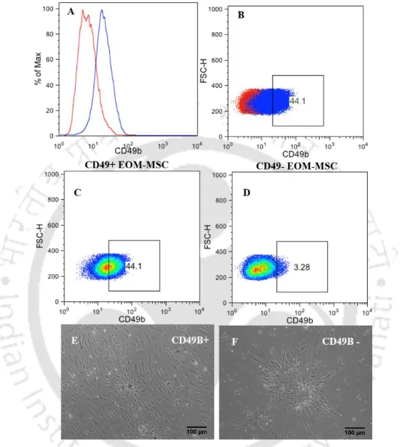
CD49B separation and neurosphere formation
The numbers (E) represent the neurospheres after 11 days in differentiation media supplemented with retinal extract and (F) neurospheres in differentiation medium without retinal extract supplement during 11 days of differentiation. Neurosphere formed both in the presence and absence of RE stained positive for GFAP by immunocytochemical analysis. Because EOM-MSC showed heterogeneous CD49B expression and CD49B-positive cells had a higher multipotent neurosphere-forming capacity (Hmjb et al., 2011).
Histogram (A) shows flow cytometry showing the expression of CD49B in EOM-MSCs, where the red line indicates the isotype control staining profile compared to the blue line indicating the specific antibody staining profile, graph (B) the percentage of cells expressed in EOM-MSCs , (C) percentage of population expressing CD49B after separation, graph (D) CD49B negative population after MACS separation (E) Neuronal differentiation of CD49B+ (F) CD49B- cells after 7 days of induction.
Morphological changes and surface markers expression in
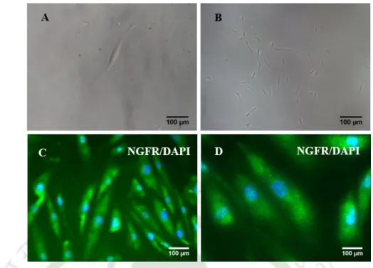
Immunofluorescence analysis of neuronal differentiated EOM-MSC showing that (A) CD90 expression was unaffected in EOM-MSC cultured in neuronal media. B) downregulation of CD146 in cells cultured neuronal media. Neuronal differentiated cells showed upregulation of neuronal lineage markers (C). expression of GD2 was detected in few differentiated cells (D) MAP2B (E) NGFR/CD271 and (F) GFAP. CD90, CD146 and GFAP staining was performed 9-10 days after addition of neuronal induction media while NGFR, MAP2B and GD2 staining was performed after 21 days of neuronal induction.
Change in gene expression during neuronal differentiation of
However, the expression of the embryonic transcription factors OCT4, SOX2 and NANOG was downregulated during neuronal differentiation, which was not observed when analyzed in the undifferentiated state. Gene expression in EOM-MSCs when differentiated into neurons detected by real-time PCR analysis. Gene expression levels were normalized to the expression level of GAPDH in the corresponding samples.
Real-time PCR analysis of OCT4, SOX2 and NANOG in EOM-MSCs cultured in growth medium and neural differentiation medium for 14 days.
Neuronal differentiation of clonal-derived EOM-MSC
Their mesenchymal differentiation capacity was significantly less compared to BM-MSC, while they had higher embryonic transcription factors and nestin gene expression and possessed high neuronal differentiation capacity making them suitable candidates for cell therapy to treat neurodegenerative diseases.
DISCUSSION
Isolation and characterization of mesenchymal stem cells from ocular
Comparative analysis of mesenchymal stem cells from bone marrow, umbilical cord blood or adipose tissue. Properties and neural-like differentiation of mesenchymal stem cells derived from fetal porcine bone marrow. Comparative study of neural differentiation of bone marrow mesenchymal stem cells by different induction methods.
Immunomodulatory effect of cultured human adipose tissue-derived stem cells: Comparison with bone marrow mesenchymal stem cells. Comparison of neural differentiation potential of human mesenchymal stem cells from amniotic fluid and adult bone marrow.
Isolation and characterization of mesenchymal stem cells from extra ocular
Differentiation of EOM-MSC into neuroectodermal lineage