I declare that the content of the research work described in this thesis entitled "Studies on the role of NGALR in triple negative breast cancer", is a presentation of my original research work carried out in the Department of Biosciences and Bioengineering, Indian Institute of Technology Guwahati, India, under the supervision of Prof. This is to certify that the work described in the thesis entitled "Studies on the role of NGALR in triple negative breast cancer" submitted by Krishan Kumar Thakur (Roll no to Indian Institute of Technology Guwahati, India, for the award of Ph.D. Philosophy is an authentic record of research work carried out under my supervision at the Department of Biosciences and Bioengineering, Indian Institute of Technology Guwahati, Guwahati, India.
Introduction and Review of Literature
Introduction
Recent studies have reported that dysregulation in iron homeostasis is associated with cancer cell proliferation and survival (Lelievre et al., 2020; Jung et al., 2019). Dysregulation of NGALR is associated with proliferation, survival, invasion and metastasis of cancer cells (Wei et al., 2020; Santiago-Sanchez et al., 2020).
Sub-types of breast cancer
- Luminal A
- Luminal B
- Triple-negative/basal-like
- HER2-enriched
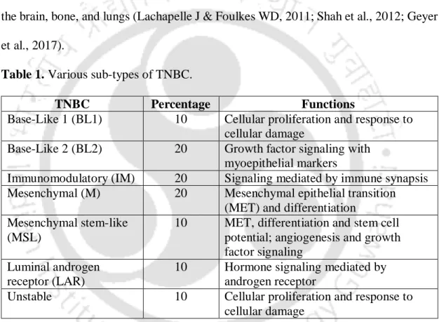
Also, luminal A tumors are more common in postmenopausal women than premenopausal women (Saha et al., 2016; . Ellingjord-Dale et al., 2017). TNBC is the most heterogeneous subtype characterized by different categories, such as clinical, morphological, genetic, proteomic, transcriptomic, epigenomic and microenvironmental (Garrido-Castro et al., 2019; Aleskandarany et al., 2018).
Tumor, lymph node, and metastasis (TNM) staging of breast cancer
It is characterized by the TP53 mutation (40-80%) and activating mutations of the p110α subunit of phosphoinositide 3-kinase (PI3K) with high expression of several genes, such as post-GPI attachment to proteins 3 (PGAP3), growth factor receptor-bound protein 7 (GRB7) and epidermal growth factor receptor (EGFR). The stage of breast cancer is described by the TNM staging system, which is overseen by the American Joint Committee on Cancer (AJCC).
Risk factors of breast cancer
- Gender
- Weight
- Diet
- Physical activity
- Alcohol consumption and smoking
- Genetic factors
- Exposure to estrogen
- Pregnancy and breastfeeding
- Other factors
However, active smoking, especially in prenatal and postmenopausal women, is associated with a high risk of breast cancer (Momenimovahed Z & Salehiniya H, 2019; Luo et al., 2011). Emerging studies have shown that the risk of breast cancer decreases with increasing parity (Kim et al., 2015; Ma et al., 2010).
Sign, symptoms, and clinical features of breast cancer
Conversely, increased incidences of abortion were associated with increased risks of breast cancer (Sun et al., 2017; . Momenimovahed Z & Salehiniya H, 2019; Bhadoria et al., 2013). Various studies have reported the protective and preventive effects of breastfeeding against the risk of breast cancer in women (Anstey et al., 2017; Holm et al., 2017).
Molecular alterations in TNBC
- BRCA1 and BRCA2
- PI3K Pathway
- Tyrosine kinase receptors
- Nuclear factor kappa-light-chain-enhancer of activated B cells (NF-κB) pathway
- JAK/STAT pathway
Poly (ADP-ribose) polymerase (PARP) inhibitors are significantly effective in patients with BRCA1/2 deficiency (Plummer R, 2011; Bartsch et al., 2010). The activated STAT enters the nucleus and modulates the transcription of several target genes (Guanizo et al., 2018).
Treatment modalities of TNBC
- Surgery and radiation
- Chemotherapy
- EGFR inhibitors
- PARP inhibitors
- Aromatase inhibitors
- Inhibition of PI3K/Akt/mTOR pathway
- Immunotherapy
- Others
Various clinical trials are ongoing with Akt inhibitors (such as ipatasertib, capivasertib) and paclitaxel for the treatment of TNBC (Kim et al., 2017; Dent et al., 2018; Schmid et al., 2020). Furthermore, induction of type I immunity microenvironment improves TNBC therapy and reduces recurrences (Alistar et al., 2014).
Challenges with TNBC Therapies
- Tumor recurrence
- Chemoresistance
Therefore, the cure for TNBC continues to elude even after the significant advances in the chemotherapeutic approaches (O'Reilly et al., 2015; Nedeljkovic M & Damjanovic A, 2019; Wein L & Loi S, 2017; Crown et al., 2012). Several mechanisms of chemoresistance associated with TNBC include; ATP-binding cassette (ABC) transporters, mutations in DNA repair enzymes (DNA mismatch repair enzymes and topoisomerase II), overexpression of β-tubulin, changes in genes related to apoptosis, aldehyde dehydrogenase 1 (ALDH1) and glutathione/glutathione- S- transferase (GST), and NF-κB signaling pathways (Zhao J, 2016; Nedeljkovic M & Damjanovic A, 2019; Yuan et al., 2018; O'Reilly et al., 2015; Bao et al., 2017) .
Neutrophil gelatinase-associated lipocalin receptor
Furthermore, the two proteins were moderately expressed in the cortex and medulla of the embryonic adrenal glands. In mouse kidneys, NGALR was expressed in the collecting ducts and apical membranes of distal tubules ( Langelueddecke et al., 2012 ).
Association of NGALR with iron transport and apoptosis
NGAL is an important biomarker of kidney injury and plays a role in the regulation of innate immunity (Goetz et al., 2002; Richardson DR, 2005). Devireddy et al demonstrated that NGAL and NGALR play a key role in iron transport and regulation of apoptosis (Devireddy.
Association of NGALR with cancers
Furthermore, NGAL and NGALR were significantly associated with poorer cellular differentiation of CRC (Lv et al., 2010). Further, Liu et al. 2011) also showed that overexpression of NGAL and NGALR was significantly associated with poor prognosis and OS in human glioma tissue samples (Liu et al., 2011). On the other hand, significant decrease of NGAL and NGALR was observed in RCC patients (Liu et al., 2018).
Importance of the study
Both NGAL and NGALR were up-regulated in HCC tissues, and up-regulation was significantly associated with poor prognosis and OS. Co-expression of NGAL and NGALR was significantly increased in colorectal carcinoma (CRC) patient samples.
Objectives
However, its role has not been deciphered in breast cancer, especially in TNBC to date. Thus, the discovery of the role of NGALR in TNBC would help us to identify new therapeutic targets for the development of highly efficient therapy for this fatal disease.
Expression of NGALR in breast cancer
Introduction
However, NGALR protein expression has not been reported in breast cancer to date. Therefore, investigating the expression of NGALR in breast cancer, especially in the TNBC subtype would help us to design a new target for TNBC treatment.
Materials and Methods 1. TCGA analysis
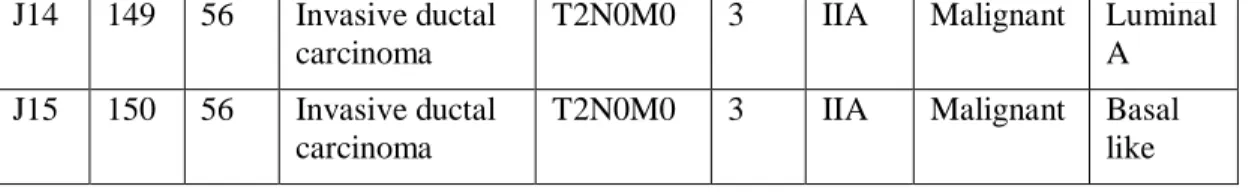
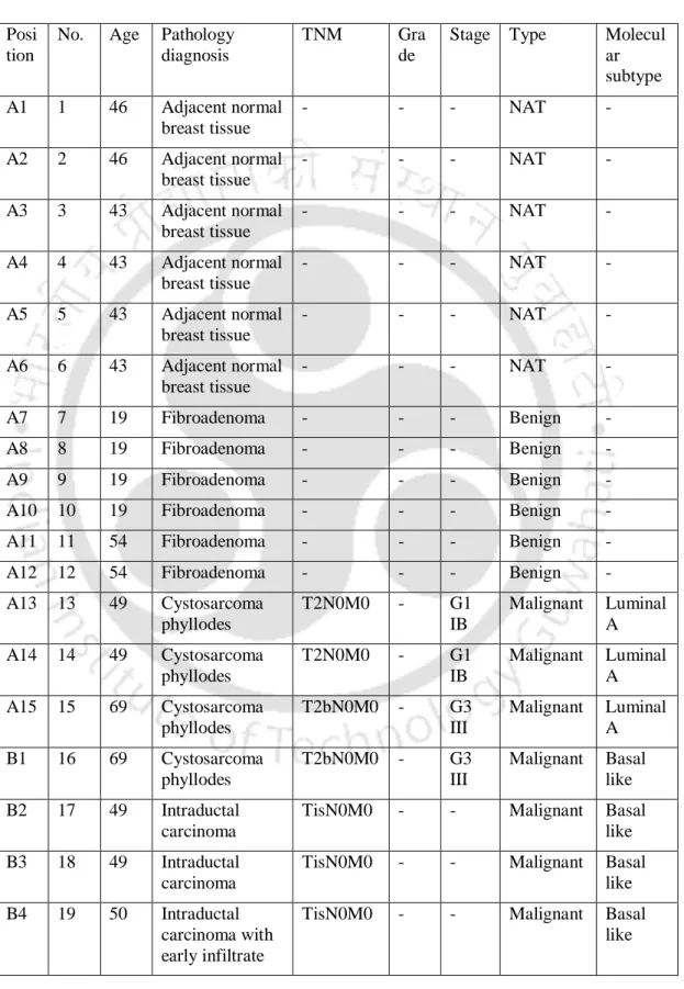
- Tissue Microarray
- Tissue Microarray details Name: BR1503e
- Immunohistochemistry (IHC)
- Scoring
- Cell culture
- Western blot analysis
- Statistical analysis
Therefore, the present chapter was designed to determine the expression of NGALR in breast cancer tissues and cell lines. NGALR expression in breast cancer tissues and different breast cancer subtypes was examined through IHC analysis.
Results and Discussion
- NGALR decreases the survival of breast cancer patients
- Overexpression of NGALR in breast cancer tissues
- Expression of NGALR in different sub-types of breast cancer
- Expression of NGALR in different age groups of TNBC patients
- Expression of NGALR in different process of the development of TNBC It has been well-known that inflammation plays a critical role in the development and
- Expression of NGALR in TNBC cell lines
In this study, NGALR expression was significantly increased by IL-1β (Mao et al., 2011). NGALR expression was determined in HaCaT (normal) and MDA-MB-231 and MDA-MB-468 (TNBC) cell lines.
Conclusion
Our results showed that NGALR was significantly upregulated in MDA-MB-231 and MDA-MB-468 cell lines than HaCaT cells (Figure 2.2 C, D). 2020) detected the expression of NGALR in CLL cells. Moreover, NGALR protein expression was significantly upregulated in TNBC cells compared to normal cells.
Effect of TNF-α and TNF-β on the regulation of NGALR in TNBC cell lines
- Introduction
- Materials and methods 1. Cell culture
- MTT assay
- Colony formation assay
- Epithelial–mesenchymal transition (EMT) assay
- Wound healing assay
- Cell death analysis
- Western blot analysis
- Statistical analysis
- Results and Discussion
- Effect of TNF-α and TNF-β on the proliferation and survival of TNBC cells
- Effect of TNF-α and TNF-β on EMT of TNBC cells
- Effect of TNF-α and TNF-β on the migration of TNBC cells
- Effect of TNF-α and TNF-β on TNBC cell death
- Effect of TNF-α and TNF-β on the expression of NGALR in TNBC cells In our earlier studies, we have demonstrated the effects of TNF-α and TNF-β on TNBC
- Conclusion
Therefore, we investigated the effect of TNF-α and TNF-β on the progression of TNBC in MDA-MB-231 and MDA-MB-468 cells. Our results also supported that TNF-α and TNF-β significantly increased the proliferation of MDA-MB-231 and MDA-MB-468 cells. We have already determined the role of TNF-α and TNF-β on survival and proliferation of MDA-MB-231 and MDA-MB-468 cells.
Our results also showed that TNF-α and TNF-β significantly induced vimentin protein expression in MDA-MB-231 and MDA-MB-468 cells. Therefore, we determined the effect of TNF-α and TNF-β on NGALR expression in MDA-MB-231 and MDA-MB-468 cells. Furthermore, TNF-α and TNF-β significantly increased NGALR expression in MDA-MB-231 and MDA-MB-468 cells.
Role of NGALR in the development of TNBC
Introduction
Furthermore, TNF-α and TNF-β were strongly associated with TNBC progression through modulation of cell proliferation, survival, EMT, migration and cell death. Moreover, NGALR expression was significantly upregulated after treatment with TNF-α and TNF-β in MDA-MB-231 and MDA-MB-468 cells compared with untreated cells. These results strongly argued that NGALR is associated with breast cancer progression and regulation, specifically TNF-α and TNF-β-mediated TNBC progression.
Therefore, in this chapter we determined the role of NGALR in the pathogenesis of TNBC. Furthermore, we determined the expression of several downstream proteins involved in the pathogenesis and progression of TNBC. In addition, we analyzed their association with TNF-α and TNF-β-mediated TNBC and deciphered the underlying mechanism of action. Notably, this is the first study to report the role of NGALR in TNBC progression.
Materials and Methods 1. Cell culture
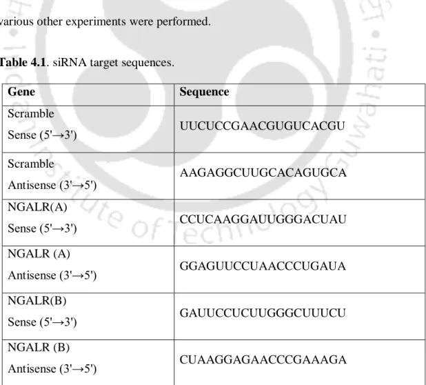
- Silencing of NGALR gene
- MTT assay
- Colony formation assay
- Cell cycle analysis
- Immunocytochemistry (ICC) analysis
- Invasion assay
- Wound healing assay
- Western blot analysis
- Statistical analysis
The effect of NGALR knockdown on the proliferation of MDA-MB-231 and MDA-MB-468 cells was determined by MTT assay. In addition, the effect of TNF-α and TNF-β on the proliferation of MDA-MB-231 and MDA-MB-468 cells transfected with Scr and siNGALR was examined by MTT assay. Clonogenicity of MDA-MB-231 and MDA-MB-468 cells transfected with Scr and siNGALR was determined by colony formation assay.
The invasiveness of Scr and siNGALR transfected MDA-MB-231 and MDA-MB-468 cells was determined by Boyden's invasion chamber assay. In addition, the effect of TNF-α and TNF-β on the migration of Scr and siNGALR-transfected MDA-MB-231 and MDA-MB-468 cells was also examined by wound healing assay. For this purpose, the post-transfected MDA-MB-231 and MDA-MB-468 cells were treated with 0.05 nM TNF-α and TNF-β for 24 hours and then lysed with lysis buffer.
Results and Discussion
- Silencing of NGALR in TNBC cells
- Silencing of NGALR decreased proliferation and survival of TNBC cells Cancer cells are strongly characterized by increased proliferation and survival and
- Silencing of NGALR induced S phase arrest of TNBC cells
- Silencing of NGALR induced autophagy in TNBC cells
- Silencing of NGALR suppressed EMT in TNBC cells
- Silencing of NGALR attenuated invasion, migration, and angiogenesis in TNBC cells
- Silencing of NGALR inhibited the Akt/mTOR and JAK/STAT pathways in TNBC cells
- Role of TNF-α and TNF-β on siNGALR knockdown TNBC cells
- Effect of TNF-α and TNF-β on the proliferation and survival of siNGALR knockdown TNBC cells
- Effect of TNF-α and TNF-β on the migration of siNGALR knockdown TNBC cells
- Effect of TNF-α and TNF-β on the regulation of Akt/mTOR and JAK/STAT pathways in siNGALR knockdown TNBC cells
We observed that NGALR silencing upregulated the expression of survivin, COX-2 and Bcl-2 in MDA-MB-231 and MDA-MB-468 cells than the Scr control (Figure 4.1 F). Furthermore, our findings showed that NGALR silencing induced autophagy by increasing LC3B protein expression in MDA-MB-231 and MDA-MB-468 cells. Therefore, we determined the expression of EMT proteins (vimentin and Snail) through ICC analysis in MDA-MB-231 and MDA-MB-468 cells after NGALR silencing.
Similarly, silencing of NGALR significantly suppressed the migration of MDA-MB-231 and MDA-MB-468 cells than Scr control. Our results showed that silencing NGALR downregulated the expression of VEGF-A protein in MDA-MB-231 and MDA-MB-468 cells than Scr control (Figure 4.5 E). We observed that silencing of NGALR reduced the expression of p-Akt (Ser473) and p-mTOR (Ser2448) in both MDA-MB-231 and MDA-MB-468 cells compared with Scr control.
Conclusion
Discussion and Conclusion
Discussion and Conclusion
Recently, a study also reported that dysregulation of NGALR was associated with poor OS in gastric cancer patients (Wei et al., 2020). However, NGALR was downregulated and associated with the poor prognosis in ccRCC patient samples (Liu et al., 2018). Recently, a study revealed the upregulated expression of NGALR in CLL cells than normal PBMCs and associated with apoptotic resistance (Bauvois et al., 2020).
Various studies have reported that inflammation is significantly associated with breast cancer progression (Mills RC, 2017; Agnoli et al., 2017). 2011) observed similar results and reported that TNF-α and TGF-β increased BCSCs by inducing EMT (Asiedu et al., 2011). In contrast, increased expression of LC3B was associated with poor prognosis and worst outcome in breast cancer (Chen et al., 2013; Bortnik et al., 2020).
Limitations and future prospective of the study