Manish Srivastava Fruits & Horticulture Technology ICAR-IARI 31. Amit Kumar Goswami Fruits & Horticulture Technology ICAR-IARI. In the last two decades, 'omics' and genomic data have enabled us to understand diverse plant-associated microbial communities, pathogens of crop plants and their behavior on plant-associated niches. Recently, Fungal Taxonomy Group at Division of Plant Pathology, ICAR-IARI has been instrumental in successfully demonstrating the genomics-assisted molecular systematic and whole genome sequencing.
Contents
Diversity and Taxonomy of Fungi
Comprehensive and detailed reviews of the use of molecular techniques in fungal systematics have been provided. Paterson and Bridge have published a compilation of the physiological techniques used in the identification of filamentous fungi.
Trends in the taxonomy of Oomycetes with reference to Phytophthora
Previous identification of Phytophthora was based solely on morphological aspects and other growth-temperature relationships. Phytophthora species identification based on analysis of restriction enzyme fragments of internal transcribed spacer regions of ribosomal RNA.
Taxonomic Characterization of Zygomycetous Fungi of Agriculture and Industrial use
- Absidia van Tieghem
- Choanephora cucurbitarum (Berkeley & Ravenel) Thaxter
- Circinella van Tieghem & Le Monnier
- Cunninghamella echinulata (Thaxter) Thaxter
- Rhizopus oryzae Went. and Prinsen Gerl
- Thamnidium Link
- Zygorhynchus Vuillemin
- Gongronella butleri (Lendner) peyronel & Dal Vesco
- Fungi in humus formation: Different groups of fungi including Zygomycetes produces humus and this is one among natural sources of plant nutrients, actually locked in plant and animal bodies in a
- Production of enzymes: Zygomycetes fungi are the source of a great diversity of enzymes, hydrolytic enzymes, such as those needed for the degradation of plant carbohydrates, including
For example, in Smittium species, germination and attachment occur within half an hour of ingestion of the spores. Their important role is in the absorption of mineral nutrients and sometimes in protection against drought or pathogenic attack. The hyphae of arbuscular mycorrhizal fungi produce the glycoprotein glomalin, which may be one of the main carbon stores in the soil.
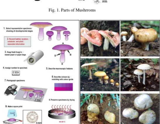
Taxonomical Studies on Macro–Fungi
K. Sharma
Sarbhoy (1997) stated that till 1979 about 450 species from the order Agaricales have been reported from India. However, Walting and Abraham (1992), were of the opinion that 650 species have been recorded from India.
Holobasidiomycetdae: Basidia have only one basal septum
- GOMPHIDIACEAE
The development of the fruiting body is hemingiocarpous, hymenium covering lamellae on the lower surface of the pileus or casing, which can be easily separated from the pileus. Characteristic feature is the presence of vertically arranged tubes on the underside of the Pileus instead of gills.

Morpho-molecular characterization of wheat fungal pathogens
Loose smut is more common in areas with a cool, moist climate during the host's flowering period. Real Time PCR has proven to be one of the promising and highly sensitive methods for the detection of plant pathogens.
Morphological Characterisation of Trichoderma species
Trichoderma citrinoviride (teleomorph Hypocrea schweinitzii, Ascomycota, Dikarya) is a very common soil fungus from the clade Longibrachiatum of the genus Trichoderma. Bluish-green compact to cottony pustules 1-2 mm in diameter form with conidial production confined to the colony margin. Phialides arise separately towards the tip of the conidiophore; each branch terminating in one or two phialides and phialides arising separately from.
Growth of 6.0-7.0 cm was observed in 4 days on PDA medium. conidial effusion covering the entire surface of the plate, or forming flat diffuse pustules, light yellow concentrated near the border, later turning dark green.
Diversity in Aspergillus
- A. niger
- A. ochraceus
- A. parasiticus
- A. versicolor
Colony: Medium growing, light bluish green to olive grey, Colorless on the back of the plate. Colony: Fast growing, dark cherry green, purplish red on the back of the plate Conidiophores: 75-100um long,. Growth on PDA & reverse of the plate, 2) Conidiophores & Conidial head, 3) Vesicle & Sterigmata, 4) Conidia.
Colony: Colony is medium growing, wine pink, yellow to red brown on the back of the plate.
Identification of commonly occurring Fusarium species
Diagnostic characters: Presence of chains of microconidia, best observed in situ, and absence of chlamydospores. Diagnostic signs: presence of chains of microconidia, distinct shape and size of spores, pigmentation. Diagnostic characters: presence of primary and secondary macroconidia, pigmentation, spore shape and presence of chlamydospores.
Chlamydospores: globose, oval to smooth, 8-12μ in diameter, intercalary, formed singly or in chains.
Taxonomy of Genus Penicillium
Penicillium aethiopicum Frisvad
Exudate droplets on CYA: Often present, abundant, yellow Inverted color on CYA: Cream, yellow, rarely brown Diffusable color: Yellow pigment is often produced. Conidium color on CYA: dull green to gray-green or blue-green at the colony edge. Exudate droplets on CYA: profuse, clear or brown. Reverse color on CYA: Cream to tan. Diffusion color: Light brown or none.
Conidium color on CYA: blue-green to green Exudate droplets on CYA: abundant, clear Reversed color: beige to grayish cream Diffusable color: none.
Talaromyces pinopilus (Hedgc.) Samson, Yilmaz, Frisvad & Seifert
Diversity in Hyphomycetous fungi
Conidiogenous cells: monoblastic, integrated, terminal, percurrent, initially clavate or subspherical, with a thin wall at the apex, later often becoming cupped. Conidiophores (phialids): macronematous, mononematous, unbranched or irregularly branched, straight or bent, hyaline or pale brown, smooth. Conidiophores: macronematous, mononematous, unbranched or branched, straight or bent, often geniculate, pale to medium brown, smooth or verruculose.
Conidia collected in mucilaginous masses, semi-endogenous or hooked, simple, allantoid, ellipsoid or cylindrical, rounded at the ends, colorless to pale brown, smooth 0-septate.
Identification of some important Coelomycetous fungi
- Botryodiplodia (Lasiodiplodia)theobromaeEll.&Ev
- Cytospora Ehrenb. ex Fr
- MacrophominaPetrak
- conidia hyaline, filiform, straight or more often hamate, eguttulate, aseptate
Conidiogenous cells: Enteroblastic, phialidic, determinate, discrete, doliiform to lageniform, hyaline, smooth, formed from the inner cells of the pycnidia wall. Conidiogenous cells : Enteroblastic, phialidic, definite, integrated, straight, hyaline, smooth, occasionally formed as very small lateral branches immediately below transverse septa, but more often as long distinct branches. Conidiophores: Hyaline, branched and septate above and at the base, smooth, cylindrical, formed by the inner cells of the pycnidia wall.
Conidiogenous cells: Enteroblastic, phialidic, determinate, rarely discrete, hyaline, cylindrical, openings apical on long or short lateral and main branches of the conidiophores.
Polyphasic Taxonomy of Endophytic Fungi
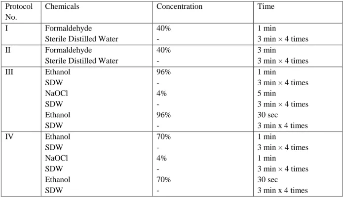
Cut into small pieces the inner tissue of the stems and inoculate on agar plate. Therefore, it is difficult to identify up to species level just based on morphology. Scrapping the mycelial mat of fungi and proceed to the genomic DNA isolation following a simple and rapid DNA extraction protocol (Aamir et al. 2015).
Check the genomic DNA on 0.8% agarose electrophoresis gel and proceed to amplify the desired gene region using appropriate primers (White et al. 1990).
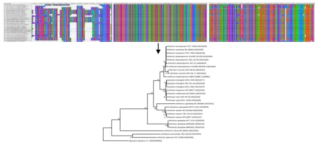
Internal Transcribed Spacer (ITS) based identification
It may be necessary to resuspend the DNA overnight at room temperature on a turntable at 20 rpm. Add a phage or other molecular weight marker DNA as a control in the adjacent well. After washing with 70% ethanol, the resulting DNA pellet should appear as a white, thick thread-like mass.
In the presence of sufficiently high NaCl, chilled absolute ethanol precipitates the DNA, the final step in a traditional DNA extraction.
Application of real time PCR technique in mycology and plant pathology
DNA is amplified using an initial denaturation at 95ºC for 3 minutes, followed by 35 cycles of 95ºC for 15 seconds, annealing for 15 seconds and extension at 72ºC for 15 seconds. Agarose gel electrophoresis of the qPCR products can be performed to confirm that the individual qPCR products correspond to a single homogeneous cDNA fragment of expected size.
Data Analysis
The real-time PCR test can simultaneously detect and quantify bacterial, fungal and viral pathogens. Real-time PCR can be a rapid diagnostic tool and may be useful as an aid in identifying potential pathogens. Objective: To detect and quantify the pathogen in infected tissues using qPCR (Case study: Optimization of real-time PCR assay for absolute quantification of Magnaporthe oryzae).
Real-time PCR monitoring of fungal development in Arabidopsis thaliana infected by Alternaria brassicicola and Botrytis cinerea.
Development of diagnostic marker for the identification of fungal pathogens
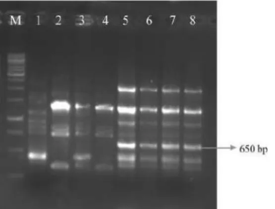
PCR amplification using URP
The concentration of PCR product is estimated by comparing with DNA molecular weight markers on an agarose gel. Optimizing PCR conditions using designed primer pairs: PCR conditions and cycle pattern are important for specificity and sensitivity of primer amplification. Sensitivity of PCR tests detected by agarose gel electrophoresis using RABSF1 and RABSR2 primers set.
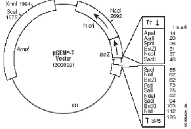
Use the following equation to calculate the appropriate amount of PCR product (insert) to include in the ligation reaction.
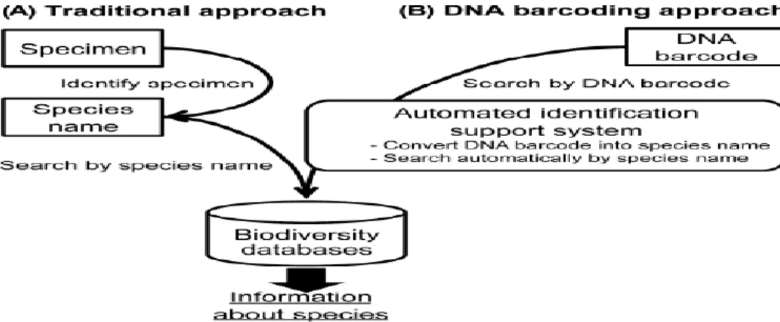
DNA Barcoding: A Moderm Tool to Explore Microbial Diversity
The Barcode of Life project was proposed to promote DNA barcoding as a global standard for sequence-based identification of eukaryotes. In 2004, this project was formally initiated by the establishment of the Consortium for the Barcode of Life (CBOL), which aims to develop a standard protocol for DNA barcoding and to build a comprehensive DNA barcode library. The Canadian Barcode of Life Network (BOLNET.ca) was the first national network for DNA barcoding.
Ten species in one: DNA barcoding reveals cryptic species in the Neotropical skipper butterfly Astraptes fulgerator.
Metagenomic approaches for studying fungal diversity
Choice of technology for fungal diversity analysis: Conventional DNA sequencing relies on dideoxy chain termination technique first described more than two decades ago (Sanger et al. 1977). Pipelines such as Community Kuber Infrastructure for Advanced Microbial Ecology Research and Analysis (KAMERA), Metagenomic-Rapid Annotation Using Subsystem Technology (MG-RAST) and Metagenome Analyzer (MEGAN) perform interactive analysis and comparison of the taxonomic and functional content of shotgun and amplicon from datasets (Glass et al., 2010; Sun et al. 2012). Markowitz VM, Chen IM, Chu K, Szeto E, Palaniappan K, et al. 2012) IMG/M: the integrated metagenome data management and comparative analysis system.
Orgiazzi A, Bianciotto V, Bonfante P, Daghino S, Ghignone S, et al. Pyrosequencing analysis of fungal assemblages from geographically distant, diverse soils reveals spatial patterning and a core mycobiome.
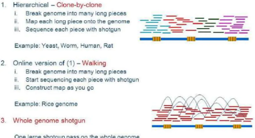
Whole Genome Sequencing: Assembly and Annotation
Whole genome shotgun sequencing
The DNA sequencing with Nanopore instrument relies on the conversion of electrical signal from nucleotides by passing through a nanopore which is an α-hemolysin pore covalently attached with cyclodextrin molecule - the binding site for nucleotides. During the sequencing process, the ionic current passing through the nanopores is blocked by the nucleotide, i.e. the one previously cleaved by exonuclease from a DNA strand interacting with cyclodextrin. One of the (relatively) early assemblers is PHRAP, which is still in use, both by itself (for small DNA sequence sets), and as a subcomponent of WGS assemblers, e.g.
Some assemblers use this data to impose additional structure on the sequence assembly (eg Gig Assembler).
Polyphasic taxonomy and Biosystematics of Fungi
- Introduction
- Why use the molecular methods for identification?
- Catalogue of gene targets for microbial identification
- Culture dependent identification of fungi A. Sequence based techniques for fungi
- PCR based finger printing techniques
- Phylogenetic information from whole genome sequences
- Culture independent identification procedures
- Polyphasic taxonomic approach: Case study
- Concluding remarks
Sequence comparison of internal transcribed spacer and D1/D2 26S rDNA spacer sequences using either primer set (Table 1) has been reported to provide a useful tool for the identification of fungi. Since the ITS regions have many restrictions, the alternative gene targets have also been proposed to be useful for fungal identification such as the D1 and D2 domains of the 28S rRNA gene, and elongation factors α-tubulin and β-tubulin ( Hall et al., 2003). One of the few studies of secondary metabolites as taxonomic markers for distinguishing Colletotrichum spp.
Comparison of the partial sequence of rpoB, sodA, groEL and gyrB genes within the genus Streptococcus.