European
Handbook of Neurological Management
Volume 1
EDITED BY
Nils Erik Gilhus MD PhD
Professor, Department of Clinical Medicine, University of Bergen and Department of Neurology, Haukeland University Hospital, Bergen, Norway
Michael P. Barnes MD, FRCP
Professor of Neurological Rehabilitation, University of Newcastle, Medical Director, Hunters Moor Neurorehabilitation Ltd, Newcastle upon Tyne, UK
Michael Brainin MD
Professor, Department of Clinical Medicine and Prevention, and Center for Clinical Neurosciences Donau-Universität Krems and Head, Department of Neurology, Landesklinikum Donauregion Tulln Tulln, Austria
SECOND EDITION
A John Wiley & Sons, Ltd., Publication
publishing program has been merged with Wiley’s global Scientifi c, Technical and Medical business to form Wiley-Blackwell.
Registered offi ce: John Wiley & Sons Ltd, The Atrium, Southern Gate, Chichester, West Sussex PO19 8SQ, UK
Editorial offi ces: 9600 Garsington Road, Oxford OX4 2DQ, UK 111 River Street, Hoboken, NJ 07030-5774, USA
The Atrium, Southern Gate, Chichester, West Sussex, PO19 8SQ, UK For details of our global editorial offi ces, for customer services and for information about how to apply for permission to reuse the copyright material in this book please see our website at www.wiley.com/wiley-blackwell
The right of the author to be identifi ed as the author of this work has been asserted in accordance with the Copyright, Designs and Patents Act 1988.
All rights reserved. No part of this publication may be reproduced, stored in a retrieval system, or transmitted, in any form or by any means, electronic, mechanical, photocopy- ing, recording or otherwise, except as permitted by the UK Copyright, Designs and Patents Act 1988, without the prior permission of the publisher.
Wiley also publishes its books in a variety of electronic formats. Some content that appears in print may not be available in electronic books.
Designations used by companies to distinguish their products are often claimed as trademarks. All brand names and product names used in this book are trade names, service marks, trademarks or registered trademarks of their respective owners. The publisher is not associated with any product or vendor mentioned in this book. This publication is designed to provide accurate and authoritative information in regard to the subject matter covered. It is sold on the understanding that the publisher is not engaged in rendering professional services. If professional advice or other expert assistance is required, the services of a competent professional should be sought.
The contents of this work are intended to further general scientifi c research, understand- ing, and discussion only and are not intended and should not be relied upon as recommending or promoting a specifi c method, diagnosis, or treatment by physicians for any particular patient. The publisher and the author make no representations or warranties with respect to the accuracy or completeness of the contents of this work and specifi cally disclaim all warranties, including without limitation any implied warranties of fi tness for a particular purpose. In view of ongoing research, equipment modifi cations, changes in governmental regulations, and the constant fl ow of information relating to the use of medicines, equipment, and devices, the reader is urged to review and evaluate the information provided in the package insert or instructions for each medicine, equipment, or device for, among other things, any changes in the instructions or indication of usage and for added warnings and precautions. Readers should consult with a specialist where appropriate. The fact that an organization or website is referred to in this work as a citation and/or a potential source of further information does not mean that the author or the publisher endorses the information the organization or website may provide or recommendations it may make. Further, readers should be aware that internet websites listed in this work may have changed or disappeared between when this work was written and when it is read. No warranty may be created or extended by any promotional statements for this work. Neither the publisher nor the author shall be liable for any damages arising herefrom.
Library of Congress Cataloging-in-Publication Data
European handbook of neurological management / edited by Nils Erik Gilhus, Michael P. Barnes, Michael Brainin. – 2nd ed.
p. ; cm.
Includes bibliographical references and index.
ISBN 978-1-4051-8533-2
1. Nervous system–Diseases–Treatment–Europe–Handbooks, manuals, etc. I. Gilhus, Nils Erik. II. Barnes, Michael P. III. Brainin, M. (Michael), 1952–
[DNLM: 1. Nervous System Diseases–therapy–Practice Guideline. WL 140 E885 2010]
RC346.E97 2010 616.8–dc22
2010023982
A catalogue record for this book is available from the British Library.
Set in 9/12 pt Minion by Toppan Best-set Premedia Limited Printed in Singapore
1 2011
iii Introduction, 1
Section 1 Investigations
1 Routine cerebrospinal fl uid (CSF) analysis, 5 2 Use of imaging in cerebrovascular disease, 19 3 Use of imaging in multiple sclerosis, 35 4 Neurophysiological tests and neuroimaging
procedures in non-acute headache, 53
5 Use of anti-interferon beta antibody measurements in multiple sclerosis, 63
6 Use of antibody testing in nervous system disorders, 75
7 Use of skin biopsy in the diagnosis of small fi bre neuropathy, 81
8 Assessment of neuropathic pain, 91
Section 2 Major Neurological Diseases 9 Ischaemic stroke and transient ischaemic
attack, 101
10 Drug treatment of migraine, 159
11 Cluster headache and other trigemino-autonomic cephalgias, 179
12 Diagnosis and treatment of primary (idiopathic) dystonia, 191
13 Mild traumatic brain injury, 207
Contents
14 Early (uncomplicated) Parkinson’s disease, 217 15 Late (complicated) Parkinson’s disease, 237 16 Alzheimer’s disease, 269
Section 3 Neuromuscular Diseases
17 Management of amyotrophic lateral sclerosis, 283 18 Post-polio syndrome, 311
19 Autoimmune neuromuscular transmission disorders, 321
20 Chronic infl ammatory demyelinating polyradiculoneuropathy, 333 21 Multifocal motor neuropathy, 343
22 Paraproteinaemic demyelinating neuropathies, 351 23 Limb girdle muscular dystrophies, 363
Section 4 Infections
24 Neurological complications of HIV infection, 373 25 Viral meningo-encephalitis, 383
Section 5 Neurological Problems 26 Treatment of neuropathic pain, 399 27 Acute relapses of multiple sclerosis, 411 28 Status epilepticus, 421
29 Alcohol-related seizures, 429
30 Brain metastases, 437
31 Paraneoplastic neurological syndromes, 447 32 Nystagmus and oscillopsia, 459
33 Orthostatic hypotension, 469
34 Cerebral venous and sinus thrombosis, 477 35 Cerebral vasculitis, 485
36 Neurological problems in liver transplantation, 491
37 Fatty acid mitochondrial disorders, 501
Section 6 Sleep Disorders
38 Management of narcolepsy in adults, 513
39 Sleep disorders in neurodegenerative disorders and stroke, 529
Section 7 Rehabilitation 40 Cognitive rehabilitation, 545
Index, 569
Introduction
N. E. Gilhus,
1M. P. Barnes,
2M. Brainin
31 University of Bergen and Department of Neurology, Haukeland University Hospital, Bergen, Norway; 2 Professor of Neurological Rehabilitation Hunters Moor Neurorehabilitation Ltd; 3 Center of Clinical Neurosciences, Donau - Universit ä t Krems, and Landesklinikum Tulln, Austria
tables 3 and 4 , for therapeutic and diagnostic measures.
For several important neurological questions, no high - class scientifi c evidence is available. The recommenda- tion of ‘ Good Practice Point ’ can be given when only Class IV evidence has been published.
The aim of developing, publishing, and disseminating European guidelines is to improve neurological practice.
The preliminary work and extensive drafting of recom- mendations, based on evaluation of clinical trials and meta - analyses by a group of leading experts is not visible in the end product. It is therefore justifi ed to note that the process of establishing high - quality guidelines is based on the right mix of experts, usually from a wider selection of European countries. This is what the Scien- tifi c Committee of the EFNS does. It reviews task forces from these panels to develop such guidelines and reviews their performance as well as their fi nal recommenda- tions. Invariably, external reviewers are also involved.
Attention is given to confl ict of interest declarations by all members of the guideline group. The product is what the current President of the EFNS, Richard Hughes, once called the ‘ fi rst peer - reviewed book in the world ’ . We cannot think of a better characterization.
This Handbook is primarily written for European neu- rologists, but should be helpful also for other groups of health professionals treating patients with disorders of the brain, spine, nerve, and muscle. Clinical experience and present practice do not always give good guidance for best treatment. The guideline chapters in this Hand- book are among the most frequently cited articles that have been published in European Journal of Neurology .
A challenge for quality control in neurology, as well as in medicine in general, is how to promote the implemen- tation of guidelines in the daily, patient - related work.
A fi rst prerequisite is the awareness of their existence, and then their availability. This second edition of the
1 This second edition of the European Handbook of Neuro-
logical Management includes peer - reviewed guidelines on topics highly relevant for all areas of neurological prac- tice. The fi rst edition of this Handbook was published in 2006. Since then there has been an ongoing development in all fi elds of neurology, making a revision necessary. All authors have revised and updated their chapters, and the newly submitted guidelines have gone through an exten- sive peer review. All guideline chapters had previously also been published in the European Journal of Neurology . Of the revised documents, only 15 will be published in the journal, having been selected mainly due to the anti- cipated interest of the topic and the extent of revision due to new developments in their fi eld. The remaining revi- sions are published in this Handbook only. Thus, the revised Handbook is the new and complete compendium for all EFNS guidelines.
The guidelines in this European Handbook of Neuro- logical Management are produced according to the rules given by EFNS and the EFNS Scientifi c Committee.
All 24 EFNS Scientist Panels ( www.efns.org/Scientist - Panels.15.0.html ) are continuously encouraged to estab- lish such task forces with the aim of producing guidelines relevant in their fi eld.
The search for and evaluation of scientifi c evidence is for all guidelines undertaken according to the EFNS guidance paper [1] . The classifi cation of evidence is explained in the enclosed tables for therapeutic interven- tion and for diagnostic measures, respectively (tables 1 and 2 ). As for the recommendations, these are graded from A to C. The scheme for this grading is explained in
European Handbook of Neurological Management: Volume 1, 2nd edition. Edited by N. E. Gilhus, M. P. Barnes and M. Brainin.
© 2011 Blackwell Publishing Ltd.
Table 1 Evidence classifi cation scheme for a therapeutic intervention.
Class I An adequately powered prospective, randomized, controlled clinical trial with masked outcome assessment in a representative population or an adequately powered systematic review of prospective randomized controlled clinical trials with masked outcome assessment in representative populations. The following are required:
(a) randomization concealment;
(b) primary outcome(s) is/are clearly defi ned;
(c) exclusion/inclusion criteria are clearly defi ned;
(d) adequate accounting for dropouts and crossovers with numbers suffi ciently low to have minimal potential for bias;
(e) relevant baseline characteristics are presented and substantially equivalent among treatment groups or there is appropriate statistical adjustment for differences.
Class II Prospective matched group cohort study in a representative population with masked outcome assessment that meets a – e above or a randomized, controlled trial in a representative population that lacks one criteria a – e.
Class III All other controlled trials (including well - defi ned natural history controls or patients serving as own controls) in a representative population, where outcome assessment is independent of patient treatment.
Class IV Evidence from uncontrolled studies, case series, case reports, or expert opinion.
Table 2 Evidence classifi cation scheme for a diagnostic measure.
Class I A prospective study in a broad spectrum of persons with the suspected condition, using a ‘ gold standard ’ for case defi nition, where the test is applied in a blinded evaluation, and enabling the assessment of appropriate tests of diagnostic accuracy.
Class II A prospective study of a narrow spectrum of persons with the suspected condition, or a well - designed retrospective study of a broad spectrum of persons with an established condition (by ‘ gold standard ’ ) compared to a broad spectrum of controls, where the test is applied in a blinded evaluation, and enabling the assessment of appropriate tests of diagnostic accuracy.
Class III Evidence provided by a retrospective study where either persons with the established condition or controls are of a narrow spectrum, and where the test is applied in a blinded evaluation.
Class IV Any design where the test is not applied in blinded evaluation or evidence is provided by expert opinion alone or in descriptive case series (without controls).
Table 3 Evidence classifi cation scheme for the rating of recommendations for a therapeutic intervention.
Level A (established as effective, ineffective, or harmful) requires at least one convincing Class I study or at least two consistent, convincing Class II studies.
Level B (probably effective, ineffective, or harmful) requires at least one convincing Class II study or overwhelming Class III evidence.
Level C (possibly effective, ineffective, or harmful) requires at least two convincing Class III studies.
Table 4 Evidence classifi cation scheme for the rating of recommendations for a diagnostic measure.
Level A (established as useful/predictive or not useful/
predictive) requires at least one convincing Class I study or at least two consistent, convincing Class II studies.
Level B (established as probably useful/predictive or not useful/
predictive) requires at least one convincing Class II study or overwhelming Class III evidence.
Level C (established as possibly useful/predictive or not useful/
predictive) requires at least two convincing Class III studies.
Handbook should help with this aspect. We recommend that the guidelines are used widely, and also for retro- spective and prospective quality control studies, to check if actual therapeutic and diagnostic procedures are undertaken in accordance with best clinical practice. Too little attention is often given to the effective dissemina- tion, adoption, and implementation strategies for clinical guidelines.
Several of the guidelines in this Handbook represent joint projects between EFNS and more disease - specifi c European neurological societies. Such co - operation between general neurological societies and disease - spe- cifi c organizations is an aim for EFNS both regarding guideline production and in general. The European Stroke Organisation has produced the guideline on stroke in this Handbook. The Movement Disorder Society – European Section and the International Peripheral Nerve Society have contributed to the guideline production. In the future, we hope for more co - operation with European and international societies regarding all aspects of the guideline production, from defi ning the topic and mem- bership to approval of the fi nal guideline.
Better treatment and more exact diagnosis is needed in European neurology. There is often a gap between best practice and what is actually carried out for the individ- ual patient. Guidelines represent an important tool to minimize this gap. Suboptimal treatment may sometimes be due to lack or resources, especially in poor countries.
In arguing for suffi cient resources, requirements as listed in the guidelines of this European Handbook may be helpful. Diseases of the brain and nervous system repre- sent one - third of the total burden of disease in Europe, and they incur huge costs on the society. The direct costs of treatment and diagnosis represent only a small part of the total cost, so that real improvements for patients ’ function and ability will usually be highly cost - effective.
These guidelines clearly demonstrate the need for more research. Even for several established treatments and diagnostic procedures there frequently is only weak scientifi c evidence. There is no doubt that practice is going on in all European departments that would not have been continued if properly controlled studies had been undertaken. Research is needed to select the best treatment and diagnostic options. Although the big breakthroughs usually come from basic research, there is room for much improvement by optimizing what is already available to most neurologists. EFNS intends to support multinational European initiatives to help in this process.
Guidelines represent recommendations. They should not have legally binding implications. In disputed cases, actual practice will be compared with approved guide- lines. For the individual patient, there may, however, be reasons to deviate from guideline recommendations.
Still, there is a clear and wanted development towards a stronger quality control of neurological practice, includ- ing the systematic use of international guidelines.
The editors thank all the authors who have contrib- uted to this second edition of the European Handbook of Neurological Management . Their work to evaluate and update all new scientifi c and controlled information in their fi eld as a joint effort within the task force is highly appreciated. Lisa M ü ller, executive director of the EFNS, has done a formidable job in following up the task forces.
We will especially thank Professor Richard Hughes, Pres- ident of EFNS, who was the leading editor of the fi rst edition of this Handbook and a key person in initiating and organizing the guideline work within EFNS.
Reference
1. Brainin M , Barnes M , Baron JC , et al . Guidance for the prep- aration of neurological management guidelines by EFNS scientifi c task forces – revised recommendations 2004 . Eur J Neurol 2004 ; 11 : 577 – 81 .
Routine c erebrospinal fl uid ( CSF ) a nalysis
F. Deisenhammer,
1A. Bartos,
2R. Egg,
1N. E. Gilhus,
3G. Giovannoni,
4S. Rauer,
5F. Sellebjerg,
6H. Tumani
71 Innsbruck Medical University, Austria; 2 Charles University, Prague, Czech Republic; 3 University of Bergen, and Haukeland University Hospital, Bergen, Norway; 4 University College London, Queen Square, London, UK; 5 Albert - Ludwigs University, Freiburg, Germany; 6 Copenhagen University Hospital, Denmark; 7 University of Ulm, Germany
conducted. Also, the key words ‘ cerebrospinal fl uid ’ or ‘ CSF ’ were cross - referenced with ‘ glucose ’ , ‘ lactate ’ , ‘ cytology ’ , ‘ cell * in title ’ excluding ‘ child * ’ . Furthermore, a search for ‘ cerebrospinal fl uid ’ and ‘ immunoglobulin ’ and ‘ diagnosis ’ and ‘ electrophoresis ’ or ‘ isoelectric focusing ’ was performed limited to the time between 1 January 1980 and 1 January 2005, and returned only items with abstracts, and English language (274 references). A search for ‘ cerebrospinal fl uid ’ AND ‘ infectious ’ limited for time (1 January 1980 until now) returned 560 abstracts.
Abstracts that primarily did not deal with diagnostic issues and infectious CSF (e.g. non - infectious infl am- matory diseases, vaccination, general CSF parameters, pathophysiology, cytokines and therapy) were excluded, resulting in 60 abstracts. Searching the items ‘ cerebrospi- nal fl uid ’ AND ‘ serology ’ limited for time (1 January 1980 until now) and excluding abstracts not directly related to the topic returned 35 abstracts and a search for ‘ cerebro- spinal fl uid ’ AND ‘ bacterial culture ’ limited for time (1 January 1980 until now) resulted in 28 abstracts.
For the current update (deadline October 2009) all the above search terms and selection criteria were applied for the time between 2005 and now.
Because this was not included in the fi rst edition an additional MEDLINE search for the items ‘ cerebrospinal fl uid analysis ’ AND ‘ quality assurance ’ from 1981 until now returned 87 references. Only 15 of these references dealt primarily with quality assurance aspects of cerebro- spinal fl uid analysis.
The abstracts were selected by the author in charge of the respective topic.
In addition, textbooks and articles identifi ed in refer- ence lists of individual papers were selected if considered appropriate.
5
Introduction
The cerebrospinal fl uid (CSF) is a dynamic, metabolically active substance that has many important functions. It is invaluable as a diagnostic aid in the evaluation of infl am- matory conditions, infectious or non - infectious, involv- ing the brain, spinal cord, and meninges, as well as in CT - negative subarachnoidal haemorrhage and in lepto- meningeal metastases. CSF is obtained with relative ease by lumbar puncture (LP). Alterations in CSF constituents may be similar in different pathologic processes and cause diffi culties in interpretation. Combining a set of CSF variables referred to as routine parameters (i.e.
determination of protein, albumin, immunoglobulin, glucose, lactate, and cellular changes, as well as specifi c antigen and antibody testing for infectious agents) will increase the diagnostic sensitivity and specifi city.
The aim of this guideline paper was to produce recom- mendations on how to use this set of CSF parameters in different clinical settings and to show how different con- stellations of these variables correlate with diseases of the nervous system (table 1.1 ) [1] .
Search s trategy
A MEDLINE search using the search terms cerebrospinal fl uid (CSF), immunoglobulin G (IgG) immunoglobulin M (IgM), immunoglobulin A (IgA), and albumin was
European Handbook of Neurological Management: Volume 1, 2nd edition. Edited by N. E. Gilhus, M. P. Barnes and M. Brainin.
© 2011 Blackwell Publishing Ltd.
levels in childhood. In adults, CSF protein concentrations increase with age [4, 5] (Class I). The CSF to serum albumin concentration quotient ( Q alb ) can also be used to evaluate blood – CSF barrier integrity [6] . The Q alb is not infl uenced by intrathecal protein synthesis, is cor- rected for the plasma concentration of albumin, and is an integral part of intrathecal immunoglobulin synthesis formulae. The Q alb is a method - independent measure, allowing the use of the same reference values in different laboratories [7, 8] . However, there are no conclusive data on how the Q alb performs compared to total protein as a measure of blood – CSF barrier function in large cohorts of unselected patients.
There is a concentration gradient for total protein and the Q alb along the neuraxis, with the lowest concentra- tions in the ventricular fl uid and the highest concentra- tions in the lumbar sac [2, 9] . A signifi cant decrease of the Q alb was observed from the fi rst 0 – 4 ml of CSF to the last 21 – 24 ml of CSF obtained by LP [7] (Class I). The Q alb is also infl uenced by body weight, sex, degenerative lower back disease, hypothyroidism, alcohol consump- tion (Class II), and smoking (Class III) [10 – 13] . Posture and physical activity may infl uence the CSF protein There are no guidelines for CSF analysis published by
the American Academy of Neurology (AAN). Individual task force members prepared draft statements for various parts of the manuscript. Evidence was classifi ed as Class I – IV and recommendations as Level A – C according to the scheme agreed for EFNS guidelines [1] . When only Class IV evidence was available but consensus could be reached, the task force has offered advice as Good Prac- tice Points (GPP) [1] . The statements were revised and adapted into a single document that was then revised until consensus was reached.
Quantitative a nalysis of t otal p rotein and a lbumin
The blood – CSF barrier is a physical barrier, consisting of different anatomical structures, for the diffusion and fi ltration of macromolecules from blood to CSF. The integrity of these barriers and CSF bulk fl ow determine the protein content of the CSF [2, 3] . In newborns, CSF protein concentrations are high, but decrease gradually during the fi rst year of life, and are maintained at low
Table 1.1 Typical constellation of CSF parameters in some neurological diseases.
Total protein (g/l) Glucose ratio Lactate (mmol/l) Cell count (per 3.2 µ l) Typical cytology
Normal values a 0.45 0.4 – 0.5 1.0 – 2.9 15 MNC
Disease
Acute bacterial meningitis
l n l 1000 PNC Viral neuro - infections
(meningo/
encephalitis)
/ l / n 10 – 1000 PNC/MNC
Autoimmune polyneuropathy
l
Infectious polyneuropathy
l l MNC
Subarachnoidal haemorrhage
l l erythrocytes,
macrophages, siderophages MNC
Multiple sclerosis / l MNC
Leptomeningeal metastases
l / n NA / l malignant cells,
mononuclears
CSF, cerebrospinal fl uid; MNC, mononuclear cells; PNC, polymorphonuclear cells. l / n , increased/decreased; , within normal limits; NA, evidence not available.
a Normal values are given for lumbar CSF in adults.
of the IgG index ( Q IgG / Q alb ) [26 – 28] . Reiber ’ s hyperbolic formula and Ö hman ’ s extended immunoglobulin indices are based on the demonstration of non - linear relation- ships between the Q alb and CSF - serum concentration quotients for IgG, IgA, and IgM [3, 29, 30] . For the detec- tion of intrathecal IgG synthesis, the detection of IgG oligoclonal bands is superior to the IgG index and the non - linear formulae both in terms of diagnostic sensitiv- ity and specifi city. However, the detection of IgG oligo- clonal bands is technically more demanding than the quantitative measures, and it has been suggested that in the setting of suspected multiple sclerosis (MS), oligoclo- nal bands analysis may be omitted in patients with an IgG - index value above 1.1, as almost 100% of such patients turn out to have intrathecally synthesized IgG oligoclonal bands (F. Deisenhammer, unpublished data).
In studies comparing CSF fi ndings in patients with MS and other neurological diseases, non - linear formulae were superior [33, 34] . Intrathecal IgA, IgG, and IgM synthesis formulae may be helpful in discriminating between different infectious diseases of the nervous system [36, 37] (Class III). However, one study suggested that increased values of the Reiber formula do not always refl ect intrathecal IgM synthesis as increased values were observed in several patients with non - infl ammatory dis- eases without IgM oligoclonal bands in CSF [38] (Class II). In conclusion, there is no evidence to support the routine use of quantitative assessment of intrathecal immunoglobulin synthesis in the diagnosis of neurologi- cal diseases, but in the setting of suspected MS, the IgG index may be used as a screening procedure to determine intrathecal IgG synthesis.
Qualitative ( o ligoclonal) i ntrathecal IgG s ynthesis
The detection of intrathecal oligoclonal IgG in the CSF is useful diagnostically, particularly as it is one of the laboratory criteria supporting the clinical diagnosis of MS [39] . In addition, it can be used to assist in the diagnosis of other putative autoimmune disorders of the CNS, such as paraneoplastic disorders and CNS infections [40 – 42] .
Using electrophoresis techniques it is possible to clas- sify the humoral responses according to the number of antibody clones produced (i.e. monoclonal, oligoclonal, concentration, resulting in higher CSF protein concen-
trations in inactive, bed - ridden patients [13] (Class III).
Elevated CSF protein concentrations can be found in the majority of patients with bacterial (0.4 – 4.4 g/l), crypto- coccal (0.3 – 3.1 g/l), tuberculous (0.2 – 1.5 g/l) meningitis and neuroborreliosis [14 – 17] (Class II). A concentration of 1.5 g/l is specifi c (99%), but insensitive (55%) for bacterial meningitis as compared to a variety of other infl ammatory diseases [18] (Class I).
In viral neuroinfections, CSF protein concentrations are raised to a lesser degree (usually 0.95 g/l) [16] (Class II). The concentration in herpes simplex virus encepha- litis is normal in half of the patients during the fi rst week of illness [19] (Class IV).
Non - infectious causes for an increased CSF protein and sometimes with an increased cell count include sub- arachnoidal haemorrhage, central nervous system (CNS) vasculitis, and CNS neoplasm [20] (Class IV). Elevated total protein concentration with normal CSF cell count (albuminocytologic dissociation) is a hallmark in acute and chronic infl ammatory demyelinating polyneuropa- thies but protein levels may be normal during the fi rst week [21, 22] (Class IV). Total CSF protein is elevated in 80% of patients with leptomeningeal metastases with a range of a median concentrations between 1 and 2.4 g/l and even wider individual ranges [23, 24] (Class III). In addition, normal pressure, hydrocephalus, spinal steno- sis, polyneuropathy, and high body weight and body mass index have been associated with increased CSF - serum albumin quotients [25] (Class III).
In conclusion, there is Class I evidence that increased Q alb and total CSF protein concentrations are mainly supportive of bacterial, cryptococcal, and tuberculous meningitis as well as leptomingeal metastases. As Q alb or protein is usually not the only CSF investigation, the combination with other CSF variables will increase the diagnostic specifi city, like albuminocytologic dissocia- tion in Gullain – Barr é syndrome.
Quantitative i ntrathecal i mmunoglobulin s ynthesis
Intrathecal Ig synthesis is found in various, mainly infl ammatory CNS diseases (table 1.2 ). There is a close correlation between the Q alb and the CSF - serum IgG con- centration quotient ( Q IgG ), which led to the development
of cases with oligoclonal bands (for a more detailed list please see [32] ). Local synthesis of oligoclonal bands is therefore not diagnostic and has to be interpreted in the clinical context. A recently published recommendation regarding detection of oligoclonal bands concluded as follows [45] :
The single most informative analysis is a qualitative assessment of CSF for IgG, best performed using IEF together with some form of immunodetection (blotting or fi xation). This qualitative analysis should be performed using unconcentrated CSF and must be compared directly with serum run simultaneously in the same assay in an adjacent track. Optimal runs utilize similar amounts of IgG from paired serum and CSF. Recognised positive and negative controls should be run with each set of samples.
In putative non - infectious infl ammatory disorders of the CNS there is Class I evidence to support the use of and polyclonal responses; fi gure 1.1 ). Earlier methods
have now been superseded by the development of the more sensitive technique of isoelectric focusing (IEF) and immunofi xation [6] .
Isoelectric focusing uses a pH gradient to separate IgG populations on the basis of charge, which are then trans- ferred onto a nitro - cellulose or other membrane before immunostaining using an anti - human immunoglobulin [43] . Some laboratories continue to use silver staining to detect oligoclonal bands (OCBs) with good results [44] . As CSF is an ultrafi ltrate of plasma, it contains immunoglobulins that are passively transferred from the plasma, as well as immunoglobulins synthesized locally.
Any systemic pattern of immunoglobulin production seen in plasma or serum will therefore be mirrored in the CSF. It is imperative that any CSF analysis for oligoclonal bands is accompanied by a paired blood analysis.
An oligoclonal intrathecal IgG antibody response is not specifi c. Table 1.3 provides a list with the proportion
Table 1.2 Percentage of patients in different categories of disease with elevated IgA - index, IgG - index, IgM - index, or non - linear intrathecal synthesis formula values (data from [31 – 35] ). Unexpected increases are more common with the IgA index, IgG index, and IgM index than with corresponding non - linear formulae.
IgG (%) IgA (%) IgM (%)
No infl ammatory and no CNS disease 5 5 5
Non - infl ammatory CNS disease
(including degenerative
and vascular diseases) 25 a 5 5
Infections of the nervous system 25 – 50 25 25
Bacterial infections 25 – 50 25 – 50 25
Viral infections 25 – 50 25 25
Lyme neuroborreliosis 25 – 50 25 75
Multiple sclerosis 70 – 80 25 25
Clinically isolated syndromes 40 – 60 10 25
Infl ammatory neuropathies 25 – 50 a 25 – 50 a 25 – 50 a
Neoplastic disorders (in general) 25 a ND ND
Paraneoplastic syndromes 25 ND ND
Meningeal carcinomatosis 25 – 50 ND ND
Other neuroinfl ammatory diseases 25 – 50 b ND c ND
CNS, central nervous system; ND, not determined in larger studies using non - linear immunoglobulin formulae.
a Usually not associated with oligoclonal bands (artefact in presence of barrier impairment);
b rare in biopsy - proven neurosarcoidosis;
c prominent IgA synthesis in adrenoleukodystrophy.
A CSF/serum glucose ratio less than 0.4 – 0.5 is considered to be pathological [48] (Class IV). CSF glucose takes several hours to equilibrate with plasma glucose; there- fore, in unusual circumstances, levels of CSF glucose can actually be higher than plasma levels for several hours.
During CSF storage glucose is degraded. Therefore, glucose determination must be performed immediately after CSF collection.
A high CSF glucose concentration has no specifi c diag- nostic importance and is related to an elevated blood glucose concentration, for example in diabetics.
The behaviour of the CSF/serum glucose ratio in dif- ferent neurological diseases is shown in table 1.1 .
The relevance of CSF lactate is similar to that of the CSF/serum glucose ratio. CSF lactate is independent of blood concentration [49] (Class IV). The normal value is considered to be 2.8 – 3.5 mmol/l [50] (Class II). Except for mitochondrial disease, CSF lactate correlates inversely with CSF/serum glucose ratio. An increased level can be detected earlier than the reduced glucose concentration.
Decreased CSF/serum glucose ratio or increased CSF lactate indicates bacterial and fungal infections or lepto- meningeal metastases.
Cytological e xamination
Cytological evaluation should be performed within 2 h after puncture, preferably within 30 min because of a lysis of both red blood cells and white blood cells [51]
(Class IV).
Cerebrospinal fl uid leukocytes are usually counted in a Fuchs - Rosenthal chamber (volume 3.2 M l) and there- fore counts are reported as ‘ /3 ’ cells to correct for a standard volume of 1 M l. A cytocentrifuge (cytospin), the Sayk sedimentation chamber, or membrane fi ltration can be used to obtain a suffi cient number of cells for cytology [52] . For cellular differentiation May – Gruen- wald – Giemsa staining is widely used but specifi c methods may be performed, especially for the detection of malig- nant cells [53, 54] (Class II).
Lymphocytes and monocytes at the resting phase and occasionally ependymal cells are found in normal CSF.
An increased number of neutrophilic granulocytes can be found in bacterial and acute viral CNS infections [54, 55] (Class II). In the postacute phase a mononuclear transformation occurs.
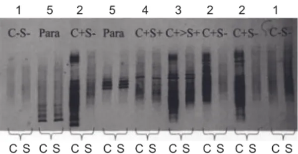
Figure 1.1. IEF immunoblots of the fi ve consensus patterns of various CSF and serum isoelectric focusing patterns for local/
systemic synthesis. The pattern number is given above the paired samples.
Type 1 (C S ): No bands in CSF and serum. Normal.
Type 2 (C + S ): Oligoclonal IgG is present in the CSF with no apparent corresponding abnormality in serum, indicating local intrathecal synthesis of IgG. Typical example: MS.
Type 3 (C + > S + ): There are IgG bands in both the CSF and serum, with additional bands present in the CSF. The oligoclonal bands that are common to both CSF and serum imply a systemic infl ammatory response, whereas the bands that are restricted to the CNS suggest that there is an additional CNS - only response.
Typical examples: MS, systemic lupus erythematosus (SLE), sarcoid, etc.
Type 4 (C + S + ): There are oligoclonal bands present in the CSF, which are identical to those in serum. This is not indicative of local synthesis, but rather, the pattern is consistent with passive transfer of oligoclonal IgG from a systemic infl ammatory response. Typical examples: Guillain – Barr é syndrome, acute disseminated encephalomyelitis (ADEM), and systemic infections.
Type 5 (Para): There is a monoclonal IgG pattern in both CSF and serum, the source of which lies outside the CNS. Typical examples:
Myeloma, monoclonal gammopathy of undetermined signifi cance (MGUS).
CSF IEF for both predictive and diagnostic testing in the diagnosis of MS. In other non - infectious infl ammatory disorders of the CNS, Class II and III evidence exists to support the use of CSF IEF to supplement other diagnos- tic tests (table 1.3 ).
CSF g lucose c oncentration,
CSF / s erum g lucose r atio and l actate
As glucose is actively transported across the blood – brain barrier the CSF glucose levels are directly proportional to the plasma levels and therefore simultaneous measure- ment in CSF and blood is required. Normal CSF glucose concentration is 50 – 60% of serum values [20] (Class IV).Eosinophils are normally not present in CSF. The pres- ence of 10 or more eosinophils/ M l in CSF or eosinophilia of at least 10% of the total CSF leukocyte count is associ- ated with a limited number of diseases, including para- sitic infections and coccidioiodomycosis. It can occur in malignancies and react to medication and ventriculo- peritoneal shunts [58] .
Malignant CSF cells indicate leptomeningeal metasta- ses. False - positive results often occur when infl am- matory cells are mistaken for tumour cells or due to contamination with peripheral blood [59] . False - negative detection of malignant cells on cytologic exam- ination of CSF is common. Factors increasing the detection rate of malignant cells include a volume of at least 10.5 ml and repeating this procedure once if the cytology is negative. The detection rate of 50 – 70%
after the fi rst investigation can be increased to 85 – 92%
after a second puncture [60] (Class III). Further LPs will only slightly increase the diagnostic sensitivity [61, 62]
(Class III).
In conclusion, cell count is generally useful because most of the indications for CSF analysis include diseases Upon activation, lymphocytes can enlarge or become
plasma cells indicating an unspecifi c infl ammatory reac- tion [54, 56] (Class IV). Resting monocytes enlarge and display vacuoles when activated. Macrophages are the most activated monocytes. These cell forms can occur in a great variety of diseases.
Erythrophages occur 12 – 18 h after haemorrhage. Sid- erophages containing haemosiderin are seen as early as 1 – 2 days after haemorrhage and may persist for weeks.
Macrophages containing haematoidin (crystallized biliru- bin) degraded from haemoglobin may appear about 2 weeks after bleeding and are a sign of a previous subarach- noid bleeding [54] (Class IV). However, spectrophotom- etry of CSF involving bilirubin quantitation has been recommended as the method of choice to prove CT - neg- ative subarachnoid bleeding up to 2 weeks after onset [57] .
Lipophages indicate CNS tissue destruction. The pres- ence of macrophages without detectable intracellular material is a non - specifi c fi nding, occurring in disc her- niation, malignant meningeal infi ltration, spinal tumours, head trauma, stroke, MS, vasculitis, infections, and sub- arachnoid haemorrhage [54] (Class IV).
Table 1.3 Infl ammatory diseases of the CNS associated with CSF oligoclonal IgG bands [32] .
Disorder Incidence of oligoclonal bands (%) Evidence
Multiple sclerosis 95 Class I a
Auto - immune
Neuro - SLE 50 Class III
Neuro - Beh ç et ’ s 20 Class II
Neuro - sarcoid 40 Class III
Harada ’ s meningitis - uveitis 60 Class III
Infectious
Acute viral encephalitis ( 7 days) 5 Class II
Acute bacterial meningitis ( 7 days) 5 Class II
Subacute sclerosing panencephalitis (SSPE) 100 Class I
Progressive rubella panencephalitis 100 Class I
Neurosyphilis 95 Class I
Neuro - AIDS 80 Class II
Neuro - borrelliosis 80 Class I
Tumour 5 Class III
Hereditary
Ataxia - telangiectasia 60 Class III
Adrenoleukodystrophy (encephalitic) 100 Class II
CNS, central nervous system; CSF, cerebrospinal fl uid; IgG, immunoglobulin G; SLE, systemic lupus erythematosus.
a This is based on studies using the Poser diagnostic criteria [46] that were validated against the original Schumacher criteria [47] . None of these criteria has been validated using population - based studies. Therefore, it could be argued that the diagnostic ‘ gold standard ’ is a fl awed standard.
that are associated with elevated numbers of various cells. Cytological staining can be helpful in distinguish- ing CNS diseases when the cell count is increased.
Investigation of i nfectious CSF
There are many small to medium - sized studies investi- gating the diagnostic sensitivity and specifi city of tests for various infectious agents but no controlled study evaluat-ing a work - up of infectious CSF in general. Therefore, there are no valid data on the indication, sensitivity, and specifi city of microbiological procedures in general (i.e.
how to proceed with CSF in obvious CNS infections).
Existing proposals for the general work - up of infectious CSF are based on clinical practice and theoretically plau- sible procedures [63 – 65] .
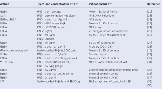
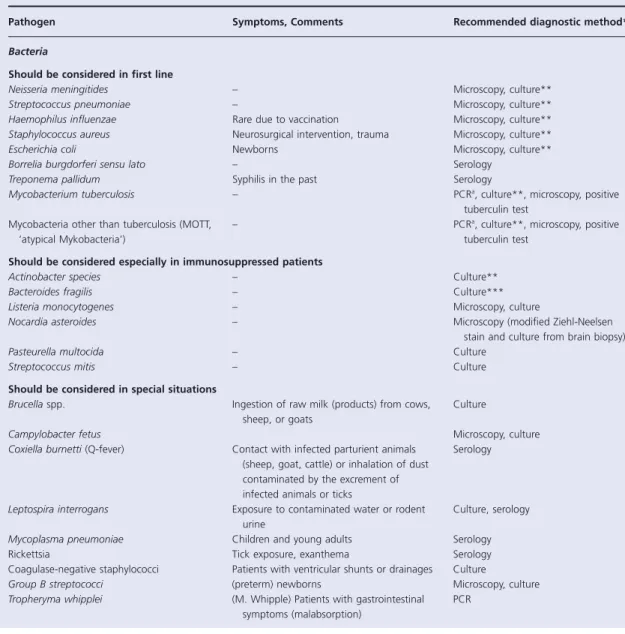
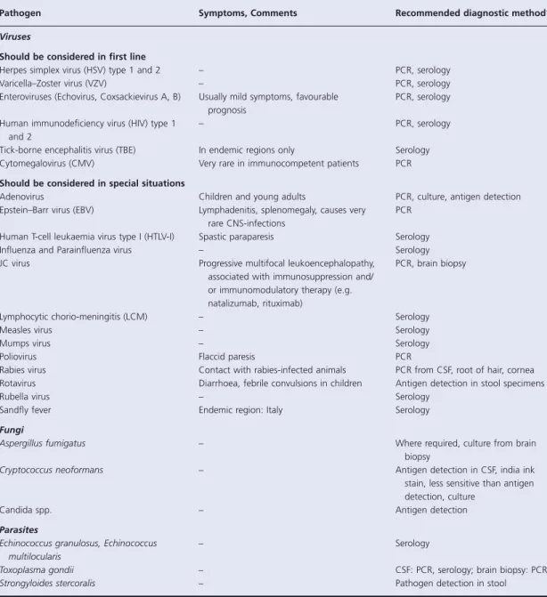
There are a great number of methods for antigen or specifi c antibody detection and their use depends mainly on the type of antigen (table 1.4 ).
Table 1.4 List of infectious agents responsible for the vast majority of infectious CNS diseases.
Pathogen Symptoms, Comments Recommended diagnostic method *
Bacteria
Should be considered in fi rst line
Neisseria meningitides – Microscopy, culture * *
Streptococcus pneumoniae – Microscopy, culture * *
Haemophilus infl uenzae Rare due to vaccination Microscopy, culture * *
Staphylococcus aureus Neurosurgical intervention, trauma Microscopy, culture * *
Escherichia coli Newborns Microscopy, culture * *
Borrelia burgdorferi sensu lato – Serology
Treponema pallidum Syphilis in the past Serology
Mycobacterium tuberculosis – PCR a , culture * * , microscopy, positive tuberculin test
Mycobacteria other than tuberculosis (MOTT, ‘ atypical Mykobacteria ’ )
– PCR a , culture * * , microscopy, positive
tuberculin test
Should be considered especially in immunosuppressed patients
Actinobacter species – Culture * *
Bacteroides fragilis – Culture * * *
Listeria monocytogenes – Microscopy, culture
Nocardia asteroides – Microscopy (modifi ed Ziehl - Neelsen
stain and culture from brain biopsy)
Pasteurella multocida – Culture
Streptococcus mitis – Culture
Should be considered in special situations
Brucella spp. Ingestion of raw milk (products) from cows, sheep, or goats
Culture
Campylobacter fetus Microscopy, culture
Coxiella burnetti (Q - fever) Contact with infected parturient animals (sheep, goat, cattle) or inhalation of dust contaminated by the excrement of infected animals or ticks
Serology
Leptospira interrogans Exposure to contaminated water or rodent urine
Culture, serology
Mycoplasma pneumoniae Children and young adults Serology
Rickettsia Tick exposure, exanthema Serology
Coagulase - negative staphylococci Patients with ventricular shunts or drainages Culture
Group B streptococci (preterm) newborns Microscopy, culture
Tropheryma whipplei (M. Whipple) Patients with gastrointestinal symptoms (malabsorption)
PCR
Pathogen Symptoms, Comments Recommended diagnostic method *
Viruses
Should be considered in fi rst line
Herpes simplex virus (HSV) type 1 and 2 – PCR, serology
Varicella – Zoster virus (VZV) – PCR, serology
Enteroviruses (Echovirus, Coxsackievirus A, B) Usually mild symptoms, favourable prognosis
PCR, serology
Human immunodefi ciency virus (HIV) type 1 and 2
– PCR, serology
Tick - borne encephalitis virus (TBE) In endemic regions only Serology Cytomegalovirus (CMV) Very rare in immunocompetent patients PCR
Should be considered in special situations
Adenovirus Children and young adults PCR, culture, antigen detection
Epstein – Barr virus (EBV) Lymphadenitis, splenomegaly, causes very rare CNS - infections
PCR
Human T - cell leukaemia virus type I (HTLV - I) Spastic paraparesis Serology
Infl uenza and Parainfl uenza virus – Serology
JC virus Progressive multifocal leukoencephalopathy,
associated with immunosuppression and/
or immunomodulatory therapy (e.g.
natalizumab, rituximab)
PCR, brain biopsy
Lymphocytic chorio - meningitis (LCM) – Serology
Measles virus – Serology
Mumps virus – Serology
Poliovirus Flaccid paresis PCR
Rabies virus Contact with rabies - infected animals PCR from CSF, root of hair, cornea Rotavirus Diarrhoea, febrile convulsions in children Antigen detection in stool specimens
Rubella virus – Serology
Sandfl y fever Endemic region: Italy Serology
Fungi
Aspergillus fumigatus – Where required, culture from brain
biopsy
Cryptococcus neoformans – Antigen detection in CSF, india ink
stain, less sensitive than antigen detection, culture
Candida spp. – Antigen detection
Parasites
Echinococcus granulosus, Echinococcus multilocularis
– Serology
Toxoplasma gondii – CSF: PCR, serology; brain biopsy: PCR
Strongyloides stercoralis – Pathogen detection in stool
The following pathogens should be considered in acute myelitis [Recommendation Level B]: HSV type 1 and 2 (PCR), VZV (PCR), enteroviruses (PCR), Borrelia burgdorferi sensu latu (serology, AI), HIV (serology), tick - borne encephalitis virus (only in endemic areas) (serology, AI).
a Nested PCR technique has been shown to be substantially more sensitive and specifi c than conventional single step PCR techniques [66] .
* * Culture from CSF and blood;
* * * aerobic and anaerobic culture from abscess aspirate, CSF, and blood.
Table 1.4 continued
Normal CSF protein concentration should be related to the patient ’ s age (higher in the neonate period and after age of 60 years) and the site of LP (Level B). Exact upper normal limits of protein concentration differ according to the technique and the examining laboratory.
The Q alb should be preferred to total protein concentrations, partly because reference levels are more clearly defi ned and partly because it is not confounded by changes in other CSF proteins (Level B).
The glucose concentration in CSF should be related to the blood concentration. Therefore CSF glucose/serum ratio is preferable. Pathological changes in this ratio or in lactate concentration are supportive for bacterial or fungal meningitis or leptomeningeal metastases (Level B).
• when microscopy, culture or serology is insensitive or inappropriate;
• when culture does not yield a result despite clinical suspicion of infectious meningitis/meningoencephalitis;
and
• in immunodefi cient patients.
Quality a ssurance in CSF d iagnostics
Some CSF quality assurance programmes have been pub- lished showing that to ensure optimal performance and results, standardized protocols should be in place for the spinal tap and sample processing [8] (Class 1). Further- more it is important to analyse the CSF in a specialized laboratory which is routinely evaluated for its perfor- mance and uses standardized analytical techniques and interpretation of the laboratory fi ndings in the clinical context [8] (Class 1); [70] (Class 4). If proteins are mea- sured that potentially originate from blood or brain com- partments, CSF and serum samples should be run in parallel in the same assay to minimize variability [8](Class I, Level A).
A cytology training programme resulted in an increase of the number of correctly identifi ed CSF cells from as low as 11% to 93% [71] . In a recent study investigating inter - laboratory variation of neurofi lament light chain detection, it turned out that the lack of preparation of accurate and consistent protein standards was the main reason for a very poor inter - laboratory accordance [72]
(Class I).
In neuroinfections specifi c antigen or antibody detec- tion should be performed depending on the clinical presentation and the results of basic CSF analysis. The formula for the estimation of the relative intrathecal synthesis of specifi c antibodies in the CSF (Antibody Index [AI] is as follows:
Estimation of intrathecal synthesis of specifi c antibodies in the CSF (Antibody Index [AI] )
Antibody ratio Antibody-concentration Antibody-concentra CSF
ttion IgG ratio = IgG-concentration
IgG-concentratio
serum CSF
n n
AI= Antibody ratio IgGratio postive>1, 5
serum
Cerebrospinal fl uid polymerase chain reaction can be performed rapidly and inexpensively and has become an integral component of diagnostic medical practice. A patient with a positive PCR result is 88 times more likely to have a defi nite diagnosis of viral infection of the CNS as compared to a patient with a negative PCR result. A negative PCR result can be used with moderate confi - dence to rule out a diagnosis of viral infection of the CNS (the probability of a defi nite viral CNS infection was 0.1 in case of a negative PCR result compared to a positive PCR result) [67] . It should be considered that false - negative results are most likely if the CSF sample is taken within the fi rst 3 days after the illness or 10 days and more after the onset of the disease [68, 69] .
In general, PCR is indicated in the following situations:
Recommendations
CSF should be analysed immediately (i.e. 1 h) after collection.
If storage is required for later investigation this can be done at 4 – 8 ° C (short term) or at 20 ° C (long term). Only protein components and RNA (after appropriate preparation) can be analysed from stored CSF (GPP).
The Level B recommendation regarding CSF partitioning and storage states that 12 ml of CSF should be partitioned into three to four sterile tubes. It is important that the CSF is not allowed to sediment before partitioning. Store 3 – 4 ml at 4 ° C for general investigations, cultivation and microscopic investigation of bacteria and fungi, antibody testing, polymerase chain reaction (PCR), and antigen detection.
Larger volumes (10 – 15 ml) are necessary for certain pathogens like Mycobacterium tuberculosis , fungi, or parasites.

![Table 1.2 Percentage of patients in different categories of disease with elevated IgA - index, IgG - index, IgM - index, or non - linear intrathecal synthesis formula values (data from [31 – 35] )](https://thumb-ap.123doks.com/thumbv2/azdoknet/10577271.0/14.803.62.693.154.463/percentage-patients-different-categories-disease-elevated-intrathecal-synthesis.webp)
![Table 1.3 Infl ammatory diseases of the CNS associated with CSF oligoclonal IgG bands [32]](https://thumb-ap.123doks.com/thumbv2/azdoknet/10577271.0/16.803.62.697.124.456/table-infl-ammatory-diseases-cns-associated-oligoclonal-bands.webp)
![Table 3.1 Diagnostic criteria for multiple sclerosis: 2005 revisions to the ‘ McDonald Criteria ’ [3]](https://thumb-ap.123doks.com/thumbv2/azdoknet/10577271.0/44.803.64.707.120.409/table-diagnostic-criteria-multiple-sclerosis-revisions-mcdonald-criteria.webp)