A Guide for Anaesthetists and Emergency
Physicians
Managing the Critically Ill Child
A Guide for Anaesthetists and Emergency Physicians
Edited by
Richard Skone
Specialist Registrar, Paediatric Intensive Care Medicine and Anaesthetics, Birmingham Children’s Hospital, Birmingham, UK
Fiona Reynolds
Paediatric Intensivist, Paediatric Intensive Care Unit, Birmingham Children’s Hospital, Birmingham, UK
Steven Cray
Consultant Paediatric Anaesthetist, Birmingham Children’s Hospital, Birmingham, UK
Oliver Bagshaw
Consultant in Anaesthesia and Intensive Care, Birmingham Children’s Hospital, Birmingham, UK
Kathleen Berry
Consultant in Paediatric Emergency Medicine, Birmingham Children’s Hospital, Birmingham, UK
Cambridge University Press
The Edinburgh Building, Cambridge CB2 8RU, UK Published in the United States of America by Cambridge University Press, New York www.cambridge.org
Information on this title: www.cambridge.org/9781107652323
© Cambridge University Press 2013
This publication is in copyright. Subject to statutory exception and to the provisions of relevant collective licensing agreements, no reproduction of any part may take place without
the written permission of Cambridge University Press.
First published 2013
Printed and bound in the United Kingdom by the MPG Books Group A catalogue record for this publication is available from the British Library
Library of Congress Cataloguing in Publication data Managing the critically ill child : a guide for anaesthetists and emergency physicians / edited by Richard Skone ... [et al.].
p. cm.
Includes index.
ISBN 978-1-107-65232-3 (Paperback)
1. Pediatric emergencies. 2. Critically ill children–Medical care.
I. Skone, Richard RJ370.M34 2013 618.9200025–dc23
2012028035 ISBN 978-1-107-65232-3 Paperback
Cambridge University Press has no responsibility for the persistence or accuracy of URLs for external or third-party internet websites referred to in this publication, and does not guarantee that any content on such websites is, or will remain, accurate or appropriate.
Every effort has been made in preparing this book to provide accurate and up-to-date information which is in accord with accepted standards and practice at the time of publication. Although case histories are drawn from actual cases, every effort has been made to disguise the identities of the individuals involved. Nevertheless, the authors, editors and publishers can make no warranties that the information contained herein is totally free from error, not least because clinical standards are constantly changing through research and regulation. The authors, editors and publishers therefore disclaim all liability for direct or con- sequential damages resulting from the use of material contained in this book. Readers are strongly advised to pay careful attention to infor- mation provided by the manufacturer of any drugs or equipment that they plan to use.
List of contributors vii
Section 1 – The District General Hospital Setting
1 Setting up a department for managing a sick child 1
Adrian P. Jennings and Julian Berlet 2 Team approach and organization 9
Richard Skone 3 Equipment 14
Fiona Reynolds
Section 2 – Clinical Conditions
4 Stabilization of the critically ill child: the initial approach 21 Richard Skone
5 The child with sepsis 33 Matthew D. Christopherson and Paul Baines
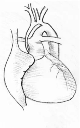
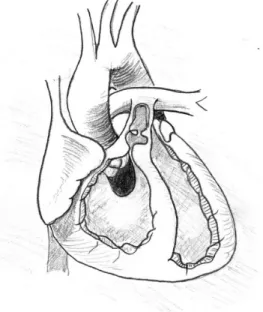
6 The child with cardiac disease 48 John Smith, with illustrations by Mazyar Kanani
7 The child with shortness of breath 64
Brian Shields and Benjamin Stanhope 8 The child with asthma 76
Josephine Langton and Stuart Hartshorn
9 The child with decreased consciousness and coma 87 Mark D. Lyttle
10 The child with seizures 95 Katie Z. Wright
11 The child with diabetic ketoacidosis 104 Ian Jenkins
12 Inherited metabolic conditions 112 Saikat Santra
13 Paediatric toxicology 122 Marius Holmes
14 The child with anaphylaxis 130 Nick Sargant
15 The child with stridor 137 Ed Carver and Tim Day-Thompson 16 The difficult paediatric
airway 146
Oliver Masters and Alistair Cranston
17 The surgical abdomen 159 Suren Arul
18 Paediatric trauma 169 Karl Thies
19 The child with raised intracranial pressure 180
Phil Hyde
20 The child with burns 192 Sapna Verma and Manu Sundaram 21 Blood product administration
in children 201 Oliver Bagshaw
v
22 The sick neonate presenting with shock 211
Fiona Reynolds
Section 3 – What You Could Be Expected to Do in a District General Hospital
23 Ongoing management of the successful arrest 219 Barney Scholefield
24 Referring-team-led transfers 228 Andrew J. Baldock and
Gareth D. Jones
25 Anaesthetizing for a surgical emergency 235
Andy Tatman and Richard Pierson
26 Managing children on an adult critical care unit 245
Helga Becker
27 Pain management in children 250 Ursula Dickson
28 Children with complex needs and disability 258
Kate Skone and Ian Wacogne
29 When a child dies 266 Fiona Reynolds
30 Child protection 270 Kate Skone and Geoff Debelle
Section 4 – The Children ’ s Hospital Setting
31 Ventilation 277 J. Nick Pratap 32 Fluid therapy 288
Adrian Plunkett
33 Pharmacology in children 300 Rhian Isaac
34 Neonates 309 Richard Skone
Section 5 – Golden Rules
35 Quick reference for emergencies 321 Kasyap Jamalapuram 36 Drug infusions 323 Kasyap Jamalapuram 37 Top tips for practical
procedures 325 Steven Cray
38 UK vaccination schedule 329 Richard Skone
39 Common syndromes in the critically ill child 330 Oliver Bagshaw
Index 340
Suren Arul
Consultant Paediatric Surgeon, Birmingham Children’s Hospital, Birmingham, UK
Oliver Bagshaw
Consultant in Anaesthesia and Intensive Care, Birmingham Children’s Hospital, Birmingham, UK
Paul Baines
Consultant in Paediatric Intensive Care, Alder Hey Children’s NHS Foundation Trust;
Wellcome Trust Biomedical Ethics Research Fellow, Department of Professional Ethics, Keele University, Staffordshire, UK
Andrew J. Baldock
Specialist Registrar in Anaesthesia, Southampton University Hospital, Southampton, UK
Helga Becker
Consultant Anaesthetist, Dudley Group NHS Foundation Trust, West Midlands, UK
Julian Berlet
Consultant Anaesthetist, Worcester Royal Hospital, Worcester, UK
Kathleen Berry
Consultant in Paediatric Emergency Medicine, Birmingham Children’s Hospital, Birmingham, UK
Ed Carver
Consultant Paediatric Anaesthetist, Birmingham Children’s Hospital, Birmingham, UK
Matthew D. Christopherson
Specialist Registrar in Paediatric Intensive Care, Alder Hey Children’s NHS
Foundation Trust, Liverpool, UK
Alistair Cranston Consultant Anaesthetist,
Birmingham Children’s Hospital, Birmingham, UK
Steven Cray
Consultant Paediatric Anaesthetist, Birmingham Children’s Hospital, Birmingham, UK
Tim Day-Thompson
Specialist Registrar in Anaesthesia, Birmingham School of Anaesthesia, Birmingham, UK
Geoff Debelle
Consultant Paediatrician, Birmingham Children’s Hospital, Birmingham, UK
Ursula Dickson
Consultant in Paediatric Anaesthesia, Birmingham Children’s Hospital, Birmingham, UK
Stuart Hartshorn
Consultant in Paediatric Emergency Medicine, Birmingham Children’s Hospital, Birmingham, UK
Marius Holmes
Specialist Registrar in Emergency Medicine, West Midlands Deanery, UK
vii
Phil Hyde
Consultant Paediatric Intensivist, Paediatric Intensive Care Unit, Southampton General Hospital, Southampton, UK
Rhian Isaac
Paediatric Pharmacist, Birmingham Children’s Hospital, Birmingham, UK
Kasyap Jamalapuram
Specialist Registrar, Paediatric Emergency Medicine, West Midlands Deanery, UK
Ian Jenkins
Consultant in Paediatric Anaesthesia and Intensive Care, Royal Hospital for Children, Bristol, UK
Adrian P. Jennings
Specialist Registrar in Paediatric Anaesthesia, Birmingham Children’s Hospital, Birmingham, UK
Gareth D. Jones
Consultant in Anaesthesia and Paediatric Intensive Care, University Hospital
Southampton NHS Trust, Southampton, UK
Mazyar Kanani
Fellow in Congenital Cardiothoracic Surgery, Great Ormond Street Hospital, London, UK
Josephine Langton
Specialist Registrar in Paediatric Emergency Medicine, Birmingham Children’s Hospital, Birmingham, UK
Mark D. Lyttle
Consultant in Paediatric Emergency Medicine, Bristol Royal Hospital for Children, Bristol, UK
Oliver Masters
Specialist Registrar in Anaesthesia, Birmingham Children’s Hospital, Birmingham, UK
Richard Pierson
Specialist Registrar in Anaesthetics, West Midlands Deanery, UK Adrian Plunkett
Consultant Paediatric Intensivist, Birmingham Children’s Hospital, Birmingham, UK
J. Nick Pratap
Assistant Professor in Anaesthesia and Paediatrics, Cincinnati Children’s Hospital and University of Cincinnati, Cincinnati, OH, USA Fiona Reynolds
Paediatric Intensivist, Paediatric Intensive Care Unit, Birmingham Children’s Hospital, Birmingham, UK Saikat Santra
Consultant in Paediatric Inherited Metabolic Disorders, Birmingham Children’s Hospital, Birmingham, UK Nick Sargant
Consultant in Paediatric Emergency Medicine, Bristol Royal Hospital for Children, Bristol, UK
Barney Scholefield
Clinical Research Fellow, Paediatric Intensive Care Unit, Birmingham Children’s Hospital, Birmingham, UK
Brian Shields
Specialist Registrar in General Paediatrics, University Hospitals Coventry and Warwickshire NHS Trust, Coventry, UK
Kate Skone
Specialist Registrar in Paediatric Neurodisability, Birmingham Children’s Hospital, Birmingham, UK
Richard Skone
Specialist Registrar, Paediatric Intensive Care Medicine and Anaesthetics, Birmingham Children’s Hospital, Birmingham, UK
John Smith
Consultant Paediatric Cardiothoracic Anaesthetist, Paediatric Intensive Care Unit, Freeman Hospital, Newcastle upon Tyne, UK
Benjamin Stanhope
Clinical Lead, Emergency Department, Birmingham Children’s Hospital, Birmingham, UK
Manu Sundaram
Specialist Registrar in Paediatric Emergency Medicine, Birmingham Children’s Hospital, Birmingham, UK Andy Tatman
Consultant Anaesthetist, Birmingham Children’s Hospital, Birmingham, UK
Karl Thies
Consultant in Paediatric Anaesthesia, Birmingham Children’s Hospital, Birmingham, UK
Sapna Verma
Consultant in Paediatric Emergency Medicine, Birmingham Children’s Hospital, Birmingham, UK Ian Wacogne
Consultant Paediatrician, Birmingham Children’s Hospital, Birmingham, UK
Katie Z. Wright
Specialist Registrar in Paediatric and Adult Emergency Medicine,
Birmingham Children’s Hospital, Birmingham, UK
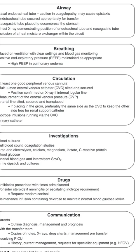
Chapter
1
Setting up a department for managing a sick child
Adrian P. Jennings and Julian Berlet
Introduction
In 2009 nearly 5000 UK children were admitted to 28 paediatric intensive care units (PICUs) from outlying hospitals, accounting for 64% of their unplanned workload.
A designated retrieval team performed the transfer to PICU in 80% of cases with a median time of arrival at the patient’s bedside of 2 hours.
Life-saving interventions required during the first few hours of stabilization remain the responsibility of the referring district general hospital (DGH) and cannot be deferred until the arrival of the retrieval team. Figure 1.1 demonstrates the frequency with which referring teams perform interventions. Any hospital that potentially manages sick children should have a series of systems in place that anticipate and ease the process of managing a critically ill child.
Endotracheal intubation
Mechanical ventilation
Central venous access
First inotropic agent
Osmolar therapy
0% 20% 40%
Percentage of interventions
60% 80% 100%
Referring hospital Retrieval team Figure 1.1. Proportion of interventions performed by referring hospitals and intensive care retrieval teams during stabilization of critically ill children. (Reproduced from S Lampariello, M Clement, AP Aralihond,et al. Stabilization of critically ill children at the district general hospital prior to intensive care retrieval: a snapshot of current practice.Arch Dis Child2010;95:681–685, with permission from BMJ Publishing Group Ltd.)
Managing the Critically Ill Child, ed. Richard Skoneet al. Published by Cambridge University Press.
© Cambridge University Press 2013.
1
Changing practice in district general hospitals (DGH) Decreasing experience
Many UK hospitals have withdrawn surgical services for children younger than 2 years of age. This leaves many non-specialist anaesthetists with a duty of care to manage sick children out of normal working hours. Nevertheless, public expectation of standards of care has increased and DGH emergency departments (EDs) have maintained open access for all children.
The Tanner report
In 2006 the Department of Health published a report entitled‘The acutely or critically sick or injured child in the district general hospital – a team response’. The working group established that concerns about deskilling in the management of critically ill children were shared by many specialties, including paediatrics and ED medicine.
The report recommended that services for the critically ill child should be planned within a network, with the local tertiary hospital acting as a source of advice and support while stabilization is performed in the local DGHs. It suggested six generic skills which can be expected of all personnel involved in the care of critically sick or injured children in the DGH:
to recognize the critically sick or injured child to initiate appropriate immediate treatment to work as part of a team
to maintain and enhance skills
to be aware of issues around safeguarding children to communicate effectively with children and carers.
There was also emphasis on the importance of considering the whole patient pathway.
Pre-hospital
The report commented that paramedic crews should transport sick children to the most appropriate paediatric facility, and make a clinical judgement to‘drive-by’a local DGH if the hospital does not have a team capable of stabilizing the critically ill child.
Resuscitation/stabilization
The report encouraged a team approach with shared responsibility. It emphasized that
‘following the initial stages of resuscitation of a critically ill⁄collapsed child, stabilization and further management should not be left solely to the anaesthetist’.
Transfer
The sickest children require transfer to the local tertiary paediatric centre and this transfer should be performed by a designated paediatric retrieval team. However, in certain cases, it may be necessary for the DGH team to facilitate transfer (see Chapter 24).
Organizing a DGH to manage a critically ill child Preparation
Relative to adult practice, the resuscitation and stabilization of critically ill children is a rare event. This increases the need for clear planning in order to ensure a smooth pathway for each patient. As a minimum each hospital should aim to have in place:
clear communication pathways within and between departments
easily available hospital protocols and guidelines for the management of common paediatric emergencies
appropriate range of drugs and equipment
adequate, ongoing training of staff in paediatric emergencies.
Location
Critically ill children usually present to the ED; however, they may also deteriorate in theatre, wards or other clinical areas. It is not practical to equip all clinical areas to stabilize a sick child. Local guidelines should be in place stating where resuscitation and stabilization should occur until the child’s condition improves or the retrieval team arrives.
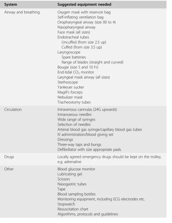
Designated areas should have appropriate neonatal and paediatric resuscitation equip- ment (see Table 1.1).
Children should ideally not be managed directly alongside adults and the need to support the family should be remembered. Parents and, where possible, children should be consulted when designing areas of care.
Equipment
Studies show that DGHs continue to perform the vast majority of key stabilization procedures on critically ill children (Figure 1.1). Therefore it is essential that paediatric and neonatal emergency equipment be readily available, well maintained and organized in a systematic fashion. Staff should undergo regular updates on its use, especially if used infrequently. A robust system should be in place to ensure that every area has rapid access to this equipment in the event of a paediatric emergency. Formal checks of drugs and equipment should be performed daily.
When equipment is procured in the DGH, it is useful to ensure suitability for use with both adult and paediatric patients as this will allow adult practitioners to use equipment with which they are familiar, and minimize cost.
The emergency equipment should be arranged in a structured manner so that staff using it infrequently can find the necessary items quickly and easily. This can be achieved in different ways:
system-based
airway equipment of all sizes in one drawer, circulation in another weight-based
one tray containing all the equipment for a child of a given weight.
Team composition
Stabilization of a critically ill child requires a team of competent individuals. As a minimum this should include:
a paediatrician or ED consultant an anaesthetist or intensivist a paediatric trained nurse.
Clinicians from other services such as surgery or radiology may also need to be involved.
All staff should be trained in the specific needs of children and their families, including child protection and consent.
It may sometimes be necessary for staff within the DGH to undertake transfer to the regional PICU if the condition is time-critical (see Chapter 24) or if the retrieval team is unavailable. Suitably trained staff to facilitate the transfer must be available at all times.
Support services
Services such as haematology, chemical pathology, radiology and blood transfusion should meet the requirements of children. Pharmacy staff with specialist paediatric knowledge should be available to ensure safe and effective drug management.
Hospitals with no acute paediatric units
Where a DGH with no on-site inpatient paediatric facilities offers children unrestricted access via the ED, the challenges of being able to manage the critically ill child are even greater. ‘Drive-by’ policies agreed with the ambulance service and close links to other hospitals with paediatric facilities are crucial.
Table 1.1. Minimum set of equipment for paediatric emergency trolley. Further equipment should be kept easily accessible if needed, e.g. ventilators.
System Suggested equipment needed
Airway and breathing Oxygen mask with reservoir bag Self-inflating ventilation bag Oropharyngeal airway (size 00 to 4) Nasopharyngeal airway
Face mask (all sizes) Endotracheal tubes
Uncuffed (from size 2.5 up) Cuffed (from size 3.5 up) Laryngoscope
Spare batteries
Range of blades (straight and curved) Bougie (size 5 and 10 Fr)
End-tidal CO2monitor
Laryngeal mask airway (all sizes) Stethoscope
Yankeuer sucker Magill’s forceps Nebulizer mask Tracheostomy tubes
Circulation Intravenous cannulas (24G upwards) Intraosseous needles
Wide range of syringes Selection of needles
Arterial blood gas syringe/capillary blood gas tubes IV administration/blood giving set
Dressings
Three-way taps and bungs
Defibrillator with size appropriate pads
Drugs Locally agreed emergency drugs should be kept on the trolley, e.g. adrenaline
Other Blood glucose monitor
Lubricating gel Scissors
Nasogastric tubes Tape
Blood sampling bottles
Monitoring equipment, including ECG electrodes etc.
Stopwatch Resuscitation chart
Algorithms, protocols and guidelines
Teamwork and training
A team approach is essential in order to utilize the skill-mix of different practitioners.
Local policies should define the members of the paediatric resuscitation team, the team leader (for example, emergency medicine consultant or paediatrician) and the roles expected to be performed by each member of the team.
Front-line medical staff should be trained in paediatric resuscitation and receive annual updates. The departments should aim to support staff as they improve their knowledge and skills through:
practising skills such as intraosseous needle insertion on purpose-built equipment completion of an accredited paediatric life-support course
repeated attendance at these courses for the purpose of recertification/revalidation secondment to a local paediatric centre
locally organized training (involving all departments) regular audit
interdepartmental morbidity/mortality meetings tutorials and e-learning modules.
Practice scenarios are a good opportunity to test equipment and review or develop local guidelines. Audit and morbidity/mortality meetings are an essential opportunity for the team to reflect on how actual cases are managed and identify areas of improvement.
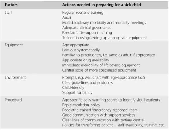
Table 1.2. Factors to be taken into consideration when planning paediatric emergency management.
Factors Actions needed in preparing for a sick child
Staff Regular scenario training
Audit
Multidisciplinary morbidity and mortality meetings Adequate clinical governance
Paediatric life-support training
Trained in using/setting up appropriate equipment
Equipment Age-appropriate
Laid out systematically
Familiar to practitioners, i.e. same as adult if appropriate Appropriate drug availability
Immediate availability of life-saving equipment Central store of more specialized equipment Environment Prompts, e.g. wall chart with age-appropriate GCS
Clear guidelines and protocols Child-friendly
Support for family
Procedural Age-specific early warning scores to identify sick inpatients Rapid escalation policy
Paediatric trained‘emergency response’team Good communication with support services Clear lines of communication with tertiary centre
Policies for transferring patient–staff availability, training, etc.
Relationship with the tertiary centre
A good relationship between a DGH and the local tertiary paediatric centre is essential if care of the acutely ill or injured child is to be well organized and effective. Links and coordination need to exist on several different levels.
Networks
The creation of paediatric service networks has helped improve communication over the last decade. In many regions, paediatricians, surgeons and paediatric anaesthetists have formed groups that create regional guidelines, conduct peer reviews of services and organize study days. There is no substitute for face-to-face meetings between clinicians as a way of developing closer links between local and tertiary centres. Outreach education and feedback sessions in local units help to foster the feeling of partnerships between DGHs and regional PICUs.
Retrieval services
In the UK, the development of regional retrieval teams has greatly improved links with DGHs. They provide facilities such as:
single point of access to PICU rapid response
advice throughout the process of managing a child web-based drug calculators
feedback sessions (two-way) teaching days
attendance at morbidity and mortality meetings.
Much of the responsibility for achieving a good relationship with the tertiary centre still lies with staff in the DGH. Liaising with specialist clinicians regarding equipment purchases, agreeing joint protocols and discussing individual patients is an effective way of forging links. Visiting the tertiary centre to observe or participate in a clinical attachment often helps to create strong links.
Summary
The burden of stabilizing critically ill children falls to all members of the ED, anaesthetic and paediatric team within a DGH. It is important that plans are put in place to help make the situation as straightforward and safe as possible.
Golden rules
Preparation is key to managing a sick child
Have appropriate equipment available and easy to access Develop clear guidelines that are easy to access
In-hospital, scenario-based training can improve teamwork and highlight potential problems
Further reading
Guidance on the provision of Paediatric Anaesthesia Services. Royal College of Anaesthetists, 2010. www.rcoa.ac.uk/docs/
GPAS-Paeds.pdf.
Lunn JN. Implications of the national confidential enquiry into perioperative deaths for paediatric anaesthesia.Paediatric Anaesthesia1992;2:69–72.
Standards for Children and Young People in Emergency Care Settings 2012: Developed by the Intercollegiate Committee for Standards
for Children and Young People in
Emergency Care Settings. www.rcpch.ac.uk/
emergencycare.
The acutely or critically sick or injured child in the District General Hospital: A team response. Department of Health, 2006.
http://www.dh.gov.uk/Consultations/
ClosedConsultations/
ClosedConsultationsArticle/fs/en?
CONTENT_ID=4124412&chk=lIVmJg.
Tomlinson A. Anaesthetists and care of the critically ill child.Anaesthesia
2003;58:309–311.
Chapter
2
Team approach and organization
Richard Skone
Introduction
A critically ill/injured child, depending on where they present, will be looked after by many of the following professionals:
paediatrician ED physician ED nurse paediatric nurse anaesthetist intensivist surgeon
operating department practitioner (ODP).
Within this team there are often two specific groups: those who have a paediatric back- ground but who do not frequently manage critically ill patients and those who are used to managing critically ill adults but have little paediatric experience. A good team will use the skills and experience of both groups and allow them to work freely and calmly.
The following chapter aims to cover the factors that make the team looking after a critically ill child function well.
Differences in paediatric emergencies
The major difference in the team composition compared to most other medical emergen- cies is the presence of paediatric doctors and nurses.
Some paediatricians will have spent a considerable amount of time on neonatal units as part of their training. They will have acquired a lot of skills such as intubation and insertion of arterial lines in babies. This can alleviate some of the pressure on anaesthetists who might otherwise feel that the burden of stabilizing small children lies with them.
It should be remembered, however, that neonatal emergencies seen in ED may not be similar to those in a maternity unit. The major difference in ED is that the children are less well‘controlled’in their presentation. A child may present at any point during their illness (rather than after birth or on a neonatal intensive care unit (NICU)). In this respect the ED doctor or anaesthetist will be more familiar with time-critical emergencies such as fulmin- ant sepsis or trauma.
Managing the Critically Ill Child, ed. Richard Skoneet al. Published by Cambridge University Press.
© Cambridge University Press 2013.
9
Organizing the team
Approaches to team management
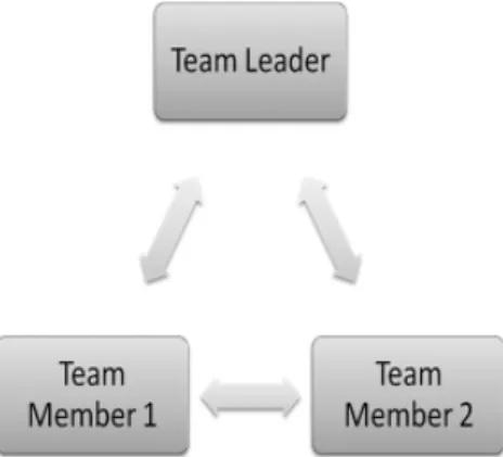
Vertical management
In the past much has been made of vertical management systems when dealing with medical emergencies (Figure 2.1). In this structure all information is relayed to the team leader, and then disseminated to the team. The team leader is responsible for all decision-making and tells the team members what to do at each stage.
Vertical management has its place. In particular, it is useful when managing an inexperi- enced team, or when a child has a single life-threatening problem that needs immediate attention.
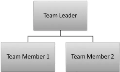
Horizontal management
When compared to vertical management, horizontal management has greater responsibility and involvement for all members (Figure 2.2). Team members make decisions, carry out tasks and communicate with each other and the team leader in a controlled, calm fashion.
The advantage of the horizontal structure is that it recognizes that people within a team have their own skills and abilities. It also recognizes that it is very rare for the management of medical emergencies to occur in a stepwise fashion. The key to this management structure is communication.
Figure 2.2. Horizontal management.
Figure 2.1. Vertical management.
Team leader
The team leader for a paediatric emergency should not be chosen purely on the basis of seniority. Experience either within the required specialty or of managing emergency teams should determine the best person to manage the situation.
This experience is essential as the team leader’s role will involve core skills such as:
keeping an overview of the situation integrating information as it feeds back prioritizing treatments
keeping everyone aware of their tasks and overall plan.
Team member
As well as having roles, team members have responsibilities. It is up to each member to:
work within their competencies take appropriate decisions listen
communicate clearly in a timely manner.
When a team member feels that they have a piece of information that needs to be raised urgently, they need to remember that they are working within a team. Other members may also have similarly urgent problems. Hence timely and concise relaying of information is essential.
If at any point in the emergency someone has been allocated a role which has no pressing problem, they should mention this at an opportune moment so that they can be reassigned further tasks within the resuscitation.
Why mistakes happen
As mentioned in Chapter 1, preparation and practice are the best way to try to improve team interaction. Errors occur for various reasons. Systems should be put in place to minimize the potential for errors. However, human factors will still contribute to problems if not addressed.
Technical skills
Technical procedures performed on small children are difficult especially if not done often.
Simulation and practical experience are important in order to stop fear of performing procedures impacting on other aspects of the team interactions. Time-critical skills should be performed by the most skilled person. The team leader also has a role in keeping the emergency room calm, as anxiety will worsen performance.
Knowledge
Like technical skills, it is up to individual doctors to make sure that their knowledge is up to date. Hospitals should facilitate relevant CPD in paediatric practice for appropriate staff members.
Team dynamic
Communication is a two-way process. Each team member needs to feel comfortable raising concerns with the team leader, while remaining clear and concise when reporting facts.
Mistakes happen within hierarchical teams when‘junior’members feel unable to raise concerns. This can be due to lack of confidence in their clinical ability or due to intimida- tion from the team leader.
When reporting a problem the team member needs to:
be clear and concise about what needs to be done emphasize importance of corrective action
escalate the issue calmly if they feel that the point has not been addressed.
At all points group responsibility needs to be emphasized by the team member so as not to seem confrontational. An example of raising a concern would therefore be:
‘We need to intubate this child urgently because the saturations are low and I’m not able to ventilate her with a mask’
Differences of opinion
Differences of opinion between specialists are difficult to manage. Should a disagreement arise the involvement of the most experienced physicians should be sought. Many PICUs have a consultant on call or a retrieval team that could provide advice. At no point should the situation be allowed to degenerate into an argument.
Before the child arrives
If the child is being brought in as an emergency it is likely that the emergency team will assemble before the patient arrives. In this instance the time should be used to:
introduce each member of the team (name and background) allocate roles appropriately
brief the team about any information given by the paramedics give initial instructions and plan (the team leader)
check equipment.
The time available to perform these tasks may vary and in cases of urgency it should be kept brief but thorough.
If the child is already present Assembling a team
A pre-hospital emergency call from an ambulance crew enables swift team assembly. It is often harder to assemble a team promptly for a patient who is already in hospital. If a child deteriorates in ED, on a ward or in theatre there needs to be a clear policy within the hospital on how to escalate the situation to have the relevant people present quickly. The escalation policy needs to make certain that rapid escalation does not have to pass through a chain of seniority. Instead, if a senior member is needed, they should be contacted as a first line.
A paediatric inpatient who is deteriorating rapidly needs the same level of attention as the child brought in by ambulance. Each hospital needs to make sure that there is an appropriate senior doctor available 24 hours a day who can make themselves available at short notice.
Integrating the team
The health professional who put out the call to assemble an emergency team for an inpatient should assume the role of the team leader initially. They will need to:
organize people as they arrive give a brief overview of the child
describe what has happened since admission explain the plan for ongoing management
hand over the team leader responsibility if appropriate.
Summary
The stress of infrequent paediatric emergencies can lead to teams underperforming. It is important that all hospitals rehearse paediatric emergency scenarios, both illness and injury, and have protocols and guidelines in place to ensure optimal management of sick and injured children.
Chapter
3
Equipment
Fiona Reynolds
Introduction
Having the correct equipment available in an emergency can make the management of a sick child much more straightforward. This chapter does not aim to tell people which particular pieces or brand of equipment to use. Instead it aims to highlight common problems in using medical equipment in children. It also offers a guide to which sizes to use in smaller children.
Airway and ventilation equipment Airway
Endotracheal (ET) tubes
There are a number of formulae for the appropriate size of ET tube in children. The most commonly used in children in the UK is:
Size of ET tube ðmmÞ ¼AgeðyrsÞ=4þ4
This, however, may either over- or underestimate the tube size so the size above and below should be immediately available. A common mistake is not to upsize a tube which is too small and has too great a leak.
Cuffed tubes have traditionally been used after puberty although more modern cuffed tubes have proved safe in children as small as term babies. These smaller cuffed tubes tend only to be available in children’s hospitals as there is little need for them in an emergency situation.
Children suffering from serious burns should have a cuffed ET tube inserted, if the appropri- ate size is available locally, as changing the tube will be difficult once facial swelling has occurred.
An uncut tube should also be used in this situation whether or not a cuffed tube is used.
Suction catheters for ET tube suctioning
Suction catheters should be available for ET tube suctioning. The appropriate size of suction catheter can be calculated using the formula:
Suction catheter sizeðFrÞ ¼ET tube sizeðmmÞ 2 ðmaximum size 14 Fr for an adolescentÞ
Managing the Critically Ill Child, ed. Richard Skoneet al. Published by Cambridge University Press.
© Cambridge University Press 2013.
14
Suction systems
Wall or portable suction may be used in children. For endotracheal suction a pressure of 60–80 mmHg (8–10 kPa) is appropriate for neonates and up to 120 mmHg (<16 kPa) for older children. It should be used for the minimum period of time.
Tracheostomy
The care-givers of children with a tracheostomy are usually expert in the manage- ment of the tracheostomy. Emergency supplies of a change of tracheostomy tube, including a smaller tube and suctioning equipment, usually accompany the child wherever they go. Most children with tracheostomies will have uncuffed tubes although occasionally a child may have a cuffed tube. Fenestrated tubes are rare before adolescence.
The care of the child with a tracheostomy mirrors that of an adult, with the understand- ing that the smaller tubes are more likely to block with secretions.
Ventilation
Mechanical ventilators
Most ICU ventilators used to ventilate adult-sized patients are capable of ventilating even the smallest of patients when used appropriately. An adult ventilator may be used with pressure control mode of ventilation, titrating the pressure to the measured tidal volume achieved or to the degree of chest movement and blood gases. Alternatively a volume control mode may be used provided the software allows a small enough tidal volume to be delivered (6–10 ml/kg).
The sedated, muscle-relaxed patient can be placed on a controlled mode of ventilation and the mechanics of triggering and synchronization are less important. Weaning from ventilation usually requires synchronization and triggering with the ventilator. Pressure triggering in children is usually not successful. Flow triggering commonly used in adult ICU is also used in paediatric practice (see Chapter 31).
Breathing circuits for ventilators
Breathing circuits come in two main sizes: 15 mm and 22 mm diameter tubing. The smaller tubing is used in children less than 20 kg.
The compression volume lost in the 15 mm ventilator tubing is smaller, i.e. the volume change of the circuit with each breath. However, some ventilators are able to compensate for any lost compression volume. In this case the size of the tubing is less of an issue.
The larger tubing often requires a catheter mount to connect from the‘Y’piece of the breathing circuit to the ET tube. The volume of the catheter mount contributes to dead space. For a child over 20 kg on mandatory ventilation this is usually not significant as the ventilator rate can be set to compensate. During the ventilator weaning phase when the patient is improving the dead space from the catheter mount can be an impediment to successful weaning.
The smaller 15 mm breathing circuits tend to have a single elbow connector. This connects directly to the ET tube without the need for a catheter mount and the attendant concerns about dead space.
Continuous positive airway pressure devices
Continuous positive airway pressure (CPAP) devices are commonly used for infants with moderate respiratory distress who do not require urgent intubation. The most common scenario is the neonate or infant with bronchiolitis. These babies may present with apnoea or moderate respiratory distress, which often improves with CPAP.
A variety of devices are used for this purpose and for the most part the paediatricians are usually familiar with the set-up in their own hospital. Anaesthetists and emergency depart- ment physicians should also make themselves familiar with the device used locally.
Most devices rely on an oxygen blender to titrate the percentage of inspired oxygen between 21% and 100%. The blender supplies a constant flow of gas maintained at a near constant pressure by an adjustable pressure valve. Alternatively the gas flow can be bubbled through an adjustable head of water (bubble CPAP). CPAP devices for infants typically use up to 15 l/min of gas and can generate pressures of up to 10 cmH2O.
Non-invasive ventilation
Non-invasive ventilation is used in children for the same indications as used in adults. The same ventilators are used in children over 10 kg as are used in adult practice. Children require smaller interfaces such as nasal, full face or face shield masks. It may be difficult to move a child requiring non-invasive ventilation between hospitals without intubation.
Humidification
Active humidification is generally achieved by a warmed water bath evaporator/humidifier.
The principles and temperatures used are identical to those used in adults. In general humidification aims at 100% relative humidity at body temperature for patients with an ET tube in situ. The humidifier dead space and resistance are not relevant in a fully ventilated patient or in the situation where a flow trigger is in use.
Passive humidification is achieved using a heat–moisture exchanger (HME). Care should be taken to use an appropriately sized HME. This is usually indicated on the device itself, giving a range of suggested tidal volumes where the device is suitable for use. An HME provides a lower level of humidity than is available using active humidification.
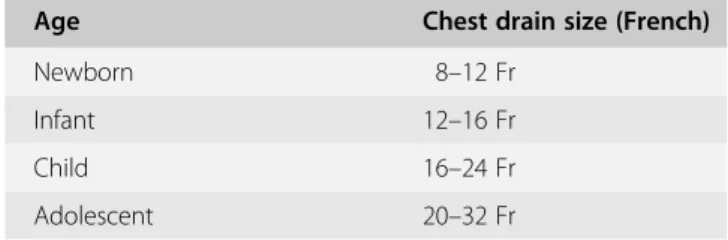
Chest drains
Insertion of a chest drain follows the same principles as in adult practice. Smaller sizes are used for draining a pneumothorax, with larger sizes used for draining fluid or blood (see Table 3.1). Underwater seals used for adults are identical to those used for children. Current guidance supports the use of ultrasound to guide the drainage of fluid.
Table 3.1. Guide to size of chest drain to use in a child.
Age Chest drain size (French)
Newborn 8–12 Fr
Infant 12–16 Fr
Child 16–24 Fr
Adolescent 20–32 Fr
Arterial and venous access Intravenous access
Peripheral lines
Peripheral lines start at 26G, which is primarily used for premature neonates under 1 kg.
24G cannulae are commonly used for term neonates although it is usually possible to insert a 22G cannula in the long saphenous vein at term.
The veins in the feet are commonly used in paediatric practice. Scalp veins are some- times used in babies, making sure the temporal artery is not cannulated. In an emergency, the external jugular vein may be useful to gain intravenous access.
Intraosseous access
If intravenous access cannot be obtained in a timely manner, intraosseous access should be secured. The most common site used is the proximal tibia, although the distal femur and proximal humerus may also be used. The hand-held needles are being replaced by battery- powered devices such as the EZ-IO®as these are associated with a higher success rate. More experienced clinicians are more likely to choose to use an intraosseous needle at an earlier stage in a patient’s treatment.
Central lines
The main sites of central venous line insertion are the internal jugular and femoral veins.
Both should be accessed under ultrasound guidance using strict aseptic technique. The subclavian route is rarely used in paediatric practice. In the neonate the jugular vein usually measures up to three times the diameter of the femoral vein, but tends only to be used by expert practitioners. Complications of femoral vein puncture are much rarer. Unlike adult data there are no paediatric data to suggest a higher infection rate in femoral central lines.
The most commonly used central lines in children from 3 to 25 kg are 4.5 or 5 Fr triple- lumen central lines, which are available from 5 to 12.5 cm in length. The 5–8 cm length can be used for most situations where central venous access is required in an emergency. Above a weight of 25 kg, a 7 Fr adult line is commonly used.
Arterial access
Arterial lines
Specific cannulae designed for use as arterial lines for adults may be used in children above 25 kg. Under 25 kg these lines may be too large in relation to the child’s artery.
In smaller children, it is common practice to use cannulae intended for intravenous insertion. Lines must be clearly marked to indicate the arterial nature of the line and any side port used for injection must be occluded by covering over with a dressing to prevent its use.
A 24G cannula is commonly used from 3 to 10 kg and a 22G above 10 kg. The radial artery and posterior tibial arteries are commonly used, with the femoral artery being used when more peripheral insertion sites prove impossible. The brachial and axillary arteries can be used. However, their use should be avoided in preterm babies due to the lack of collateral arterial flow.
Blood gas sampling devices
Arterial blood samples are taken in the same way as in adults. In-line sampling devices used for adults may be used in children. It is common practice to return the dead-space sample through either the inline sampling device or a syringe if an in-line sampling device is not used.
The minimum volume of blood should be taken; most modern blood gas machines need less than 1 ml per sample.
Monitoring and defibrillators
Monitoring
Minor modifications of the interfaces for physiological monitoring are necessary depending on the size of the patient. These modifications are smaller ECG stickers, pulse oximeter probes, blood pressure cuffs and temperature probes.
Invasive monitoring may rely on the insertion of smaller arterial and central lines but the transducers used in paediatric practice are the same as used in adult practice. In children less than 10 kg, many PICUs will use a flush of 1 ml per hour connected to a syringe driver by a ‘Y’ connector rather than the pressure transducer’s default 3 ml/h flush. In the emergency situation the default 3 ml/h flush is entirely appropriate for a term baby or older child.
Defibrillators
Manual defibrillators have a range of energy levels that are suitable for even the smallest neonate. The energy required is dependent on the rhythm to be treated and is calculated from the weight of the child.
Two sizes of hands-free stick-on pads are available: small for children less than 10 kg and adult size for use in adults and children greater than 10 kg. The small pads are applied in the same position used in adults. If the only pads available are adult pads, they may be used in an emergency on the anterior and posterior chest wall using the same energy calculated according to the weight of the child and rhythm to be treated.
External pacing
Many external defibrillators have an external pacing mode which may be used for bradyar- rhythmias. External pacing uses the same principles as in adults. The capture current is relatively lower and the paced rate should be higher according to the normal heart rate for the age of child.
Other equipment
Nasogastric tubes
Nasogastric tube length is measured from the nose to ear then to the stomach. Insertion in a child is usually simpler than in an adult. The site of placement must be confirmed by pH testing or X-ray. The size of nasogastric tube is usually:
6–8 Fr in a neonate 10–12 Fr in a toddler 14–16 Fr in an adolescent.
A nasogastric tube may be used to deflate the stomach when ventilating a child using a face mask and may be crucial to the ease of bagging in an emergency. For this reason, a nasogastric tube should always be available when intubating a paediatric patient.
Cervical hard collars
A properly sized hard collar can be used for cervical spine immobilization along with blocks and tapes for children over the age of 3 years. Single-piece hard collars used by the ambulance service and EDs do not fit most children under the age of 3 years.
There are hard two-piece collars designed for this younger age group, e.g. Miami Jr
®
from Ossur, but these tend to only be available in children’s hospitals. In the absence of an appropriately sized hard collar, manual in-line immobilization should be maintained. If a small enough hard collar is not available for a toddler, after intubation and ventilation the sedated and muscle-relaxed patient may be immobilized using blocks and tapes pending admission to PICU. Care must be taken to minimize flexion and extension of the neck.
Summary
It is important that the right equipment is available to team members as they strive to stabilize a child. Delays in finding the right equipment can make an already fraught situation more stressful. As always, preparation and planning can make the process of stabilizing a sick patient more efficient.
Golden rules
Most equipment used in adult practice is suitable for use with appropriate adjustments for size
Equipment should be stored in a systematic way so that it is immediately available Advice on equipment can be obtained from the local PICU
Chapter
4
Stabilization of the critically ill child: the initial approach
Richard Skone
Introduction
Most acutely critically ill children present first to their local DGH. However, they still remain a rare occurrence outside paediatric intensive care. The chances are slim therefore of a child presenting to a team with regular experience of resuscitating critically ill children.
There is plenty of knowledge, expertise and experience available amongst the team members outside specialized centres that can be applied to managing the critically ill child.
This chapter aims to help to overcome some of the most common hurdles that adult physicians face, and address the most common fears.
All doctors in a specialty that may manage sick children should already be familiar with the treatment algorithms provided by resuscitation groups. This chapter will focus on the emergency care provided by DGH ED physicians and anaesthetists.
Differences between children and adults
When sitting exams from medical school to postgraduate training the list of differences between adults and children is usually learnt as in Table 4.1. In reality these differences make little difference if you:
stick meticulously to the same gold standard principles of care delivered to adults, i.e.
optimize oxygen delivery, fluid balance, electrolytes and temperature
refer to reliable reference sources to remind you of normal values and drug doses ask for help from regional PICU retrieval teams early.
Managing the Critically Ill Child, ed. Richard Skoneet al. Published by Cambridge University Press.
© Cambridge University Press 2013.
21
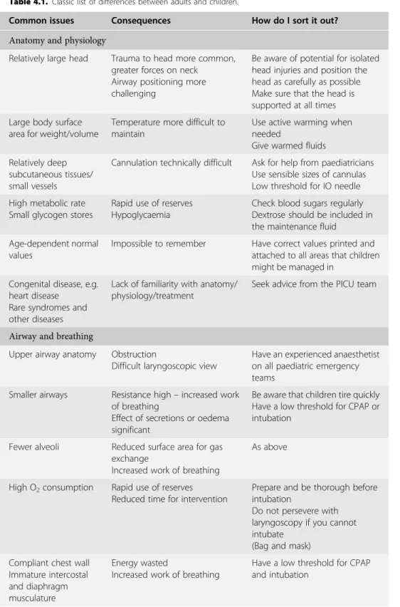
Table 4.1. Classic list of differences between adults and children.
Common issues Consequences How do I sort it out?
Anatomy and physiology
Relatively large head Trauma to head more common, greater forces on neck
Airway positioning more challenging
Be aware of potential for isolated head injuries and position the head as carefully as possible Make sure that the head is supported at all times Large body surface
area for weight/volume
Temperature more difficult to maintain
Use active warming when needed
Give warmed fluids Relatively deep
subcutaneous tissues/
small vessels
Cannulation technically difficult Ask for help from paediatricians Use sensible sizes of cannulas Low threshold for IO needle High metabolic rate
Small glycogen stores
Rapid use of reserves Hypoglycaemia
Check blood sugars regularly Dextrose should be included in the maintenance fluid
Age-dependent normal values
Impossible to remember Have correct values printed and attached to all areas that children might be managed in
Congenital disease, e.g.
heart disease Rare syndromes and other diseases
Lack of familiarity with anatomy/
physiology/treatment
Seek advice from the PICU team
Airway and breathing
Upper airway anatomy Obstruction
Difficult laryngoscopic view
Have an experienced anaesthetist on all paediatric emergency teams
Smaller airways Resistance high–increased work of breathing
Effect of secretions or oedema significant
Be aware that children tire quickly Have a low threshold for CPAP or intubation
Fewer alveoli Reduced surface area for gas exchange
Increased work of breathing
As above
High O2consumption Rapid use of reserves Reduced time for intervention
Prepare and be thorough before intubation
Do not persevere with laryngoscopy if you cannot intubate
(Bag and mask) Compliant chest wall
Immature intercostal and diaphragm musculature
Energy wasted
Increased work of breathing
Have a low threshold for CPAP and intubation
By focussing on the differences between adults and children, many teams forget that they have the skills at hand to manage children well. Working calmly and in an organized fashion can compensate, to a degree, for a lack of familiarity. This is best achieved as described in Table 4.2.
Cardiovascular
Left ventricle less compliant Less responsive stroke volume
Tachycardia in an attempt to increase cardiac output
Pay close attention to response to fluid challenges
Optimize electrolytes (especially calcium in neonates)
Myocardial failure/
vasoconstriction in response to sepsis
Blood pressure not useful to predict cardiac output Vasodilatory agents precipitate significant hypotension
Same as in adults
Relatively high circulating blood volume
Small total blood volume
Small losses have significant cardiovascular effect
Fluid challenges form the mainstay of assessing fluid balance
Be aware of significance of small volume losses
Emotional
Parents History has to be obtained from
care-givers
Care-giver emotional upset affects team
Paediatricians on team will be useful
Emotional impact of resuscitating sick child
Team performance may be affected by fear and upset
Regular debriefing sessions
Table 4.1. (cont.)
Common issues Consequences How do I sort it out?
Table 4.2. Broad outline of approach to a sick child.
The essence of care for children is:
Don’t make it more complicated than it has to be Be systematic
Use basic principles
Use/listen to the parents/carers
Access help/expertise from your team and PICU
Acknowledge fear and upset–in yourself and others–then put it aside
Concentrate on making the team work–remember human factors have a huge impact on ability to perform
Prepare for eventualities (see Chapters 1 and 2) Display visible normal values charts
Display charts for ET tube size/length and other parameters Advanced paediatric resuscitation course–staff up-to-date
Debrief each significant episode with learning points–not just those with poor outcomes!
Assessing the critically ill child – when to intervene Approach to the child
How you approach a sick child will depend on the severity of their illness. If the child arrives in cardiac arrest then a team approach where each member assesses, reports and acts on their designated role should be used. It is always worth taking 5 seconds when the patient arrives for the team leader to make a quick assessment, even if it is to confirm a lack of cardiac output.
If a child comes in seriously unwell, but awake, a single person examining the child may be appropriate in order to not upset the child. The team may arrive in a staggered fashion and the approach can quickly fragment. It is worth the team leader periodically repeating the management plan and allocation of roles in order to maintain order.
Airway assessment
The narrow airway of a child makes obstruction more likely when conditions such as inflammation and oedema of the trachea occur. If any of the signs seen in Table 4.3 occur then it is important to find out whether it is normal for that child. If it is not then the anaesthetist should be prepared to intubate the child (see Chapter 15).
Indications for intubation
There are many indications for intubation in a child. Most are the same as in adults, but some differ. Typical indications for intubation are detailed in Table 4.4.
Table 4.3. Signs of potential airway obstruction.
Stridor–biphasic (inspiration and expiration) stridor being more serious Stertor/snoring
Position of child–the child may sit in a‘tripod’position (leaning forward) to maintain optimum airway position
Accessory muscle use in the presence of other upper airway signs
Tracheal tug–indrawing of skin above sternal notch–especially if stridor present
Table 4.4. Indications for ventilation.
Respiratory failure
Cardiovascular instability–fluid resuscitation greater than 40 ml/kg with no response should prompt consideration
Active significant bleeding An unconscious child (GCS<9)
Head injury with a need for control of CO2/neuroprotection, etc.
Airway burns
Cardiac/respiratory arrest Significant invasive procedures
It is usually necessary to intubate and ventilate small children if you are performing invasive procedures, such as a jugular central line or chest drain insertion, as these are tolerated less well. The threshold for ventilation usually favours earlier intubation.
Breathing assessment
Children have a very compliant chest wall, and respiratory muscles that fatigue quickly. Their work of breathing, even at rest, is high compared to adults. The cartilaginous component of the ribs in children means that there is less opposition to the chest wall elastance. This results in a high closing capacity and a tendency towards airway collapse and atelectasis.
However, children are also very good at maintaining‘normal values’, such as saturations and PaCO2. They may not always look as if they’re about to decompensate. This might go some way to explaining why paediatricians seem to have a low threshold for requesting intubation and ventilation in their patients.
By the time children do look as if they are in need of support, their ability to continue to maintain ‘normal values’ including any semblance of cardiovascular stability may be significantly compromised. Clinical signs that would make you worry about a child’s respiratory effort include those seen in Table 4.5.
Intercostal recession is a common finding in many children with chest infections. On its own it may not point towards respiratory compromise. However, when present with some of the other signs from Table 4.5 it may become significant.
Capillary gases can be used in the same way as arterial blood gases (ABG) when deciding on management plans. The PaCO2 will equate to that of an ABG. See below for further information about capillary gases.
When making a decision about intervening, waiting for a child to become ‘sick enough to need intubation’ can turn a straightforward procedure into a highly pressured risky event.
Cardiovascular assessment
The circulating volume of small children is large relative to their size. However, the absolute volume is easy to overestimate. Fluid losses (GI, skin, respiratory, urine, third-spacing) have a significant impact. For example, a baby with a circulating volume of 80 ml/kg who weighs 4 kg will have a circulating volume of 320 ml. A loss of 80 ml equates to 25% of their circulating volume.
Blood pressure is well maintained despite significant illness in children. It should always be measured but never relied on as a single parameter to guide therapy. Capillary refill time
Table 4.5. Clinical signs of significant respiratory pathology.
Respiratory rate–tachypnoea is one of the most sensitive signs Grunting–an effort to generate positive end-expiratory pressure (PEEP) Intercostal and subcostal recession
Appearing drowsy or tired Apnoeas–especially in babies
A slowing of the respiratory rate and bradycardia are potentially preterminal events.
(CRT) is commonly used in paediatric practice to aid assessment of cardiovascular status.
As a single parameter it is not useful either–however, CRT can be used along with other parameters such as:
heart rate pulse quality blood pressure lactic acidosis
response to therapies (e.g. volume bolus) urine output.
Children have a huge capacity for vasoconstriction in the face of a low cardiac output.
When examining a child in shock, feel along the arms and legs. There is often a cut-off point at which the limb becomes cool. The higher up the level, the more the child is compensating for a poor cardiac output.
The‘cold shock’response to sepsis seen in smaller children differs from the more usual adult model of ‘high output’ shock as it can occur very early in the disease process.
Myocardial failure and intense vasoconstriction is a difficult situation to manage. It needs early recognition and aggressive management (see below and Chapter 5).
Electrolytes and temperature
Electrolytes and blood sugar
The advent of blood gas machines that measure electrolytes and blood sugars in addition to the usual parameters has made their management very straightforward. A separate blood sugar measurement should be performed immediately on arrival for any sick child. It should then be noted on every blood gas from there on, at regular intervals.
Temperature
Core temperature should also be measured on arrival and, as with blood sugars, should be monitored at regular intervals. Again, management is straightforward; it is forgetting to measure that forms the main stumbling block.
As well as hypothermia in sick children, pyrexia can cause significant problems. Pyrexia can make children appear disproportionately unwell with lethargy and tachycardia. Pyrexia can also worsen outcome in head injuries. It should be treated by:
Table 4.6. Signs of a child with cardiovascular compromise.
Tachycardia–a child may look well, but have an elevated heart rate (the cause of the tachycardia needs to be treated)
Weak pulses
Cool peripheries–the level at which a limb becomes cool may extend proximally as the child becomes sicker
Mottling–assuming the child is not cold
Hepatomegaly–this is a sign of heart failure, e.g. congenital heart disease or severe sepsis Decreased conscious level
removing the child’s clothes
giving antipyretics such as paracetamol keeping the environment cool
placing icepacks (head, axillae and over femoral arteries).
Carrying out the interventions – what to do when you are the specialist help
As mentioned above, anxiety can seriously affect performance. It is better to acknowledge fear and to deal with it than to drown under the pressure of expectation. If you have been called to manage a child, it is because you have the necessary skills that others in the team do not.
The major advantage of paediatric emergencies is that they attract a large team of people who are desperate to help. Use your team. Take a deep breath and manage the situation with the same calm reasoning as you would an adult.
Airway
As children get older the airway becomes more and more familiar to those working in adult medicine. By the time they reach the age of 1–2 years, the larynx is not as anterior, the tongue relatively smaller and the FRC greater. For children closer to the neonatal age paediatricians may become more helpful for practical procedures. Some will be very familiar with intubating and siting arterial lines on neonates.
Bag-mask ventilation
When bagging small children, the biggest problem by far is the anxiety-driven airway grip.
Pressing on the soft tissue under the mandible easily obstructs the airways of children.
Managing the airway in babies should be done by fingertip pressure on bony prominences, with the head in a neutral position.
The second biggest problem is gastric distension and is caused by even experienced paediatric anaesthetists. Air forced into the stomach during bagging can compromise ventilation. An early NG tube can make the situation much better. In fact it is commonplace to put in an NG tube for induction of anaesthesia in critically ill children.
Intubation
In small children, the occiput forms the biggest problem. The right size of roll under- neath/between the shoulder blades can make a huge difference. If your intubation has failed, stop early, bag the patient and reposition (as you would in adults). Always intubate orally first. The retrieval team can make a decision on nasal intubation for transfer.
Important things to consider when intubating critically ill children are mentioned in Table 4.7.
There are situations where a gas induction in theatre should be the method of choice for induction. These are discussed in Chapters 15 and 16. Endotracheal intubation is covered in Chapter 37.
Breathing
It has already been mentioned that smaller patients have to work harder to maintain a
‘normal state’. They also fatigue more quickly. The relatively immature chest wall of small children means that they have a high closing capacity and a tendency towards airway collapse and atelectasis.
Pressure-control ventilation
Once intubated the settings for the ventilator need to be set. In general, pressure-control ventilation will compensate for leaks around the ET tube. The pressures should be the same as in adults. Because the tidal volume measurements become less reliable as children become smaller, always look at the movement of the child’s chest, to check that it is sufficient (but not too much).
Positive end-expiratory pressure (PEEP)
The tendency towards airway collapse means that PEEP is essential when setting a ventila- tor. Start off with the same pressure as you would in adults (about 5 cmH2O) and titrate as needed. The biggest problem in small children is that as the PEEP increases, the venous return becomes compromised. If this occurs, first give a fluid bolus then back off the pressure (back to 5 cmH2O).
Difficulty ventilating
If you are having difficulty in ventilating a child, the commonest problems are:
the ET tube is not in the trachea–use end-tidal CO2monitoring
the ET tube is too far in (very common)–clinical examination and a CXR will confirm this the ET tube is blocked–pass a suction catheter
the ET tube is too small and there is a large leak
the child has significant lung pathology or fluid overload there is a pneumothorax
the child has pulmonary hypertension.
Table 4.7. Tips for intubating a sick child.
If possible place a tube for gastric decompression prior to induction–expect gastric distension and high risk of aspiration
Have access secured (peripheral or IO under local anaesthetic) Have volume already given and/or available to give during induction Have inotrope attached and/or running (depending on cardiovascular status)
Consider carefully the choice of induction agents (ketamine 1–2 mg/kg is the default choice for most retrieval teams)
Use a cuffed ET tube for burns, and if high airway pressures are anticipated, e.g. asthma Be ready to abandon laryngoscopy and attempts to intubate at any stage and sooner
than expected–have a timekeeper and agree time allowed for each attempt (usually 30 seconds)
Do not cut the length of an ET tube until its position has been checked on a CXR