저작자표시-비영리-변경금지 2.0 대한민국 이용자는 아래의 조건을 따르는 경우에 한하여 자유롭게
l 이 저작물을 복제, 배포, 전송, 전시, 공연 및 방송할 수 있습니다. 다음과 같은 조건을 따라야 합니다:
l 귀하는, 이 저작물의 재이용이나 배포의 경우, 이 저작물에 적용된 이용허락조건 을 명확하게 나타내어야 합니다.
l 저작권자로부터 별도의 허가를 받으면 이러한 조건들은 적용되지 않습니다.
저작권법에 따른 이용자의 권리는 위의 내용에 의하여 영향을 받지 않습니다. 이것은 이용허락규약(Legal Code)을 이해하기 쉽게 요약한 것입니다.
Disclaimer
저작자표시. 귀하는 원저작자를 표시하여야 합니다.
비영리. 귀하는 이 저작물을 영리 목적으로 이용할 수 없습니다.
변경금지. 귀하는 이 저작물을 개작, 변형 또는 가공할 수 없습니다.
의학박사 학위논문
비소세포폐암에서 EMT 관련 EGFR 표적치료 내성 극복을 위한 CDK7 억제제의 적용
The application of CDK7 inhibitors to overcome EMT- associated EGFR-TKIs resistance in non-small cell lung cancer
울 산 대 학 교 대 학 원
의 학 과 지 원 준
[UCI]I804:48009-200000288270 [UCI]I804:48009-200000288270
비소세포폐암에서 EMT 관련 EGFR 표적치료 내성 극복을 위한 CDK7 억제제의 적용
지 도 교 수 이 재 철 , 최 창 민
이 논문을 의학박사 학위 논문으로 제출함
2020 년 2 월
울 산 대 학 교 대 학 원 의 학 과
지 원 준
지원준의 의학박사학위 논문을 인준함
심사위원 김형렬 ( 인 ) 심사위원 이재철 ( 인 ) 심사위원 장승훈 ( 인 ) 심사위원 최창민 ( 인) 심사위원 황대욱 ( 인 )
울 산 대 학 교 대 학 원
2020 년 2 월
국문 요약
연구 목적: EMT (Epithelial to mesenchymal transition) 현상은 EGFR 표적치료제에 대한
내성과 관련 있는 기전으로 알려져 있다. 하지만 3 세대 EGFR 타이로신 키나제
억제제에 대한 내성 기전으로써 EMT 현상을 극복하기 위한 방안에 대한 자료는 매우
미흡한 상태이다. 본 연구에서는 비소페포폐암 세포주에서 EMT 현상과 관련한 3세대
EGFR 타이로신 키나제 억제제 내성 극복을 위한 CDK7 (Cyclin dependent kinase 7)
억제제의 효과에 대하여 확인해 보고자 하였다.
연구재료와 연구방법: 본 연구를 위하여 H1975 세포주를 활용하여 Osimertinib 과
WZ4002 에 대한 EGFR 표적치료제 내성 세포주를 구축하였다. EMT 발현 여부를
확인하기 위한 형태학적 변화를 확인하고, 침습/이동성 분석 및 면역탁본검사를
시행하였다. 또한 EMT 로 인한 유전적 변화를 확인하기 위하여 RNA 분석을
시행하였으며, CDK7 억제제로는 THZ1와 QS1189를 사용하였다.
연구결과: 실험을 통하여 구축된 H1975 내성 세포주는 형태학적으로 방추형 모양 및
위족 형성을 포함한 EMT 변화가 관찰되었다. 면역탁본검사에서 E-cadherin의 감소와
Vimentin 의 증가가 관찰되었으며, RNA 분석에서 EPCAM 과 CDH1 의 감소가
확인되었다. 침습 및 이동성 분석에서도 현저히 증가된 침습성과 이동성을 확인할 수
있었다. 이러한 EMT 변화가 나타난 내성 세포주는 모세포보다 THZ1 에 보다 민감한
약제 반응을 보여주었다. 이러한 현상은 TGF-ß 에 의하여 유도된 EMT 세포주에서도
유사하게 관찰되었으며, HCC827 과 PC9 세포주를 이용하여 구축한 gefinitib 내성
세포주에서도 같은 현상을 확인하였다.
결론: EMT현상은 EGFR타이로신 키나제 억제제에 대한 약제 감수성 감소와 관련이
있는 것으로 보이며, 이러한 EMT 에 의하여 생성된 내성세포주는 CDK7 억제제에
보다 높은 민감도를 보여주었다. 이러한 결과는 비소세포폐암에서 EMT관련 3세대
EGFR 표적치료제 내성을 극복하기 위한 전략으로서 CDK7 억제제의 활용 가능성을
시사한다.
중심단어: 비소세포폐암, EGFR 타이로신 키나제 억제제 획득 내성, EMT, CDK7
억제제
Index
Korean Abstract ··· i
Tables and Figures Legends ··· v
Introduction ··· 1
Materials and Methods ··· 5
1. Cell culture and reagents ··· 5
2. Establishment of a 3rd generation EGFR-TKI resistant cell line ··· 5
3. Immunoblot analysis ··· 6
4. Invasion and Migration assays ··· 7
5. RNA sequencing ··· 8
6. Cellular viability assays ··· 9
7. Statistics ··· 9
Results ··· 10
1. The induction of EMT in acquired resistance to 3rd generation EGFR-TKIs ··· 10
2. Results of immunoblot analysis used to evaluate EMT in the acquired resistance to 3rd generation EGFR TKIs ··· 10
3. Results of Invasion and Migration assays of EMT associated acquired resistance cell lines to 3rd generation EGFR-TKIs ··· 11
4. Results of RNA sequencing for the evaluation of genetic changes attributed to EMT during acquired resistance to 3rd generation EGFR-TKIs ··· 12
5. Efficacy of CDK7 inhibitors on the EMT associated acquired resistance cell lines
for the 3rd generation EGFR-TKIs ··· 12
6. Efficacy of CDK7 inhibitors on the TGF-β induced EMT cell lines ··· 13
7. Efficacy of CDK7 inhibitors on the TGF-β induced EMT using T790M naïve cell lines ··· 14
Discussion ··· 15
Conclusions ··· 20
References ··· 21
English Abstract ··· 42
Tables and Figure Legends
Table 1. Results of drug sensitivity to the 3rd generation EGFR-TKIs ··· 30 Table 2. Top 15 up & down regulated genes in the EMT induced EGFR-TKI resistance cell
lines ··· 31
Table 3. Results of drug sensitivity to CDK7 inhibitors in the EMT related EGFR- TKI resistance cell lines ··· 33
Figure 1. Establishment of the 3rd generation EGFR-TKI resistance cell line ··· 34
Figure 2. Phenotypic changes between the mother cell and the EMT induced EGFR-TKI resistance cell lines ··· 35 Figure 3. Immunoblot analysis of EMT expression ··· 36
Figure 4. Invasion and Migration analysis on the EMT related EGFR-TKI resistance cell lines ··· 37
Figure 5. Effect of CDK7 inhibitors on the EMT induced EGFR-TKI resistance cell lines ··· 38
Figure 6. Immunoblot analysis according to CDK7 inhibitor treatment on the EMT induced
EGFR-TKI resistance cell lines ··· 39 Figure 7. Effects of THZ1 on the TGF-β induced EMT cell lines ··· 40 Figure 8. Effects of THZ1 on the TGF-β induced EMT in T790M negative EGFR
mutant cell lines ··· 41
Introduction
Lung cancer is the leading cause of cancer mortalities globally1, 2). Resistance to lung
cancer therapies contributes to higher death rates in these patients. A targeted therapy
utilizing an Epidermal Growth Factor Receptor (EGFR) mutation improved the
survival rate of patients until resistance began to develop3). First-generation EGFR-
tyrosine kinases (TKIs) therapy exhibits efficacy for a short time (<2 years); however,
inevitably, resistance gradually develops, leading to disease progression4). Various
resistance mechanisms for EGFR-TKIs have been reported in previous research,
including T790M point mutations, EGFR amplification, ERBB2 amplification, small
cell lung cancer transformation, MET amplification, PI3K mutation, epithelial
mesenchymal transition (EMT), BRAF mutation, and KRAS mutation4-7). The T790
mutation is considered the most prevalent type (40~50% of cases) of resistance to the
first-generation EGFR-TKIs. Recently, a third generation drug osimertinib has been
developed to overcome T790M associated resistance8). However, most of the
mechanisms involved in third-generation EGFR-TKI resistance have not yet been
elucidated. As such, various attempts to overcome acquired resistance to 3rd
generation EGFR inhibitors are critical for improving the prognosis of patients.
EMT is a biological process via which cells undergo a switch from the polarized
epithelial phenotype to the mesenchymal fibroblastoid phenotype and considered to
be one of the resistance mechanisms. EMT is involved in several diverse processes,
including embryonic development, chronic inflammation, fibrosis9, 10), tumorigenesis,
invasion, metastasis and drug resistance11-13). As a result of EMT, cells downregulate
the expression of epithelial proteins containing E-cadherin and upregulate the
expression of mesenchymal proteins, including vimentin. In addition, cells
undergoing EMT are characterized by the loss of apico-basal polarity and intact cell-
cell junctions followed by the acquisition of front-rear polarity and morphologic
change to a spindle shape with remodeling of the cytoskeleton14). This EMT program
enhances the invasive capacity, therapeutic resistance, and cancer stem-cell-like
properties13). EMT has also been reported as one of the mechanisms of EGFR-TKI
resistance7). However, studies to overcome EMT related EGFR-TKI resistance are
still insufficient.
Cyclin-dependent kinases (CDKs) are a family of serine-threonine kinases that play
an important role in cell cycle progression. Over 90% of tumors have been found to
be upregulated in the CDKs due to changes in the expression and genetic variation of
CDKIs. Based on this, CDKs inhibitors are likely to be developed as anticancer
agents that inhibit the growth of various cancers. In particular, CDK7 acts as a master
regulator of transcription and is known to modulate RNA polymerase II activity15).
Recently, CDK7 inhibitors have been reported to inhibit abnormal cell growth
associated with hematologic malignancy16), breast17), esophageal18), and small cell
lung cancer19). Specifically, small cell lung cancer studies have shown that the action
of THZ1 significantly reduces the activity of super-enhancers and associated
oncogene transcription factors19), which suggests that CDK inhibitors could
potentially be used as anticancer agents.
This study evaluated whether EMT was expressed in the resistant cell lines generated
by treatment with 3rd generation EGFR-TKIs, and analyzes the effect of CDK7
inhibitors on EMT-associated resistance cells to evaluate a potential therapeutic
strategy to overcome EMT related EGFR-TKI resistance.
Materials and Methods
2.1. Cell culture and reagents
The H1975 and HCC827 cell lines were obtained from the American Type Culture
Collection (Rockville, MD), and the PC-9 cell line was provided by Dr. Kazuto
Nishio (National Cancer Center Hospital, Tokyo, Japan). Cells were cultured in 10%
fetal bovine serum (FBS), 100 U/mL penicillin, and 100 mg/mL streptomycin
(Invitrogen, Carlsbad, CA) at 37°C in an atmosphere with 5% CO2. Osimertinib,
WZ4002, and THZ1 were purchased from Selleck Chemicals (Houston, TX). The 3-
(4,5-dimethylthiazo-2-yl)-2,5-diphenyltetrazolium bromide (MTT) solution and
TGF-β1 was purchased from Sigma (St. Louis, MO) and R&D Systems
(Minneapolis, MN), respectively. QS1189 was kindly provided by Orient Bio
(Seongnam, Korea).
2.2. Establishment of a 3rd generation EGFR-TKI resistant cell line
H1975/WR and H1975/OR cells were subjected to repeated exposure to WZ4002 or
osimertinib, as reported in the previous studies20). In all studies, resistant cells were
then cultured in a drug-free medium for >1 week to eliminate the effects of each drug.
The resistant cell lines were authenticated using STR analysis and confirmed to be
mycoplasma free using standard methods.
2.3. Immunoblot analysis
Whole-cell lysates were prepared using EBC lysis buffer (50 mM Tris-HCl [pH, 8.0],
120 mM NaCl, 1% Triton X-100, 1 mM EDTA, 1 mM EGTA, 0.3 mM
phenylmethylsulfonyl fluoride, 0.2 mM sodium orthovanadate, 0.5% NP-40, and 5
U/mL aprotinin) and then centrifuged. The resultant supernatant (20 μg) was
separated on an 8% to 12% sodium dodecyl sulfate-polyacrylamide gel
electrophoresis and transferred onto polyvinylidene difluoride membranes
(Invitrogen). The membranes were probed with antibodies against p-EGFR
(Tyr1173), p-Erk (Thr202/Tyr204), Erk, Akt, E-cadherin, β-actin (all from Santa
Cruz Biotechnology, Santa Cruz, CA), p-Akt (Ser473), β-catenin, EpCAM,
Desmoplakin, cytokeratin-8/18, (all from Cell Signaling Technology, Beverly, MA),
and vimentin was used as the first antibody (Cell Signaling, Beverly, MA). Then the
membranes were treated with horseradish peroxidase-conjugated secondary antibody.
All membranes were developed using ECL kits (PerkinElmer, Waltham, Mass).
2.4. Invasion and Migration assays
The cell migration and invasion assays were done using Transwell (6.5 mm diameter,
8 mm pore size polycarbonate membrane), which was obtained from Corning
(Cambridge, MA). The cells (1 × 10⁵) in 200 μL medium were placed in the upper
chamber, and the lower chamber was filled with 1 mL of serum-free media
supplemented with 0.1% bovine serum albumin. Following incubation for 24 h, the
cells that migrated to the lower surface of the filters were stained with a Diff-Quick
kit (Fisher Scientific, Pittsburgh, PA), and then they were counted under a
microscope. The migration assay was conducted using the same procedure with
filters that were coated with extracellular matrix on the upper surface (BD
Biosciences, Bedford, MA). The mean represents the results in triplicate (standard
deviation).
2.5. RNA sequencing
Each cell was pelleted, and total RNA was extracted using an RNeasy Mini Kit
(Qiagen, Hilden, Germany). Genomic DNA was eliminated, and RNA integrity was
verified using the Agilent 2100 Bioanalyzer (Agilent Technologies, Santa Clara, CA,
USA). RNA Sequencing was performed by a specialized company (Macrogen, Seoul,
Korea) using the TruSeq RNA Sample Prep Kit v2 and HiSeq. 2500 platform
(Illumina, San Diego, CA, USA). Sequenced reads were trimmed using
TrimMomatic 0.32. The trimmed reads were mapped to the hg19 reference genomes
using HISAT (version 2.0.5) and Bowtie2. Expression levels were measured as
fragments per kilobase for map reads (transcript per million) using StringTie (version
1.3.3b).
2.6. Cellular viability assays
Cells (1 × 10⁴) were seeded into 96-well sterile plastic plates overnight and then
treated with the relevant drugs. Following 72 h, 15 μL of MTT solution (5 mg/mL)
was added to each well, and the plates were incubated for 4 h. Crystalline formazan
was solubilized in 100 μL of 10% (w/v) SDS solution for 24 h. Absorbance at 595
nm was read spectrophotometrically using a microplate reader. Cell counting was
determined using an ADAM-MC automatic cell counter (NanoEnTek, Seoul, Korea) according to the manufacturer’s instructions. The results represent at least three
independent experiments, and the error bars signified the standard deviation from the
mean. The IC50 values were determined using GraphPad Prism software (GraphPad,
Inc., La Jolla, CA).
2.7. Statistics
Data are presented as the mean ± standard deviation. P values ≤0.05 were determined using unpaired or paired t-tests between groups using the GraphPad Prism software.
Results
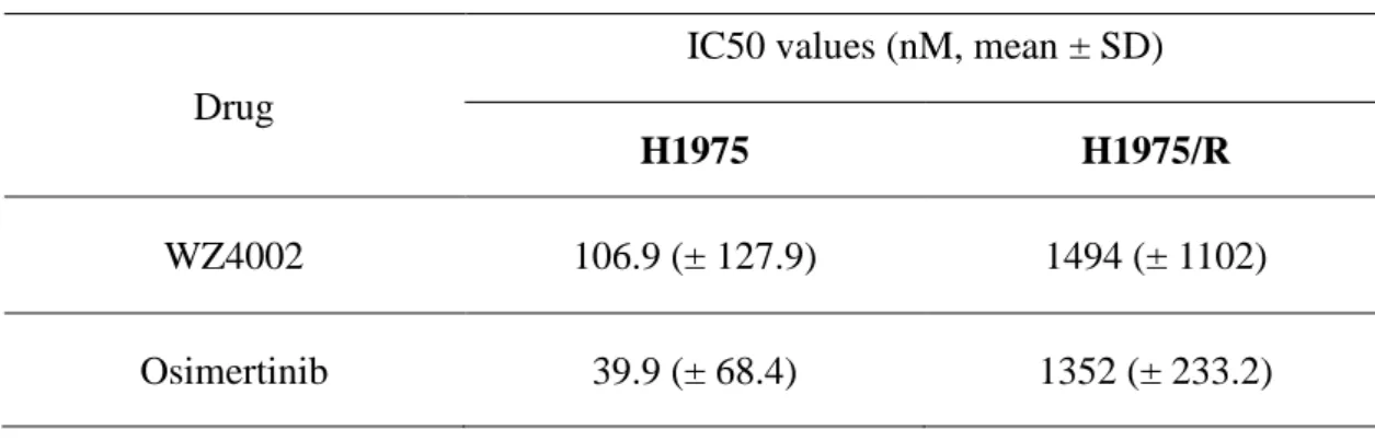
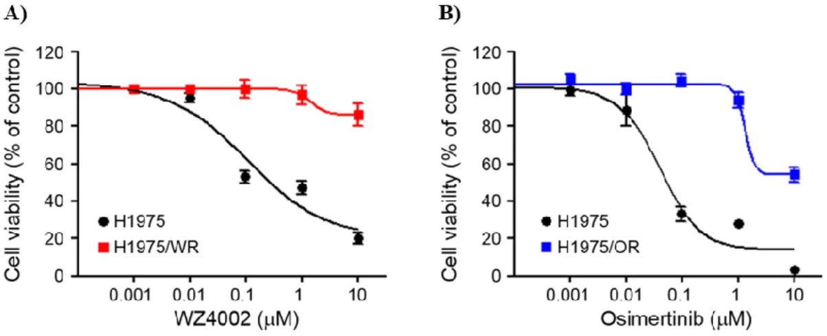
3.1. The induction of EMT in acquired resistance to 3rd-generation EGFR-TKIs
To investigate the mechanisms of acquired resistance to 3rd-generation EGFR-TKIs,
we established H1975/WR and H1975/OR through stepwise selection in WZ4002 or
osimertinib, as described in previous studies20). Both types of cells exhibited
resistance more than 10 times to each drug compared with the parent cells (WZ4002
IC50 = 106.9 nM in H1975 and 1494 nM in H1975/WR, osimertinib IC50 = 39.9 nM
in H1975 and 1352 in H1975/OR; Figure 1 and Table 1). These resistant cells
exhibited an increase in spindle-shaped cells that were similar to the spindle-shaped
cells produced by EMT (Figure 2).
3.2. Results of immunoblot analysis used to evaluate EMT in the acquired resistance
to 3rd-generation EGFR-TKIs
In order to evaluate the induction of EMT in both of the resistant cells, we analyzed
the expression of marker proteins for epithelial or mesenchymal phenotypes by using
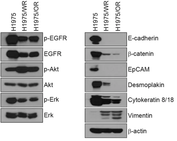
western blotting (Figure 3). Compared with the H1975 cells, epithelial marker
proteins containing E-cadherin, β-catenin, EpCAM, desmoplakin, and cytokeratin-
8/18 were significantly reduced in both of the resistant cells, whereas vimentin
expression was increased. Moreover, the expression and activity of EGFR were
reduced in both of the resistant cells; while the activity of Akt was significantly
upregulated.
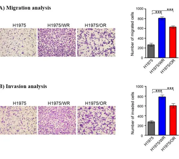
3.3 Results of Invasion and Migration assays of EMT associated acquired resistance
cell lines to 3rd-generation EGFR-TKIs
We investigated the ability of migration and invasion, which are considered
functional hallmarks of EMT. We found that the abilities of migration and invasion
were significantly enhanced in both of the resistant cells (Figure 4). Thus, these data
suggested that the acquisition of resistance to 3rd-generation EGFR-TKIs induced
molecular changes that were consistent with EMT.
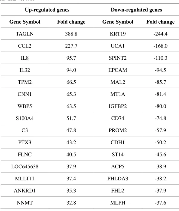
3.4. Results of RNA sequencing for the evaluation of genetic changes attributed to
EMT during acquired resistance to 3rd-generation EGFR-TKIs
To determine the genetic changes caused by EMT associated markers in the resistant
cell lines, we performed RNA sequencing. The top 15 genes with increasing or
decreasing patterns at the RNA level are shown in Table 2. EPCAM and CDH1 were
significantly reduced, and this suggested that EMT was induced at the genetic level.
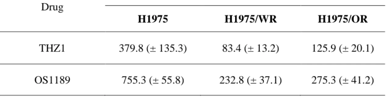
3.5. Efficacy of CDK7 inhibitors on the EMT acquired resistance cell lines for the
3rd-generation EGFR-TKIs
Previous studies showed that CDK7 was associated with EMT; however, it was
controversial whether targeting CDK7 could overcome EMT 21-24). Therefore, to
determine the effect of CDK7 inhibition in both types of resistant cells, we used
THZ1 and QS1189 as CDK7 inhibitors. QS1189 was developed as a novel CDK7
inhibitor in a previous study16). As shown in Figure 5 and Table 3, both resistant cells
were more sensitive to CDK7 inhibitors than the parental cells (THZ1 IC50 = 379 nM
in H1975, 83.4 nM in H1975/WR, 125.9 nM in H1975/OR; QS1189 IC50 = 755.3 nM
in H1975, 232.8 nM in H1975/WR, 275.3 nM in H1975/OR). Next, CDK7 kinase
activity has been involved in phosphorylation of the CTD of RNAPII, which played
a role in transcription initiation and RNAPII procession15, 25, 26). We performed
western blotting following treatment of THZ1 (Figure 6) to evaluate the inhibitory
effect of CDK7 substrate in the parental and resistant cells,. The inhibitory effect of
THZ1 on the activity of RNAPII-CTD was similar for H1975 and H1975/OR cells.
However, H1975/WR cells showed the inhibition of RNAPII-CTD phosphorylation
at Ser2, Ser5, and Ser7 at a lower concentration of THZ1. In addition, THZ1
treatment failed to inhibit the activity of EGFR and Akt, while dose-dependent
induction activity was associated with Erk.
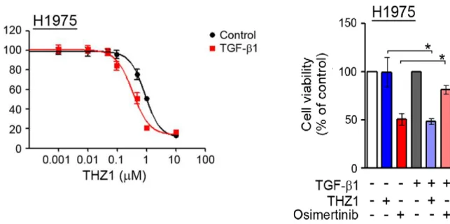
3.6. Efficacy of CDK7 inhibitor on the TGF-ß induced EMT cell lines
To determine whether induction of EMT could affect sensitivity to the CDK7
inhibitor, we assessed the response of THZ1 to TGF-β1-induced EMT. When cells
were treated with TGF-β1 for 24 h, cells with mutant EGFR were scattered with an
observed loss of E-cadherin and increased vimentin. The sensitivity to THZ1 was
increased in cells with TGF-β1-induced EMT (Figure 7). In addition, TGF-β1
treatment did not affect the inhibition of RNAPII-CTD phosphorylation by THZ1.
Therefore, the induction of EMT enhanced sensitivity to the CDK7 inhibitor.
3.7. Efficacy of CDK7 inhibitor on the TGF-ß induced EMT using T790M naïve cell
lines
Following induction of EMT using TGF-ß in T790M negative cell lines (HCC827
and PC-9), the effects of CDK inhibitors were evaluated. The sensitivity to THZ1
increased in the cells with TGF-β1-induced EMT, while the sensitivity to osimertinib
had decreased (Figure 8). This pattern was consistent with the experimental results
for the efficacy of CDK7 inhibitors on EMT associated resistant cell lines created
with 3rd-generation EGFR-TKIs.
Discussion
We established two 3rd generation EGFR-TKI resistant cell lines (H1975/WR,
H1975/OR) via repeated exposure to Osimertinib and WZ4002. The resistant cells
showed phenotypical changes similar to EMT, including a spindle-cell shape and
increased pseudopodia formation. Decreased E-cadherin and increased vimentin
were observed in the immunoblot assay, and decreased EPCAM and CDH1 were
found during RNA sequencing. The abilities of invasion and migration were
increased in the resistant cells. The EMT related resistance cells showed higher
sensitivity to THZ1 than the mother cells. This phenomenon was observed in the
TGF-ß induced EMT cell lines and the T790M negative resistant cell lines (HCC827,
PC-9).
EMT is associated with poor prognosis for non-small cell lung cancer patients 27)28),
and previous studies have shown that the mesenchymal phenotype was more resistant
to the 1st generation EGFR-TKI treatment than the epithelial phenotype29-31).
Previous studies have revealed that EMT was a potential mechanism involved in
first-generation EGFR-TKI resistance. Moreover, there have been a few reports of
EMT related third-generation EGFR-TKI resistance32). Li X et al. reported that
C797S was the most common cause of third-generation EGFR-TKI resistance, but
EMT is also a possible cause of acquired resistance33). This study found that EMT
was related to acquired resistance caused by 3rd generation EGFR-TKI. These
phenomena were confirmed by (a) the alteration of morphology such as formation of
spindle-shaped cells and pseudopodia, (b) changes in the molecular marker proteins
loss of E-cadherin and gain of vimentin, (c) increased invasive and migratory ability,
and (d) decreased RNA levels which were related to the epithelial markers.
Previous studies have reported that continuous exposure of tumor cells to TGF-ß
induced EMT through the SMAD and MAPK pathways11, 34). Therefore, to determine
whether EMT led to increased sensitivity to the CDK7 inhibitors apart from EGFR-
TKI exposure, we established TGF-ß induced EMT cell lines and then measured the
response to the CKD7 inhibitors and EGFR-TKIs. In the current study, we found that
drug sensitivity for EGFR-TKIs decreased, while sensitivity to CDK7 inhibitors
increased for the TGF-induced EMT cells. These data suggested that EMT might be
a potential mechanism of acquired resistance to the 3rd generation EGFR-TKIs in
non-small cell lung cancer.
An interesting part of this study was that the drug sensitivity to CDK7 inhibitors was
higher in the EMT associated EGFR-TKI resistant cell lines than in the parental cells.
Cyclin-dependent kinase (CDK)7 is necessary for transcription and acts by
phosphorylating the C-terminal domain (CTD) for RNA polymerase II (PolII), which
enables transcription initiation. Increases in the transcription factors have been
reported for various carcinomas, and there were a number of studies that reported
pharmacological modulation of the CDK7 kinase activity, which might provide
potential anti-cancer activities 17, 18, 35-37). In this study, we found that treatment using
CDK7 inhibitors inhibited the RNA polymerase II activity in EMT associated EGFR-
TKI resistance cells. This suggested that EMT induced resistant cells might have
transcriptional addiction, which increased the sensitivity of the CDK7 inhibitors.
These results suggested a potential utility of CDK7 inhibitors as a strategy to
overcome 3rd generation EGFR-TKI resistance.
A few limitations of this study should be taken into consideration. Firstly, while our
study found that EMT was correlated with acquired resistance to EGFR-TKI, the
potential mechanism involved is not yet clear. Various extracellular signal factor
stimuli and the activation of corresponding intracellular signaling pathways
ultimately resulted in the downregulation of E-cadherin, which is considered as the
hallmark of EMT14). Further research is required to identify the specific mechanisms
of action involved. Secondly, it is still unclear what proportion of EGFR-TKI
resistance was associated with EMT expression in the patients who used 3rd
generation EGFR-TKIs. In a previous study on acquired resistance mechanisms to 1st
generation EGFR-TKIs, EMT associated drug resistance was reported to be about 1-
2%4). However, the proportion of resistance mechanisms by EMT expression is
unclear when 3rd generation EGFR-TKI were used. This could be evaluated by using
patient samples obtained in clinical practice settings. Thirdly, specific mechanisms
could not be confirmed as to whether EMT induced EGFR-TKI resistant cell lines
produced higher sensitivity to the CDK7 inhibitors. Our results suggested that EMT
induced resistant cells showed higher transcriptional activity and might affect
increased sensitivity to CDK7 inhibitors; further research is required to elucidate this
mechanism.
Conclusions
In conclusion, EMT was associated with decreased drug sensitivity to EGFR-KTI,
and EMT related resistance cells showed increased sensitivity to the CDK7
inhibitors. This suggested that CKD7 inhibitors could potentially be used as a
therapeutic strategy to overcome EMT associated EGFR-TKI resistance in NSCLC.
References
1. Siegel RL, Miller KD, Jemal A. Cancer statistics, 2018. CA: a cancer journal
for clinicians. 2018;68(1):7-30.
2. Jung KW, Won YJ, Kong HJ, Lee ES. Cancer Statistics in Korea: Incidence,
Mortality, Survival, and Prevalence in 2015. Cancer research and treatment :
official journal of Korean Cancer Association. 2018;50(2):303-16.
3. Greenhalgh J, Dwan K, Boland A, Bates V, Vecchio F, Dundar Y, et al. First-
line treatment of advanced epidermal growth factor receptor (EGFR)
mutation positive non-squamous non-small cell lung cancer. Cochrane
Database Syst Rev. 2016. doi:
10.1002/14651858.CD010383.pub2.(5):Cd010383.
4. Westover D, Zugazagoitia J, Cho BC, Lovly CM, Paz-Ares L. Mechanisms of
acquired resistance to first- and second-generation EGFR tyrosine kinase
inhibitors. Ann Oncol. 2018;29(suppl_1):i10-i9.
5. Yu HA, Arcila ME, Rekhtman N, Sima CS, Zakowski MF, Pao W, et al.
Analysis of tumor specimens at the time of acquired resistance to EGFR-TKI
therapy in 155 patients with EGFR-mutant lung cancers. Clin Cancer Res.
2013;19(8):2240-7.
6. Sequist LV, Waltman BA, Dias-Santagata D, Digumarthy S, Turke AB, Fidias
P, et al. Genotypic and histological evolution of lung cancers acquiring
resistance to EGFR inhibitors. Sci Transl Med. 2011;3(75):75ra26.
7. Zhu X, Chen L, Liu L, Niu X. EMT-Mediated Acquired EGFR-TKI
Resistance in NSCLC: Mechanisms and Strategies. Front Oncol. 2019;9:1044.
8. Lamb YN, Scott LJ. Osimertinib: A Review in T790M-Positive Advanced
Non-Small Cell Lung Cancer. Target Oncol. 2017;12(4):555-62.
9. Grunert S, Jechlinger M, Beug H. Diverse cellular and molecular mechanisms
contribute to epithelial plasticity and metastasis. Nat Rev Mol Cell Biol.
2003;4(8):657-65.
10. Kalluri R, Neilson EG. Epithelial-mesenchymal transition and its
implications for fibrosis. J Clin Invest. 2003;112(12):1776-84.
11. Buonato JM, Lazzara MJ. ERK1/2 blockade prevents epithelial-mesenchymal
transition in lung cancer cells and promotes their sensitivity to EGFR
inhibition. Cancer research. 2014;74(1):309-19.
12. Shibue T, Weinberg RA. EMT, CSCs, and drug resistance: the mechanistic
link and clinical implications. Nat Rev Clin Oncol. 2017;14(10):611-29.
13. Ye X, Weinberg RA. Epithelial-Mesenchymal Plasticity: A Central Regulator
of Cancer Progression. Trends Cell Biol. 2015;25(11):675-86.
14. Lamouille S, Xu J, Derynck R. Molecular mechanisms of epithelial-
mesenchymal transition. Nat Rev Mol Cell Biol. 2014;15(3):178-96.
15. Glover-Cutter K, Larochelle S, Erickson B, Zhang C, Shokat K, Fisher RP, et
al. TFIIH-associated Cdk7 kinase functions in phosphorylation of C-terminal
domain Ser7 residues, promoter-proximal pausing, and termination by RNA
polymerase II. Mol Cell Biol. 2009;29(20):5455-64.
16. Choi YJ, Kim DH, Yoon DH, Suh C, Choi CM, Lee JC, et al. Efficacy of the
novel CDK7 inhibitor QS1189 in mantle cell lymphoma. Sci Rep.
2019;9(1):7193.
17. Li B, Ni Chonghaile T, Fan Y, Madden SF, Klinger R, O'Connor AE, et al.
Therapeutic Rationale to Target Highly Expressed CDK7 Conferring Poor
Outcomes in Triple-Negative Breast Cancer. Cancer research.
2017;77(14):3834-45.
18. Jiang YY, Lin DC, Mayakonda A, Hazawa M, Ding LW, Chien WW, et al.
Targeting super-enhancer-associated oncogenes in oesophageal squamous cell
carcinoma. Gut. 2017;66(8):1358-68.
19. Christensen CL, Kwiatkowski N, Abraham BJ, Carretero J, Al-Shahrour F,
Zhang T, et al. Targeting transcriptional addictions in small cell lung cancer
with a covalent CDK7 inhibitor. Cancer cell. 2014;26(6):909-22.
20. Rho JK, Choi YJ, Lee JK, Ryoo BY, Na, II, Yang SH, et al. Epithelial to
mesenchymal transition derived from repeated exposure to gefitinib
determines the sensitivity to EGFR inhibitors in A549, a non-small cell lung
cancer cell line. Lung cancer (Amsterdam, Netherlands). 2009;63(2):219-26.
21. Zhou Y, Lu L, Jiang G, Chen Z, Li J, An P, et al. Targeting CDK7 increases
the stability of Snail to promote the dissemination of colorectal cancer. Cell
Death Differ. 2019;26(8):1442-52.
22. Ning J, Ma X, Long C, Mao Y, Kuang X, Huang Z, et al. Anti-tumor Drug
THZ1 Suppresses TGFbeta2-mediated EMT in Lens Epithelial Cells via
Notch and TGFbeta/Smad Signaling Pathway. J Cancer. 2019;10(16):3778-88.
23. Zhang J, Zhu J, Yang L, Guan C, Ni R, Wang Y, et al. Interaction with
CCNH/CDK7 facilitates CtBP2 promoting esophageal squamous cell
carcinoma (ESCC) metastasis via upregulating epithelial-mesenchymal
transition (EMT) progression. Tumour biology : the journal of the
International Society for Oncodevelopmental Biology and Medicine.
2015;36(9):6701-14.
24. Wang Y, Liu F, Mao F, Hang Q, Huang X, He S, et al. Interaction with cyclin
H/cyclin-dependent kinase 7 (CCNH/CDK7) stabilizes C-terminal binding
protein 2 (CtBP2) and promotes cancer cell migration. The Journal of
biological chemistry. 2013;288(13):9028-34.
25. Akhtar MS, Heidemann M, Tietjen JR, Zhang DW, Chapman RD, Eick D, et
al. TFIIH kinase places bivalent marks on the carboxy-terminal domain of
RNA polymerase II. Molecular cell. 2009;34(3):387-93.
26. Roy R, Adamczewski JP, Seroz T, Vermeulen W, Tassan JP, Schaeffer L, et al.
The MO15 cell cycle kinase is associated with the TFIIH transcription-DNA
repair factor. Cell. 1994;79(6):1093-101.
27. Bremnes RM, Veve R, Gabrielson E, Hirsch FR, Baron A, Bemis L, et al.
High-throughput tissue microarray analysis used to evaluate biology and
prognostic significance of the E-cadherin pathway in non-small-cell lung
cancer. J Clin Oncol. 2002;20(10):2417-28.
28. Deeb G, Wang J, Ramnath N, Slocum HK, Wiseman S, Beck A, et al. Altered
E-cadherin and epidermal growth factor receptor expressions are associated
with patient survival in lung cancer: a study utilizing high-density tissue
microarray and immunohistochemistry. Mod Pathol. 2004;17(4):430-9.
29. Thomson S, Buck E, Petti F, Griffin G, Brown E, Ramnarine N, et al.
Epithelial to mesenchymal transition is a determinant of sensitivity of non-
small-cell lung carcinoma cell lines and xenografts to epidermal growth
factor receptor inhibition. Cancer research. 2005;65(20):9455-62.
30. Yauch RL, Januario T, Eberhard DA, Cavet G, Zhu W, Fu L, et al. Epithelial
versus mesenchymal phenotype determines in vitro sensitivity and predicts
clinical activity of erlotinib in lung cancer patients. Clin Cancer Res.
2005;11(24 Pt 1):8686-98.
31. Weng CH, Chen LY, Lin YC, Shih JY, Lin YC, Tseng RY, et al. Epithelial-
mesenchymal transition (EMT) beyond EGFR mutations per se is a common
mechanism for acquired resistance to EGFR TKI. Oncogene. 2019;38(4):455-
68.
32. Uchibori K, Inase N, Nishio M, Fujita N, Katayama R. Identification of
Mutation Accumulation as Resistance Mechanism Emerging in First-Line
Osimertinib Treatment. J Thorac Oncol. 2018;13(7):915-25.
33. Le X, Puri S, Negrao MV, Nilsson MB, Robichaux J, Boyle T, et al.
Landscape of EGFR-Dependent and -Independent Resistance Mechanisms to
Osimertinib and Continuation Therapy Beyond Progression in EGFR-Mutant
NSCLC. Clin Cancer Res. 2018;24(24):6195-203.
34. Yoshida T, Song L, Bai Y, Kinose F, Li J, Ohaegbulam KC, et al. ZEB1
Mediates Acquired Resistance to the Epidermal Growth Factor Receptor-
Tyrosine Kinase Inhibitors in Non-Small Cell Lung Cancer. PLoS One.
2016;11(1):e0147344.
35. Kwiatkowski N, Zhang T, Rahl PB, Abraham BJ, Reddy J, Ficarro SB, et al.
Targeting transcription regulation in cancer with a covalent CDK7 inhibitor.
Nature. 2014;511(7511):616-20.
36. Augert A, MacPherson D. Treating transcriptional addiction in small cell lung
cancer. Cancer cell. 2014;26(6):783-4.
37. Ali S, Heathcote DA, Kroll SH, Jogalekar AS, Scheiper B, Patel H, et al. The
development of a selective cyclin-dependent kinase inhibitor that shows
antitumor activity. Cancer research. 2009;69(15):6208-15.
Tables
Table 1. Results of drug sensitivity to the 3rd generation EGFR-TKIs Drug
IC50 values (nM, mean ± SD)
H1975 H1975/R
WZ4002 106.9 (± 127.9) 1494 (± 1102)
Osimertinib 39.9 (± 68.4) 1352 (± 233.2)
Table 2. Top 15 up & down regulated genes in the EMT induced EGFR-TKI resistance cell lines
A) H1975/WR
Up-regulated genes Down-regulated genes Gene Symbol Fold change Gene Symbol Fold change
TAGLN 388.8 KRT19 -244.4
CCL2 227.7 UCA1 -168.0
IL8 95.7 SPINT2 -110.3
IL32 94.0 EPCAM -94.5
TPM2 66.5 MAL2 -85.7
CNN1 65.3 MT1A -81.4
WBP5 63.5 IGFBP2 -80.0
S100A4 51.7 CD74 -74.8
C3 47.8 PROM2 -57.9
PTX3 43.2 CDH1 -50.2
FLNC 40.5 ST14 -45.6
LOC645638 37.9 ACP5 -38.9
MLLT11 37.4 PHLDA3 -38.2
ANKRD1 35.3 FHL2 -37.9
NNMT 32.8 MLPH -37.6
B) H1975/AR
Up-regulated genes Down-regulated genes Gene Symbol Fold change Gene Symbol Fold change
TAGLN 125.0 EPCAM -260.1
NNMT 71.6 UCA1 -194.5
GNG11 70.5 CLDN4 -134.5
C3 63.3 SPINT2 -109.2
COL12A1 56.2 MAL2 -90.0
ACTG2 53.5 CDH3 -83.0
CCL2 40.0 CD74 -77.5
PLAC8 37.7 CLDN7 -75.5
LARP6 37.4 GPNMB -62.0
TPM2 36.2 CDH1 -61.1
QPRT 26.6 PHLDA3 -50.4
IGFBP7 25.1 IGFBP2 -47.3
ZEB1 22.4 ST14 -46.6
ACTA2 21.5 F11R -45.6
MYL9 21.0 ACP5 -44.4
Table 3. Results of drug sensitivity to CDK7 inhibitors in the EMT related EGFR-TKI resistance cell lines
Drug
IC50 values (nM, mean ± SD)
H1975 H1975/WR H1975/OR
THZ1 379.8 (± 135.3) 83.4 (± 13.2) 125.9 (± 20.1) OS1189 755.3 (± 55.8) 232.8 (± 37.1) 275.3 (± 41.2)
Figures
Figure 1. Establishment of the 3rd generation EGFR-TKI resistance cell line
Induction of EMT in cells with acquired resistance to WZ4002 or osimertinib. Cells
were treated with the indicated doses of WZ4002 or osimertinib for 72 h, and cell
viability was determined by the MTT assay. IC50 values were calculated with
GraphPad software through three independent experiments. Both resistant cells
exhibited the resistance more than 10 times to each drug compared with parent cells.
A) WZ4002 IC50 = 106.9 nM in H1975 and 1494 nM in H1975/WR, B) osimertinib
IC50 = 39.9 nM in H1975 and 1352 in H1975/OR.
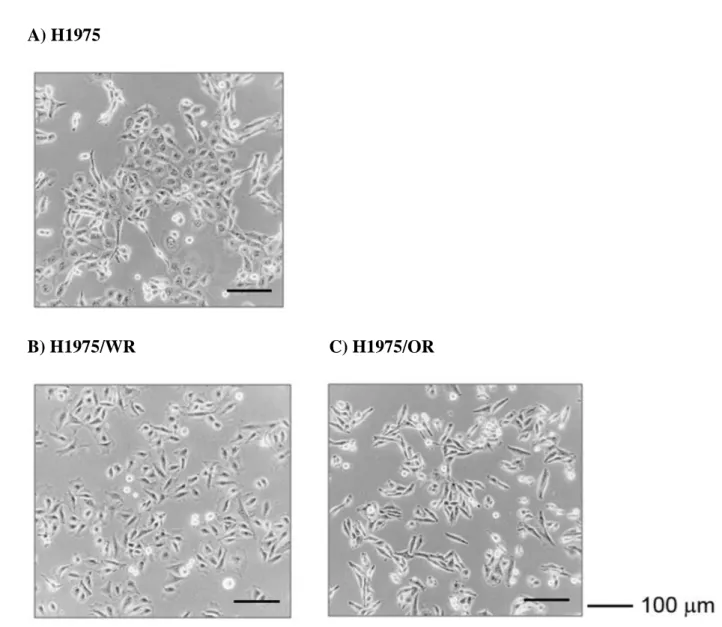
Figure 2. Phenotypic changes between the mother cell and the EMT induced EGFR-TKI resistance cell lines
A) H1975
B) H1975/WR C) H1975/OR
H1975 parental cells and both resistant cells (H1975/WR and H1975/OR) were
evaluated for morphologic changes that were consistent with EMT using a light
microscope. B), C) The resistant cells showed the increase of spindle-shaped cells
that was like to EMT-like changes.
Figure 3. Immunoblot analysis of EMT expression
EGFR signaling and EMT-related molecules were analyzed by Western blotting.
Epithelial marker proteins containing E-cadherin, β-catenin, EpCAM, desmoplakin
and cytokeratin-8/18 were significantly reduced in both resistant cells (H1975/WR
and H1975/OR), whereas vimentin expression was increased.
Figure 4. Invasion and Migration analysis on the EMT related EGFR-TKI resistance cell lines
Cells were seeded onto either collagen or Matrigel-coated polycarbonate filters to determine their migratory and invasive potentials. Cells were incubated in modified Boyden chambers for 24 h, and the cells that penetrated the filter were stained and counted using a light microscope. Experiments were repeated in triplicate. Bars represent the standard deviations. ***P < 0.0005 compared with H1975 cells. A) migration and B) invasion were significantly enhanced in both resistant cells.
Figure 5. Effect of CDK7 inhibitors on the EMT induced EGFR-TKI resistance cell lines
Cells were treated with the indicated doses of THZ1 and QS1189, CDK7 inhibitors for 72 h, and cell viability was determined by the MTT assay. Both resistant cells were more sensitive to CDK7 inhibitors than parental cells. A) THZ1 IC50 = 379 nM in H1975, 83.4 nM in H1975/WR, 125.9 nM in H1975/OR, B)QS1189 IC50 = 755.3 nM in H1975, 232.8 nM in H1975/WR, 275.3 nM in H1975/OR).
Figure 6. Immunoblot analysis according to CDK7 inhibitor treatment on the EMT induced EGFR-TKI resistance cell lines
EGFR signaling and CDK7 related molecules were analyzed by Western blotting.
The inhibitory effect of THZ1 on activity of RNAPII-CTD was similar in H1975 and
H1975/OR cells. However, H1975/WR cells showed the inhibition of RNAPII-CTD
phosphorylation at Ser2, Ser5 and Ser7 at lower concentration of THZ1.
Figure 7. Effects of THZ1 on the TGF-β induced EMT A) B)
Cells were treated with the indicated doses of THZ1, CDK7 inhibitors for 72 h, and cell viability was determined by the MTT assay. A) & B) the sensitivity to THZ1 was increased in cells with TGF-β1-induced EMT. Bars represent the standard deviations.
*P < 0.05 compared to pre-exposure of TGF-β.
Figure 8. Effects of THZ1 on the TGF-β induced EMT in T790M negative EGFR mutant cell lines
A)
B)
Cells were treated with the indicated doses of THZ1 and Osimertinib for 72 h, and cell viability was determined by the MTT assay. A) & B) The sensitivity to THZ1 was increased in cells with TGF-β1-induced EMT compared to the sensitivity to osimertinib was decreased. Bars represent the standard deviations. *P < 0.05 and **P
< 0.005 compared to pre-exposure of TGF-β.
영문요약
Title:
The application of CDK7 inhibitors to overcome EMT-associated EGFR-TKIs
resistance in non-small cell lung cancer
Name and Affiliation:
Wonjun Ji
Department of Pulmonary and Critical Care Medicine, Asan Medical Center,
University of Ulsan College of Medicine, Seoul, Korea.
Background: Epithelial to mesenchymal transition (EMT) is associated with
resistance during EGFR tyrosine kinase inhibitor (EGFR-TKI) therapy. This study
investigated the effects of cyclin-dependent kinase 7 (CDK7) inhibitors on EMT-
associated EGFR-TKI resistance in non-small cell lung cancer (NSCLC).
Methods: In this study, we established an EGFR-TKI resistant cell line (H1975/WR,
H1975/OR) via repeated exposure to Osimertinib and WZ4002. Morphologic
analysis, invasion and migration assays, and immunoblot analysis were performed to
evaluate the changes that occur during EMT. RNA sequencing was used to assess
genetic changes caused by EMT. THZ1 was used as a CDK7 inhibitor.
Results: The established EGFR-TKI resistant cell lines (H1975/WR, H1975/OR)
showed phenotypic changes in EMT, including a spindle-cell shape and increased
pseudopodia formation. Decreased E-cadherin and increased vimentin were observed
in the immunoblot assay. RNA sequencing revealed decreased EPCAM and CDH1.
Invasion and migration increased in the resistance cells. The EMT related resistance
cells showed higher sensitivity to THZ1 than the mother cells. This phenomenon was
observed in TGF-ß induced EMT cell lines and gefitinib resistant cell lines (HCC827,
PC-9)
Conclusion: In conclusion, EMT was associated with decreased drug sensitivity to
EGFR-KTI; while, EMT related resistance cells were more sensitive to CDK7
inhibitors. This suggested that CKD7 inhibitors could potentially be used as a
therapeutic strategy to overcome EMT associated EGFR-TKI resistance in NSCLC.
Key words: Non-small cell lung cancer, EGFR-TKI acquired resistance, EMT,
CDK7 inhibitor