저작자표시-비영리-변경금지 2.0 대한민국 이용자는 아래의 조건을 따르는 경우에 한하여 자유롭게
l 이 저작물을 복제, 배포, 전송, 전시, 공연 및 방송할 수 있습니다. 다음과 같은 조건을 따라야 합니다:
l 귀하는, 이 저작물의 재이용이나 배포의 경우, 이 저작물에 적용된 이용허락조건 을 명확하게 나타내어야 합니다.
l 저작권자로부터 별도의 허가를 받으면 이러한 조건들은 적용되지 않습니다.
저작권법에 따른 이용자의 권리는 위의 내용에 의하여 영향을 받지 않습니다. 이것은 이용허락규약(Legal Code)을 이해하기 쉽게 요약한 것입니다.
Disclaimer
저작자표시. 귀하는 원저작자를 표시하여야 합니다.
비영리. 귀하는 이 저작물을 영리 목적으로 이용할 수 없습니다.
변경금지. 귀하는 이 저작물을 개작, 변형 또는 가공할 수 없습니다.
의학박사 학위논문
심장이식 수혜자에서 Everolimus의 심장 동종이식 혈관병에 대한 효과: Cyclosporine 기반 및 Tacrolimus 기반
protocol과 비교 연구
The Efficacy of Everolimus to Prevent Cardiac Allograft Vasculopathy in Heart Transplant Recipients:
Comparison With Cyclosporine-Based and Tacrolimus- Based Protocols
울 산 대 학 교 대 학 원 의 학 과
최 효 인
[UCI]I804:48009-200000175259
[UCI]I804:48009-200000175259
[UCI]I804:48009-200000175259
[UCI]I804:48009-200000175259
[UCI]I804:48009-200000175259
[UCI]I804:48009-200000175259
[UCI]I804:48009-200000175259
[UCI]I804:48009-200000175259
[UCI]I804:48009-200000175259
[UCI]I804:48009-200000175259
[UCI]I804:48009-200000175259
[UCI]I804:48009-200000175259
[UCI]I804:48009-200000175259
[UCI]I804:48009-200000175259
[UCI]I804:48009-200000175259
[UCI]I804:48009-200000175259
[UCI]I804:48009-200000175259
[UCI]I804:48009-200000175259
The Efficacy of Everolimus to Prevent Cardiac Allograft Vasculopathy in Heart Transplant Recipients:
Comparison With Cyclosporine-Based and Tacrolimus- Based Protocols
지 도 교 수 김 재 중
이 논문을 의학박사 학위 논문으로 제출함 2018년 12월
울 산 대 학 교 대 학 원 의 학 과
최 효 인
최효인의 의학박사학위 논문을 인준함
심사위원 김 재 중 인 심사위원 김 영 학 인 심사위원 박 덕 우 인 심사위원 이 상 언 인 심사위원 이 종 영 인
울 산 대 학 교 대 학 원
2018년 12월
Abstract
The Efficacy of Everolimus to Prevent Cardiac Allograft Vasculopathy in Heart Transplant Recipients: Comparison With Cyclosporine-Based and Tacrolimus-Based Protocols
Hyo-In Choi
Division of Cardiology and Department of Internal Medicine Asan Medical Center, University of Ulsan College of Medicine
Background
Previous studies have reported the superiority of everolimus (EVL) over antimetabolites in mitigating cardiac allograft vasculopathy (CAV) after heart transplantation (HT). However, data on the long-term effect of de novo EVL immunosuppression on CAV progression and clinical outcomes are lacking.
Objective
The aim of this study was to determine the long-term safety and efficacy of EVL as de novo immunosuppressant therapy on CAV progression and outcomes after HT.
Methods
We retrospectively reviewed the medical records of 144 HT recipients (EVL group = 24, Cyclosporine (CSA) group = 48, Tacrolimus (TAC) group = 72) who survived at least at 1 year after HT. This study evaluated treatment failure defined as all-cause death, graft failure, retransplantation and treatment requiring rejection. CAV progression was assessed using serial coronary intravascular ultrasound (IVUS) in recipients who underwent at least 2 IVUS studies.
Results
A significant attenuation in percent atheroma volume progression was observed with EVL (1.2%) compared with CSA (7.3%; p = 0.005 vs EVL) or TAC (6.6%; p = 0.0052 vs EVL) at 1 year after HT, and this trend has remained unchanged for 3 years (4.7% vs 12.4% vs 12.5%
for EVL vs CSA vs TAC respectively, p = 0.006) and 5 years (7.9% vs 14.9% vs 14.9% for EVL vs CSA vs TAC respectively, p = 0.02) after HT. The remodeling index was greater in the EVL (1.08) group than in CSA (0.23) or TAC (-0.25) groups at 1 year after HT. Kaplan-Meier analysis over a median follow-up period of 8 years did not show a statistical difference in primary endpoint event-free survival between the three groups. No death or re-transplantation occurred in the EVL group while 10 (21.8%) and 14 (20.6%) occurred in CSA and TAC group respectively.
Conclusions
De novoimmunosuppression with EVL is associated with attenuated CAV progression during 5 years of IVUS follow up and with comparable long-term clinical outcomes compared with CSA or -TAC based protocols.
Key Words:heart transplantation, cardiac allograft vasculopathy, immunosuppressive therapy, intravascular ultrasound, treatment outcome
Contents
Abstract... i
Contents... iii
List of tables...iv
Lists of figure...v
Abbreviations...vi
Introduction...1
Methods...3
Study population...3
Immunosuppression and management ...3
CAV and IVUS assessment ...4
Outcome endpoints ...5
Statistical analysis...6
Results...7
Patient characteristics...7
Effect of immunosuppression on plaque progression and vascular geometry...7
Effect of immunosuppression regimens on clinical outcomes ...27
Discussion...31
Limitations...34
Conclusion...35
References...36
Korean abstracts……….……41
List of tables
Table 1. Baseline characteristics of the patients according to immunosuppressant regimen ...8 Table 2. Calcineurin inhibitor trough level during follow up ...10 Table 3. Everolimus dose and trough level during follow-up...11 Table 4. Assessment of cardiac allograft vasculopathy progression by IVUS during follow-up ...13 Table 5. Clinical endpoints at 8-year follow up according to groups...29
Lists of figure
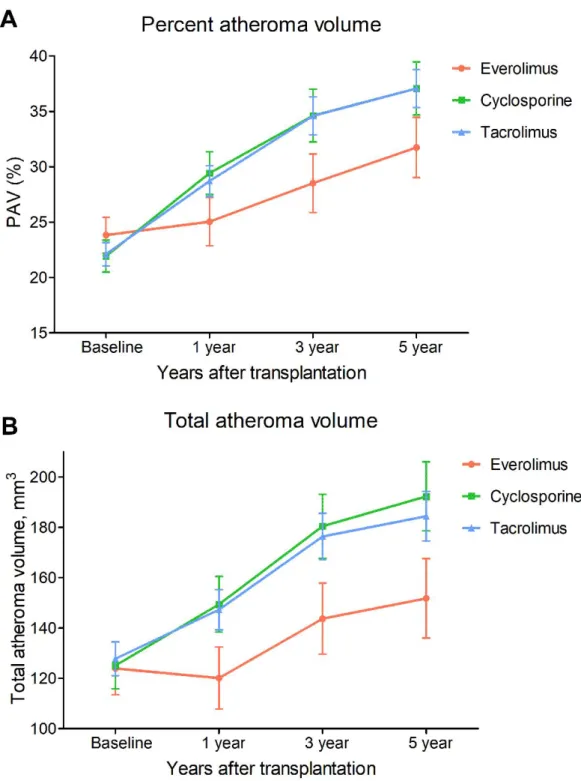
Figure 1. Changes in PAV (A) and TAV (B) as assessed by serial IVUS examinations during follow-up, stratified by type of immunosuppressive regimen. Values are mean ± SEM for each
treatment group………...16
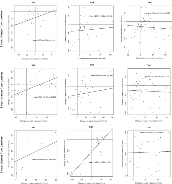
Figure 2A Remodeling patterns (Vessel changes in response to intimal changes) during follow up according to immunosuppressant regimen………17
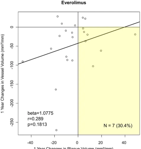
Figure 2B. 1 year change from baseline in everolimus group………18
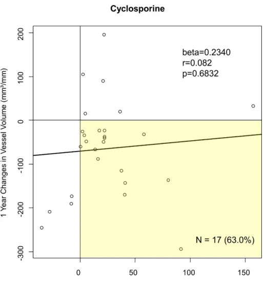
Figure 2C. 1 year change from baseline in cyclosporine group……….19
Figure 2D. 1 year change from baseline in tacrolimus group……….20
Figure 2E. 3 year change from baseline in everolimus group………21
Figure 2F. 3 year change from baseline in cyclosporine group………22
Figure 2G. 3 year change from baseline in tacrolimus group………..23
Figure 2H. 5 year change from baseline in everolimus group……….24
Figure 2I. 5 year change from baseline in cyclosporine group………25
Figure 2J. 5 year change from baseline in tacrolimus group………26
Figure 3 Event free survival in heart transplant recipients according to immunosuppressive regimen. ………..28
Abbreviations CAV = cardiac allograft vasculopathy
CNI = calcineurin inhibitor CSA = cyclosporine EVL = everolimus
HT = heart transplantation
ISHLT = International Society for Heart & Lung Transplantation IVUS = intravascular ultrasound
LV = lumen volume
MMF = mycophenolate mofetil
mTOR = mammalian target of rapamycin PAV = percent atheroma volume
SRL = sirolimus TAC = tacrolimus
TAV = total atheroma volume VV = vessel volume
Introduction
Over the past 50 years, heart transplantation (HT) has been consolidated as the therapy of choice for patients with end-stage heart disease. In HT recipients, cardiac allograft vasculopathy (CAV) is the leading cause of late morbidity and mortality, accounting for one- third of all-cause mortality at 5 years 1). CAV mechanisms are not fully understood, but probably driven by immune and nonimmune mechanisms that cause inflammation, endothelial injury, and smooth muscle cell hyperplasia in the epicardial vessels as well as in the microvasculature 2). CAV is characterized by endothelial injury and exaggerated repair response, leading to diffuse intimal hyperplasia and luminal stenosis that can involve the entire coronary arterial tree. Given the negative impact of CAV on the duration of HT graft survival, prevention remains the most effective strategy for optimizing long-term outcomes.
To prevent CAV, various medications have been studied. Studies have demonstrated the benefits of statins in improving patient survival and reduced the incidence and severity of CAV and allograft rejection 3). Among immunosuppressants, mammalian target of rapamycin (mTOR) inhibitors has antiproliferative effects on fibroblasts and smooth muscle cells, thus they hold the potential of reducing intimal hyperplasia in coronary arteries, preventing CAV and its progression 4). Previous studies have shown mTOR inhibitors to attenuate the progression of CAV when used as secondary immunosuppression in place of either azathioprine or mycophenolate mofetil (MMF) and in combination with a reduced dose of calcineurin inhibitor (CNI) as primary immunosuppression 5-8). However, intravascular ultrasound (IVUS) data from large randomized trials present short to mid-term follow-up and
8, 9), there are limited long-term data regarding these patients. Also, published experience with de novo use of EVL in HT in routine clinical practice is currently relatively limited 10, 11). Moreover, as far as we know, there has been no study regarding the effect and safety of de novoEVL based protocol in the Asian population.
The purposes of this study were to investigate the long-term effect of everolimus (EVL) as primary immunosuppressant on CAV progression as assessed by serial IVUS examinations
and to investigate the long-term safety and efficacy of an EVL-based regimen on clinical events compared with maintenance on a cyclosporine (CSA) -based and tacrolimus (TAC) - based regimen.
Methods Study population
This study included patients ≥ 18 years who underwent HT at Asan Medical Center from 2005 to 2015 and survived for at least one year. Multiorgan transplant recipients were excluded.
Since 2005, a total of 24 patients used the EVL-based protocol as a de novo immunosuppressant, and although all patients who used EVL as ade novoimmunosuppressive regimen also received a calcineurin inhibitor (CNI), we will refer to this group of patients as the EVL group. Forty-eight and seventy-two patients, two and three times the number of patients in the EVL group respectively, were consecutively included from the patients using the CSA-based protocol (CSA group) and the TAC based protocol (TAC group).
Demographic, clinical follow-up, and laboratory data were obtained by review of the patients’
medical records and from a prospectively collected clinical database. Immunosuppressive medications were reviewed and recorded at each outpatient visit post-HT.
Immunosuppression and management
All HT recipients received induction therapy with interleukin-2 monoclonal antibody (rituximab) as part of a standard induction protocol and a 3-drug maintenance immunosuppressive regimen consisting of a CNI (CSA or TAC), an antimetabolite agent (MMF), and tapering doses of prednisone post-HT. Withdrawal or continuance of the steroid (at the maintenance dose) was based on the attending physician’s discretion and the patient’s clinical status. Patients in stable condition at least 2 weeks post-HT, without evidence of rejection, or infection, MMF was replaced with EVL (trough level, 3-8 ng/mL) 0.5 mg bid orally. CSA was the major CNI used until 2007, and TAC was predominantly used after 2007.
The CNI target trough concentration in first 3 months after HT was 300-350 ng/mL for CSA and 10-15 ng/mL for TAC. The target trough concentration was adjusted between 100 and 200 ng/mL for CSA and between 5 and 10 ng/mL for TAC 12 months after HT. Trough levels of CSA, TAC, and EVL were measured using high- performance liquid chromatography with tandem mass spectroscopy (XEVO TQ-S, Waters, Milford, USA) or microparticle enzyme
immunoassay (Dimension® EXL™ 200 Integrated Chemistry System; SIEMENS, Munich, Germany) and adjusted according to institutional protocols.
All patients underwent serial endomyocardial biopsies at regular intervals until 1 year after HT. An International Society of Heart and Lung Transplantation (ISHLT) grade of 2R or greater acute cellular rejection on routine endomyocardial biopsy was treated with augmented immunosuppression and intravenous steroids 12).
Statin therapy was generally initiated within 2 weeks after HT for all post-HT recipients, regardless of cholesterol level, except for recipients who experienced adverse effects due to statin therapy. Lipid-lowering therapy with statins (initial daily dose, 10 mg of pravastatin) was mandatory 13), but if the lipid profile worsened with conventional statin use, the pravastatin dosage was increased, or pravastatin was exchanged for a stronger statin. “Intensive statin therapy” was defined as follows: (1) increasing the statin dosage during the study period, and (2) converting to a strong statin from pravastatin during the study period.
The Cytomegalovirus (CMV) antigenemia assay was performed weekly during hospitalization and during every visit at the outpatient clinic until 1 yr after HT. Gancyclovir prophylaxis was prescribed for all patients. Trimethoprim/sulfamethoxazole prophylaxis was prescribed for all patients for 1 yr after HT.
CAV and IVUS assessment
Coronary angiography with 3-dimensional IVUS as part of the surveillance for CAV progression has been performed since 2004 in most HT recipients. Baseline coronary angiography was performed within 3 months after HT unless patients had contraindications to coronary angiography. And 1, 3, 5, 8, 10 years after HT, or with any change in clinical status, follow-up coronary angiography was performed as routine surveillance. CAV was classified according to the ISHLT criteria 14). For assessment of CAV progression, as measured by IVUS, patients with 2 or more IVUS examinations were included for analysis. IVUS was performed during routine coronary angiography after intracoronary administration of 100 to 200 mg nitroglycerin. Core laboratory analysts at the Asan Medical Center, blinded to treatment status
and sequencing of imaging studies (baseline vs. follow-up) defined the leading edges of the lumen and outer vessel wall by manual planimetry in images spaced 1-mm apart and where there is no artifact obscuring >90 º of the contiguous outer vessel wall, with reproducibility as previously reported 15, 16). A number of measures of plaque burden were determined. Percent atheroma volume (PAV) was calculated as follows:
S( )
S × 100
where EEMarea was the cross-sectional area of the external elastic membrane and Lumenarea
was the cross-sectional area of the lumen.Normalized total atheroma volume (TAV) was calculated as follows to compensate for differences in the examined vessel to the examined segment length:
S( )
× Median number of images in a group
where the average plaque area in each image was multiplied by the median number of images analyzed in each group to compensate for differences in segment length between patients. The vessel volume (VV) and lumen volume (LV) were also calculated to normalized values. CAV progression was assessed by calculating the changes in PAV, TAV, VV, and LV between the baseline and 1, 3, and 5 years of follow-up IVUS examinations for each patient. The remodeling index, a quantitative measure of extent and direction of remodeling, was calculated as the ratio of the change in the vascular area at the lesion site relative to the change in intimal area 17). An index ≥ 1 indicates positive remodeling that adequately compensated (=1) or overcompensated (>1) for the intimal growth, whereas a remodeling index >0 and <1 indicates positive remodeling inadequate for intimal growth. An index <=0 indicates negative remodeling with no compensation (=0) or shrinkage (<0).
Outcome endpoints
The primary outcome endpoint was treatment failure defined by the composite of all-cause death, graft failure, retransplantation, and treatment requiring rejection in median follow-up.
Biopsy-proven rejection of at least grade 2R or any episode of rejection associated with hemodynamic compromise was defined as treatment requiring rejection.
Post-transplant diabetes mellitus (PTDM) was diagnosed using the American Diabetes Association criteria 18). Post transplant hypertension was defined as systolic blood pressure >
140/diastolic blood pressusre > 90 mmHg/or use of antihypertensive medication. Patients receiving intensive statin therapy was defined to have hyperlipidemia. CMV antigemia was defined as > 50 CMV-positive cell per 200,000 leukocytes and CMV treatment with ganciclovir was started when > 100 CMV-positive cells per 200,000 and/or CMV disease was diagnosed. The study outcome “non-CMV infection” was defined as any infectious disease needing hospitalization and/or more than 2 weeks of treatment.
Statistical analysis
Quantitative data are summarized as mean standard deviation (SD) unless heavily skewed, in which case we used the median and interquartile range (IQR). Categorical data are expressed as counts and percentages. Baseline variables were compared using Student’s t-test, Wilcoxon test, or chi-square test as appropriate. We estimated Kaplan-Meier survival curves and the log- rank test was used to compare survival function and clinical event incidence. Some adverse events were evaluated using a logistic regression model. A linear mixed model was used to assess the absolute change in PAV, TAV, VV, and LV between treatment groups. All P values were two-sided, and a value of P < 0.05 was considered significant. All analyses were performed using SAS version 9.4 (SAS Institute, Cary, North Carolina).
Results Patient characteristics
Table 1shows baseline demographics, medications, and laboratory data for the three groups.
Mean recipient age at HT was 48 years old, and most patients were male. Median follow duration was 8.3 years (8.0 years for EVL group, 11.1 years for CSA group and 7.9 years in TAC group). At the time of HT, baseline characteristics were not significantly different between the three groups. Sixty percent of patients were diagnosed with dilated cardiomyopathy before transplantation, and 18% were diagnosed with ischemic cardiomyopathy. Patients in the TAC group had more preoperative mechanical circulatory support use, but there was no statistically significant difference between the other groups. The TAC group showed a slight difference from the CSA group, with fewer female to male transplants and lesser use of statins. Our post-HT management protocol recommended to use statin in every patient except those with contraindications; most of the patients used statin at 5 years after HT. In the TAC group, 80% of patients used statins at 5 years of follow-up, which was statistically less than the CSA group. The average trough level of CSA and TAC during the follow-up period was significantly lower in the EVL group than in the CSA group or TAC group (Table 2). EVL trough levels remained within the target range (3– 8 ng/mL) throughout the study and the mean dose at 5 years was 0.9 ± 0.3 mg/day (Table 3).
Effect of immunosuppression on plaque progression and vascular geometry
For the analysis of volumetric measures and progression of CAV, we identified 107 patients with a baseline and at least one more IVUS examination after HT (23 patients in the EVL group, 29 patients in the CSA group, and 55 patients in the TAC group). We excluded poor- quality images or images which could not be analyzed from the IVUS analysis. A total of 376 coronary IVUS examinations (median 4 [range: 3.4 to 4.6] per patient) were included for analysis of CAV progression in each group of patients. Volumetric measurements of plaque progression by 3-dimensional IVUS at baseline and at 1, 3, and 5- year follow-up IVUS are summarized in Table 4. At baseline, left anterior descending artery normalized TAV, VV, PV,
Table 1. Baseline characteristics of the patients according to immunosuppressant regimen
Characteristics Everolimus
(N=24)
Cyclosporine (N=48)
Tacrolimus (N=72) Recipient characteristics
Age, years 48.9 ± 13.0 47.0 ± 8.93 48.4 ± 13.2
Male (no. %) 20 (83.3%) 40 (83.3%) 45 (62.5%)
BMI, kg/m² 22.6 ± 4.30 22.2 ± 3.22 22.5 ± 3.60
Comorbidities
CMV IgG-positive 24 (100%) 48 (100%) 71 (98.6%)
Diabetes 4 (16.7%) 4 (8.33%) 8 (11.1%)
Diagnosis
DCMP 17 (70.8%) 25 (52.1%) 44 (61.1%)
ICMP 1 (4.17%) 11 (22.9%) 14 (19.4%)
Others 6 (25.0%) 12 (25.0%) 14 (19.4%)
Preoperative MCS 0 (0.00%) 1 (2.08%) 7 (9.72%)
Laboratory data
Creatinine, mg/dl 1.03 ± 0.42 1.30 ± 0.50 1.39 ± 1.50
Bilirubin, mg/dl 1.17 ± 0.75 1.72 ± 1.01 1.73 ± 1.36
Donor characteristics
Age of donor, years 34.7 ± 9.56 31.4 ± 9.31 34.4 ± 11.0
Donor male 18 (75.0%) 39 (81.2%) 57 (79.2%)
Female to Male 2 (8.33%) 7 (14.6%) 1 (1.39%)*
Weight difference -9.09 ± 19.2 -7.78 ± 19.6 -10.83 ± 20.9 Total ischemic time, minutes 169 ± 72.3 163 ± 59.4 155 ± 53.1 Medications at 5 years after HT
Statin 22 (91.7%) 47 (97.9%) 58 (80.6%)†
Aspirin 14 (58.3%) 37 (77.1%) 42 (58.3%)
Steroid 8 (33.3%) 11 (22.9%) 15 (21.1%)
* p = 0.02 for the comparison with the cyclosporine group.
† p = 0.03 for the comparison with the cyclosporine group
BMI, body mass index; CMV, cytomegalo virus; IgG, immunoglobulin G; DCMP, dilated cardiomyopathy; ICMP, ischemic cardiomyopathy; MCS, mechanical circulatory support; HT, heart transplantation
Table 2. Calcineurin inhibitor trough level during follow up Everolimus
(N=24)
Cyclosporine (N=48)
Tacrolimus
(N=72) P
Tacrolimus trough level during follow up, ng/ml
At 1 month 11.0 - 11.5 ± 3.65 NS
At 1 year 4.15 ± 2.05 - 8.08 ± 5.51 NS
At 3 years 4.60 ± 0.71 - 7.41 ± 2.33 NS
At 5 years - - 5.95 ± 2.20 NS
Cyclosporine trough level during follow up, ug/L
At 1 month 290 ± 60.8 327 ± 110 - NS
At 1 year 113 ± 36.6 268 ± 119 - <0.001, vs CSA
At 3 years 105 ± 152 304 ± 273 - 0.01, vs CSA
At 5 years 94.4 ± 149 217 ± 146 - NS
CSA, cyclosporine
Table 3. Everolimus dose and trough level during follow-up
Everolimus (N=24) Everolimus dose during follow up (mg/day)
At 1 month 1.08 ± 0.34
At 1 year 1.07 ± 0.50
At 3 years 1.00 ± 0.48
At 5 years 0.90 ± 0.28
Everolimus trough level during follow up (ng/ml)
At 1 month 4.75 ± 1.97
At 1 year 5.70 ± 2.18
At 3 years 5.24 ± 1.83
At 5 years 4.28 ± 1.03
patient) were included for analysis of CAV progression in each group of patients. Volumetric measurements of plaque progression by 3-dimensional IVUS at baseline and at 1, 3, and 5- year follow-up IVUS are summarized in Table 4. At baseline, left anterior descending artery normalized TAV, VV, PV, LV and PAV were not significantly different between groups. During follow-up, PAV was significantly increased in all three groups, but the change between 1, 3, and 5-year IVUS and the baseline measurement was significantly lower in the EVL group compared with those patients with CSA or TAC. A significant attenuation in PAV progression was observed with EVL (+1.2%) compared with CSA (+7.3%; p = 0.005) or TAC (+6.6%; p
= 0.0052) at 1 year after HT, and this trend has remained unchanged for 3 years (+4.7% vs +12.4% vs +12.5% for EVL vs CSA vs TAC respectively, p = 0.006) and 5 years (+7.9% vs +14.9% vs +14.9% for EVL vs CSA vs TAC respectively, p = 0.02) after HT. Interestingly, after 1 year of treatment, EVL decreased TAV (-3.8 mm³) while treatment with CSA (+24.2 mm³; P = 0.005) or TAC (+19.3 mm³; p = 0.007) showed an increase in TAV. Three and five years after HT, the TAV in the EVL group also increased, but the level of increase was statistically less than that of the other two. The efficacy of EVL to attenuate the progression of CAV was maintained at 5 years as presented by changes in PAV (Figure 1A) and TAV (Figure 1B) as compared with CSA and TAC groups.
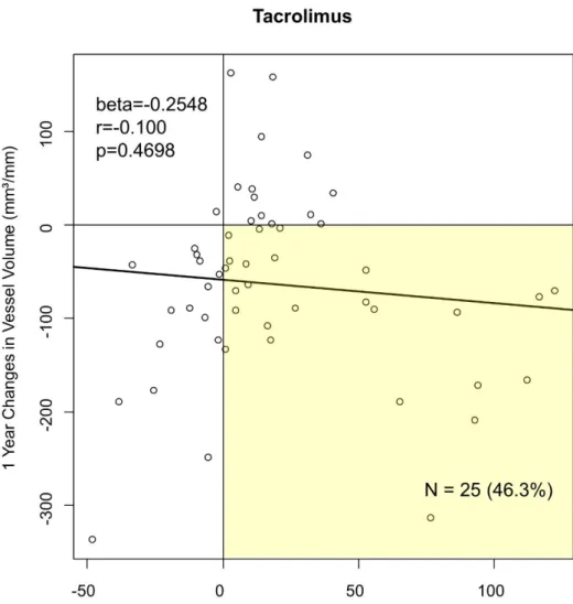
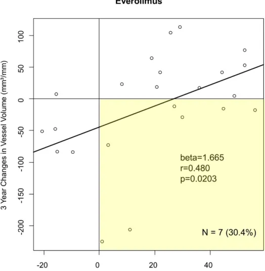
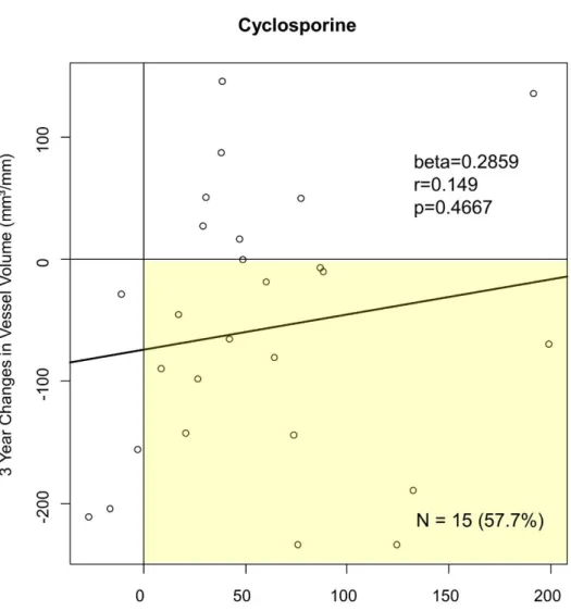
Figure 2 shows the remodeling patterns (vessel changes in response to intimal changes) observed over the 1, 3, and 5-year follow up. In the EVL group, positive correlations between the changes in the vessel and atheroma volumes (i.e., adaptive vessel response) were observed through the entire period. However, the changes in VV were only weakly, inconsistently, or even inversely correlated to the changes in TAV in the CSA or TAC groups throughout the follow-up periods. The remodeling index was greater in the EVL (1.08) group than in CSA (0.23) or TAC (-0.25) groups. At 1-year post-HT, negative vessel remodeling (intimal increase with negative vessel remodeling) was observed in 30.4 %, 63.0% and 46.3 % for the EVL, CSA, and TAC group respectively.
Table 4. Assessment of cardiac allograft vasculopathy progression by IVUS during follow-up Everolimus Cyclosporin
e
Tacrolimus Overall P Pbetween EVL-CSA
Pbetween EVL-TAC
Pbetween CSA-TAC
% atheroma volume change from baseline
Baseline 23.8 ± 1.6 21.9 ± 1.4 22.1 ± 1.1 0.62 0.39 0.37 0.92
1 year after HT 25 ± 2.2 29.4 ± 1.9 28.7 ± 1.4 0.27 0.13 0.16 0.76
1 year change from baseline 1.2 ± 1.6 7.3 ± 1.4 6.6 ± 1 0.008 0.005 0.005 0.68
p value 0.45 <0.001 <0.001
3 years after HT 28.5 ± 2.6 34.6 ± 2.4 34.6 ± 1.7 0.13 0.09 0.05 0.99
3 year change from baseline 4.7 ± 2.1 12.4 ± 1.9 12.5 ± 1.4 0.006 0.01 0.003 0.97
p value 0.03 <0.001 <0.001
5 years after HT 31.7 ± 2.7 37.1 ± 2.4 37.1 ± 1.7 0.22 0.14 0.1002 0.95
5 year change from baseline 7.9 ± 2.2 14.9 ± 1.9 14.9 ± 1.3 0.018 0.02 0.0072 0.99
p value <0.001 <0.001 <0.001
Total atheroma volume change from baseline
Baseline 123.9 ± 10.4 125.1 ± 9.3 127.8 ± 6.7 0.94 0.94 0.7568 0.81
1 year after HT 120.1 ± 12.4 149.4 ± 11.1 147.3 ± 8 0.14 0.08 0.068 0.88
1 year change from baseline -3.8 ± 7.3 24.2 ± 6.5 19.3 ± 4.7 0.01 0.005 0.0088 0.55
p value 0.60 <0.001 <0.001
3 years after HT 143.7 ± 14.1 180.5 ± 12.7 176.3 ± 9.2 0.10 0.06 0.05 0.794
3 year change from baseline 19.8 ± 9.5 55.1 ± 8.5 48.3 ± 6.2 0.02 0.007 0.01 0.52
p value 0.04 <0.001 <0.001
5 years after HT 151.8 ± 15.8 192.3 ± 13.7 184.5 ± 9.8 0.13 0.05 0.08 0.64
5 year change from baseline 26.9 ± 11.4 67 ± 9.5 56.3 ± 6.8 0.02 0.01 0.03 0.36
p value 0.01 <0.001 <0.001
Total vessel volume change from baseline
Baseline 518.6 ± 26.1 576.3 ± 23.2 582.3 ± 16.9 0.11 0.10 0.04 0.84
1 year after HT 471.9 ± 24.5 515 ± 22.1 518.5 ± 15.9 0.26 0.19 0.11 0.89
1 year change from baseline -46.7 ± 19.9 -59.8 ± 18.0 -64.1 ± 12.9 0.77 0.63 0.47 0.84
p value 0.02 0.001 <.0001
3 years after HT 506.8 ± 23.7 519.9 ± 21.7 523.7 ± 15.5 0.84 0.68 0.55 0.89
3 year change from baseline -11.9 ± 22.1 -55.0 ± 20.1 -58.7 ± 14.5 0.19 0.15 0.08 0.88
p value 0.59 0.006 <0.001
5 years after HT 471.2 ± 26.7 517 ± 22.7 506.6 ± 16.2 0.40 0.19 0.26 0.71
5 year change from baseline -39.7 ± 26.5 -59.5 ± 22.4 -75.2 ± 16.0 0.51 0.57 0.26 0.57
p value 0.07 0.009 <0.001
Total lumen volume change from baseline
Baseline 394.7 ± 23.1 451.3 ± 20.6 454.5 ± 15 0.08 0.07 0.03 0.90
1 year after HT 351.8 ± 22.1 365.8 ± 20 371.5 ± 14.3 0.76 0.64 0.46 0.82
1 year change from baseline -42.9 ± 21.3 -83.7 ± 19.3 -83.5 ± 13.8 0.25 0.16 0.11 0.99
p value 0.05 <0.001 <0.001
3 years after HT 363.1 ± 22.9 339.5 ± 21 347 ± 15 0.74 0.45 0.56 0.77
3 year change from baseline -31.6 ± 23.5 -110.4 ± 21.3 -107.0 ± 15.4 0.02 0.01 0.008 0.90
p value 0.18 <0.001 <0.001
5 years after HT 319.9 ± 22.9 326.8 ± 19.6 322.9 ± 14 0.97 0.82 0.91 0.87
5 year change from baseline -64.3 ± 25.3 -127.4 ± 21.7 -131.6 ± 15.5 0.07 0.06 0.02 0.87
p value 0.003 <0.001 <0.001
EVL, everolimus; CSA, cyclosporine; TAC, tacrolimus; HT, heart transplantation
Figure 1.Changes in PAV (A) and TAV (B) as assessed by serial IVUS examinations during follow-up, stratified by type of immunosuppressive regimen. Values are mean ± SEM for each treatment group.
Figure 2ARemodeling patterns (Vessel changes in response to intimal changes) during follow up according to immunosuppressant regimen.
Figure 2B.1 year change from baseline in everolimus group.
Figure 2C. 1 year change from baseline in cyclosporine group.
Figure 2D.1 year change from baseline in tacrolimus group.
Figure 2E.3 year change from baseline in everolimus group.
Figure 2F.3 year change from baseline in cyclosporine group.
Figure 2G.3 year change from baseline in tacrolimus group.
Figure 2H.5 year change from baseline in everolimus group.
Figure 2I.5 year change from baseline in cyclosporine group.
Figure 2J.5 year change from baseline in tacrolimus group.
Effect of immunosuppression regimens on clinical outcomes
Kaplan-Meier analysis over a median follow-up period of 8 years did not show a statistical difference in event-free survival between the three groups (Figure 3). No death or re- transplantation occurred in the EVL group during the follow-up period while 10 (21.8%) and 14 (20.6%) occurred in the CSA and TAC group respectively (Table 5). We did not identify any difference in rates of treatment requiring rejection among patients treated with EVL-based protocol and those treated with CSA-based or TAC-based protocol during follow-up (0% vs 19.9% vs 19.4 in EVL, CSAand TAC group respectively; p = 0.1). In the CSA group, 9 patients (19.7%) had moderate-to-severe (ISHLT grade 2R or 3R) cellular rejection and in the TAC group, 7 patients (11.1%) had moderate-to-severe cellular rejection, while no patients in the EVL group had moderate-to-severe cellular rejection. Regarding the metabolic effects of immunosuppressant protocols, the prevalence of PTDM was not significantly different between groups at baseline and at 8 years of follow-up. A patient who received intensive statin therapy was numerically higher in the EVL group compared to the CSA or TAC group (87.5%
vs. 69.9% vs. 54.8% in EVL, CSA, and TAC group respectively) but statistical significance was not made. The incidence of CMV infections was less with EVL-based protocol treatment (0 (0%)) than treatment with either CSA-based protocol (9 (18.8%), p=0.27) or TAC-based protocol (13 (18.8%), p = 0.16); the incidence of non-CMV infections did not differ between groups. The patients treated with EVL had less development in moderate-to-severe CAV (as assessed with the ISHLT nomenclature) compared to the other two groups. Changes in serum creatinine did not differ significantly over 5 years of follow up between the groups. However, the number of patients who ever had more than 2 mg/dl of serum creatinine was greater in the EVL group.
Figure 3.Event free survival in heart transplant recipients according to immunosuppressive regimen.
Table 5. Clinical endpoints at 8-year follow up according to groups
Everolimus Cyclosporine Tacrolimus Overall P Pbetween EVL-CSA
Pbetween EVL-TAC
Pbetween CSA-TAC
Primary endpoint 0 (0%) 10 (21.8%) 14 (20.6%) 0.07 0.14 0.24 0.99
Death 0 (0%) 3 (6.3%) 6 (9%) 0.40 0.68 0.46 0.94
Retransplantation Treatment requiring rejection
2 (8.3%) 9 (19.7%) 13 (19.4%) 0.30 0.33 0.32 0.99
PTDM 5 (20.8%) 14 (29.4%) 30 (42.1%) 0.22 0.95 0.19 0.46
Hypertension 17 (72.7%) 26 (58.8%) 26 (37.9%) 0.003 0.43 0.003 0.23
Hyperlipidemia 21 (87.5%) 33 (69.9%) 39 (54.8%) 0.001 0.33 0.002 0.26
CMV infection 0 (0%) 9 (18.8%) 13 (18.8%) 0.09 0.18 0.21 0.99
Treatment 0 (0%) 8 (16.8%) 13 (18.6%) 0.1 0.27 0.16 0.93
Viremia 0 (0%) 7 (14.6%) 6 (8.8%) 0.14 0.12 0.79 0.59
Non-CMV infection 10 (44.6%) 25 (52.1%) 40 (56.6%) 0.32 0.55 0.36 0.92
ISHLT CAV grade 1 (4.2%) 8 (19%) 17 (25.5%) 0.05 0.07 0.07 0.93
0 23 (95.8%) 37 (77.1%) 55 (76.4%)
1 1 (4.2%) 6 (12.5%) 14 (19.4%)
2 0 (0%) 3 (6.3%) 1 (1.4%)
3 0 (0%) 2 (4.2%) 2 (2.8%)
Changes of serum creatinine
At 1 month -0.05 ± 0.22 -0.17 ± 0.16 -0.29 ± 0.13 0.63 0.68 0.36 0.55
At 1 year 0.13 ± 0.24 -0.08 ± 0.17 0.04 ± 0.14 0.74 0.47 0.75 0.57
At 3 years 0.10 ± 0.24 0.11 ± 0.17 0.01 ± 0.14 0.88 0.99 0.72 0.64
At 5 years 0.14 ± 0.24 0.31 ± 0.17 -0.08 ± 0.14 0.19 0.57 0.41 0.07
EVL, everolimus; CSA, cyclosporine; TAC, tacrolimus; PTDM, post transplant diabetes mellitus
Discussion
The main findings of this study can be summarized as follows: 1) de novo use of EVL as secondary immunosuppression combined with reduced dose CNI was associated with attenuated progression of CAV to 5 years after HT; 2) EVL was not associated with significantly different all-cause mortality, graft failure, retransplantation, and treatment requiring rejection after 8 years of follow-up, although the trend of clinical benefit appeared in the EVL group patients; and 3) early adoption of EVL-based immunosuppression was safe and well tolerated for the long term compared with conventional CNI-based regimens. To the best of our knowledge, this is the first study to demonstrate long-term IVUS results of de novo EVL use and efficacy and safety in the Asian population.
In our study, the progression of CAV has been attenuated with the introduction of EVL, and these results can be sustained over 5 years after HT. EVL has been demonstrated to reduce first-year intimal thickening by IVUS in several clinical trials. The current study demonstrated that significant increases in plaque volume and vessel shrinkage were observed in the CSA and TAC group, resulting in a significant increase in PAV and reduced LV at the 5-year follow- up, whereas these worsening changes in the IVUS indices indicating CAV progression were attenuated in the EVL group. Our findings were consistent with the previously reported beneficial effects of mTOR inhibitors, including sirolimus (SRL)7, 19)and its derivative EVL
8, 20) on the progression of CAV compared with azathioprine or MMF among de novo HT patients on full- or reduced-dose CNI. The IVUS substudy of A2310 (Everolimus Versus Mycophenolate Mofetil in HT: A Randomized Multicenter Trial) found that the increase in maximal intimal thickness 12 months post-HT and the incidence of CAV were significantly lower in the EVL and reduced-dose CSA group compared with the MMF and standard-dose CSA group 8). Recently, Asleh et al 21)showed that primary immunosuppression with SRL with complete withdrawal of CNI was related to significant attenuation of plaque volume progression (SRL: 2.8 ± 2.3; CNI: 0.46 ± 1.8; p <0.0001) and plaque index (SRL: 12.2 ± 9.6%;
CNI: 1.1 ± 7.9%; p <0.0001), compared to the CNI group. Interesting findings of our study
include the decrease in TAV at 1 year in follow-up IVUS compared to baseline IVUS and positive remodeling appeared with EVL-based immunosuppression. A growing body of evidence supports mTOR inhibitor-mediated mechanisms of CAV attenuation beyond its immunosuppressive properties, and suggests that the primary mechanism is derived from its antiproliferative and anti-migratory effects on vascular smooth muscle cells, as demonstrated with in vitroand in vivostudies 14, 22). The mTOR inhibitors also reduce extracellular matrix accumulation and fibrosis 23)and induce production of nitric oxide 24), both of which can result in positive vascular remodeling and less obliteration of the coronary artery lumen. Regarding remodeling of the coronary artery, vessel responses of heart transplants seem to be different from native coronary arteries. 25-27)Based on these results, EVL is thought to cause positive remodeling in the coronary arteries of HT patients.
In our study, the primary composite endpoint of all-cause death, retransplantation, and treatments requiring rejection were not different between EVL-based protocols and CSA or TAC-based protocols. Individual variables responsible for the benefit were the reduction in all-cause mortality and treatment-requiring rejection, although statistical significance was not reached. Previous randomized trials on mTOR inhibitors showed favorable rejection and efficacy results in EVL combined with standard- or reduced dose CNI 10, 20, 28). Two randomized trials have assessed the use of EVL with reduced-dose CSA versus MMF with standard-dose CSA in de novo HT populations 5, 10) showed than EVL with reduced-dose CSA offers equivalent efficacy to standard-dose CSA. Using an EVL target range of 3-8 ng/mL, the primary composite efficacy endpoint and the incidence of biopsy-proven acute rejection were similar in the EVL and MMF treatment arms at 12 months post-tranplant in each study.
However, complete withdrawal of CNI should be carefully considered since studies on CNI- free regimen demonstrated conflicting results on allograft rejection 29, 30). Although a recent large retrospective study 21)showed lower all-cause mortality (adjusted hazard ratio [HR]: 0.46;
95% confidence interval [CI]: 0.30 to 0.68; p = 0.0001), and fewer CAV-related events (adjusted HR: 0.35; 95% CI: 0.21 to 0.59; p <0.0001) in complete withdrawal cohort, routine
use after HT to reduce mortality is not supported by a preponderance of the evidence 20, 31). Despite a trough level of CNI in the EVL group that was lower than comparable groups, there were no significant differences in changes of serum creatinine. Recent evidences relating to a renal benefit of EVL with reduced CNI in HT recipient is less convincing. Most of the patients (22, 91.7%) were using CSA as the conjunctive primary immunosuppressant. During the study period, the trough level of CSA was maintained at about half that of the CSA group. A recent study with Asleh et al 21)had shown that an mTOR inhibitor, SRL did not cause nephrotoxicity compared to CNIs. These results suggest that the level of CNI used in combination with EVL mainly influenced renal function rather than EVL itself and nephrotoxicity can be prevented by reducing CNI trough level. In addition to the well-established manifestations of CMV syndrome and potentially organ-invasive CMV disease, CMV infection increases the risk of acute rejection 32)and is associated with accelerated development of CAV 33),with an increased risk for secondary infections 34). The mTOR inhibitors appear to inhibit CMV amplification by blocking the phosphatidylinositol 3-kinase pathway, a critical step for viral signaling and replication 35, 36). The most robust data relating to an effect of EVL on CMV infection and CMV-related events following HT are derived from three randomized studies of de novoEVL therapy with standard CNI 20), reduced-dose CNI 5, 10), or reduced CNI with early CNI withdrawal 30). EVL was again observed to reduce the incidence of CMV infection may reduce CAV development. CNIs are known to promote hypertension because they increase oxidative stress and sympathetic activation, which may cause afferent arteriolar vasoconstriction37). CNI minimization with mTOR inhibition may reduce the incidence of hypertension compared with standard-dose CNI, but data are conflicting. In our result, hypertension was more common in the EVL group. Although hypertension is a common side effect of mTOR inhibitors 38-40), further studies are needed to confirm this effect. In the EVL group, MMF was replaced with 0.5mg bid EVL at 2 to 4 weeks after HT. A previous systematic review of randomized controlled trials of either SRL or EVL concluded that the risk of wound complications is increased in patients receiving an mTOR inhibitor with CNI therapy 41), but included early
trials in which large SRL loading doses and high exposure levels were used with standard- exposure CSA. Randomized trials of delayed initiation of EVL and reduced dose CSA with MMF as a bridge appears to provide a better safety profile than immediate initiation, by reducing the incidence of pericardial effusions, especially those requiring pericardiocentesis, and by improving overall drug tolerability, with less adverse event-driven discontinuations, without compromising antirejection efficacy42).
Limitations
The main limitations of this study were the small sample size and the observational, retrospective design without randomization. Patients were switched to EVL only when stable, for example, when not actively undergoing rejection, though they could be subsequently converted to EVL. Also, some patients could not convert to EVL because of side effects were excluded. Patients who did not undergo serial 3D IVUS were excluded from IVUS analysis.
Furthermore, patients with rapidly progressive CAV without serial IVUS examinations were excluded from the analysis.
Conclusion
De novoimmunosuppression with EVL is associated with attenuated CAV progression during 5 years of IVUS follow up and with comparable long-term clinical outcomes compared with CSA- or TAC-based protocols. In addition, this 3D-IVUS study suggests that the suppressive effects of EVL on CAV progression may be induced not only by reducing plaque progression but also by suppressing vessel shrinkage. Early introduction of EVL can provide adequate immunosuppressive potency in selected patients, but it should be borne in mind that numerous exclusion criteria were applied.
References
1. Lund LH, Edwards LB, Kucheryavaya AY, Dipchand AI, Benden C, Christie JD, et al.
The Registry of the International Society for Heart and Lung Transplantation:
Thirtieth Official Adult Heart Transplant Report—2013; Focus Theme: Age. The Journal of Heart and Lung Transplantation 2013;32(10):951-64.
2. Arora S, Gullestad L. The challenge of allograft vasculopathy in cardiac transplantation. Curr Opin Organ Transplant 2014;19(5):508-14.
3. Wenke K, Meiser B, Thiery J, Nagel D, von Scheidt W, Krobot K, et al. Simvastatin Initiated Early After Heart Transplantation. Circulation 2003;107(1):93-7.
4. Shipkova M, Hesselink DA, Holt DW, Billaud EM, van Gelder T, Kunicki PK, et al.
Therapeutic Drug Monitoring of Everolimus: A Consensus Report. Ther Drug Monit 2016;38(2):143-69.
5. Eisen HJ, Kobashigawa J, Starling RC, Pauly DF, Kfoury A, Ross H, et al. Everolimus versus mycophenolate mofetil in heart transplantation: a randomized, multicenter trial.
Am J Transplant 2013;13(5):1203-16.
6. Mancini D, Pinney S, Burkhoff D, LaManca J, Itescu S, Burke E, et al. Use of Rapamycin Slows Progression of Cardiac Transplantation Vasculopathy.
2003;108(1):48-53.
7. Keogh A, Richardson M, Ruygrok P, Spratt P, Galbraith A, O’Driscoll G, et al.
Sirolimus in De Novo Heart Transplant Recipients Reduces Acute Rejection and Prevents Coronary Artery Disease at 2 Years. 2004;110(17):2694-700.
8. Kobashigawa JA, Pauly DF, Starling RC, Eisen H, Ross H, Wang S-S, et al. Cardiac Allograft Vasculopathy by Intravascular Ultrasound in Heart Transplant Patients:
Substudy From the Everolimus Versus Mycophenolate Mofetil Randomized, Multicenter Trial. JACC: Heart Failure 2013;1(5):389-99.
9. Eisen HJ, Kobashigawa J, Starling RC, Pauly DF, Kfoury A, Ross H, et al. Everolimus Versus Mycophenolate Mofetil in Heart Transplantation: A Randomized, Multicenter Trial. American Journal of Transplantation 2013;13(5):1203-16.
10. Lehmkuhl HB, Arizon J, Vigano M, Almenar L, Gerosa G, Maccherini M, et al.
Everolimus with reduced cyclosporine versus MMF with standard cyclosporine in de novo heart transplant recipients. Transplantation 2009;88(1):115-22.
11. Lehmkuhl HB, Mai D, Dandel M, Knosalla C, Hiemann NE, Grauhan O, et al.
Observational study with everolimus (Certican) in combination with low-dose cyclosporine in de novo heart transplant recipients. J Heart Lung Transplant 2007;26(7):700-4.
12. Stewart S, Winters GL, Fishbein MC, Tazelaar HD, Kobashigawa J, Abrams J, et al.
Revision of the 1990 Working Formulation for the Standardization of Nomenclature in the Diagnosis of Heart Rejection. The Journal of Heart and Lung Transplantation 2005;24(11):1710-20.
13. Kobashigawa JA, Katznelson S, Laks H, Johnson JA, Yeatman L, Wang XM, et al.
Effect of pravastatin on outcomes after cardiac transplantation. N Engl J Med 1995;333(10):621-7.
14. Mehra MR, Crespo-Leiro MG, Dipchand A, Ensminger SM, Hiemann NE, Kobashigawa JA, et al. International Society for Heart and Lung Transplantation working formulation of a standardized nomenclature for cardiac allograft vasculopathy-2010. J Heart Lung Transplant 2010;29(7):717-27.
15. Nicholls SJ, Ballantyne CM, Barter PJ, Chapman MJ, Erbel RM, Libby P, et al. Effect of Two Intensive Statin Regimens on Progression of Coronary Disease.
2011;365(22):2078-87.
16. Nissen SE, Nicholls SJ, Sipahi I, et al. Effect of very high-intensity statin therapy on regression of coronary atherosclerosis: The asteroid trial. JAMA 2006;295(13):1556- 65.
17. Lim TT, Liang DH, Botas J, Schroeder JS, Oesterle SN, Yeung AC. Role of compensatory enlargement and shrinkage in transplant coronary artery disease. Serial intravascular ultrasound study. Circulation 1997;95(4):855-9.
18. Diagnosis and classification of diabetes mellitus. Diabetes Care 2011;34 Suppl 1:S62- 9.
19. Mancini D, Pinney S, Burkhoff D, LaManca J, Itescu S, Burke E, et al. Use of
rapamycin slows progression of cardiac transplantation vasculopathy. Circulation 2003;108(1):48-53.
20. Howard J. Eisen MD, E. Murat Tuzcu, M.D., Richard Dorent, M.D., Jon Kobashigawa, M.D., Donna Mancini, M.D.,, Hannah A. Valantine-von Kaeppler MD, Randall C.
Starling, M.D., M.P.H., Keld Sørensen, M.D., Manfred Hummel, M.D., Joan M. Lind, B.S., Kamal H. Abeywickrama, Ph.D., and Peter Bernhardt, Ph.D., for the RAD B253 Study Group. Everolimus for the Prevention of Allograft Rejection and Vasculopathy in CardiacTransplant Recipients. N Engl J Med 2003;349:847-58.
21. Asleh R, Briasoulis A, Kremers WK, Adigun R, Boilson BA, Pereira NL, et al. Long- Term Sirolimus for Primary Immunosuppression in Heart Transplant Recipients. J Am Coll Cardiol 2018;71(6):636-50.
22. Chaves ÁJ, Sousa AGMR, Mattos LA, Abizaid A, Staico R, Feres F, et al. Volumetric Analysis of In-Stent Intimal Hyperplasia in Diabetic Patients Treated With or Without Abciximab. 2004;109(7):861-6.
23. Azzola A, Havryk A, Chhajed P, Hostettler K, Black J, Johnson P, et al. Everolimus and mycophenolate mofetil are potent inhibitors of fibroblast proliferation after lung transplantation. Transplantation 2004;77(2):275-80.
24. Cheng C, Tempel D, Oostlander A, Helderman F, Gijsen F, Wentzel J, et al. Rapamycin modulates the eNOS vs. shear stress relationship. Cardiovascular Research 2008;78(1):123-9.
25. Mainigi SK, Goldberg LR, Sasseen BM, See VY, Wilensky RL. Relative contributions of intimal hyperplasia and vascular remodeling in early cardiac transplant-mediated coronary artery disease. The American Journal of Cardiology 2003;91(3):293-6.
26. Lim TT, Liang DH, Botas J, Schroeder JS, Oesterle SN, Yeung AC. Role of Compensatory Enlargement and Shrinkage in Transplant Coronary Artery Disease.
1997;95(4):855-9.
27. Kobashigawa J, Wener L, Johnson J, Currier JW, Yeatman L, Cassem J, et al.
Longitudinal study of vascular remodeling in coronary arteries after heart transplantation. The Journal of Heart and Lung Transplantation 2000;19(6):546-50.
28. Zuckermann A, Wang SS, Ross H, Frigerio M, Eisen HJ, Bara C, et al. Efficacy and Safety of Low-Dose Cyclosporine with Everolimus and Steroids in de novo Heart Transplant Patients: A Multicentre, Randomized Trial. J Transplant 2011;2011:535983.
29. Kobashigawa JA, Zuckermann AO. Everolimus in heart transplantation: does it finally have a home? Am J Transplant 2014;14(8):1719-20.
30. Andreassen AK, Andersson B, Gustafsson F, Eiskjaer H, Radegran G, Gude E, et al.
Everolimus initiation and early calcineurin inhibitor withdrawal in heart transplant recipients: a randomized trial. Am J Transplant 2014;14(8):1828-38.
31. Vigano M, Tuzcu M, Benza R, Boissonnat P, Haverich A, Hill J, et al. Prevention of acute rejection and allograft vasculopathy by everolimus in cardiac transplants recipients: a 24-month analysis. J Heart Lung Transplant 2007;26(6):584-92.
32. Stern M, Hirsch H, Cusini A, van Delden C, Manuel O, Meylan P, et al.
Cytomegalovirus serology and replication remain associated with solid organ graft rejection and graft loss in the era of prophylactic treatment. Transplantation 2014;98(9):1013-8.
33. Koskinen PK, Kallio EA, Tikkanen JM, Sihvola RK, Häyry PJ, Lemström KB.
Cytomegalovirus infection and cardiac allograft vasculopathy. 1999;1(2):115-26.
34. Snydman DR, Limaye AP, Potena L, Zamora MR. Update and review: state-of-the-art management of cytomegalovirus infection and disease following thoracic organ transplantation. Transplant Proc 2011;43(3 Suppl):S1-S17.
35. Ozaki KS, Câmara NOS, Galante NZ, Camargo LFA, Pacheco-Silva A. Decreased Cytomegalovirus infection after antilymphocyte therapy in sirolimus-treated renal transplant patients. International Immunopharmacology 2005;5(1):103-6.
36. Marty FM, Bryar J, Browne SK, Schwarzberg T, Ho VT, Bassett IV, et al. Sirolimus- based graft-versus-host disease prophylaxis protects against cytomegalovirus reactivation after allogeneic hematopoietic stem cell transplantation: a cohort analysis.
2007;110(2):490-500.
37. Zeier M, Van Der Giet M. Calcineurin inhibitor sparing regimens using m-target of rapamycin inhibitors: an opportunity to improve cardiovascular risk following kidney
transplantation? 2011;24(1):30-42.
38. Kahan BD. Efficacy of sirolimus compared with azathioprine for reduction of acute renal allograft rejection: a randomised multicentre study. The Lancet 2000;356(9225):194-202.
39. De Simone P, Nevens F, De Carlis L, Metselaar HJ, Beckebaum S, Saliba F, et al.
Everolimus With Reduced Tacrolimus Improves Renal Function in De Novo Liver Transplant Recipients: A Randomized Controlled Trial. 2012;12(11):3008-20.
40. Silva Jr. HT, Cibrik D, Johnston T, Lackova E, Mange K, Panis C, et al. Everolimus Plus Reduced-Exposure CsA versus Mycophenolic Acid Plus Standard-Exposure CsA in Renal-Transplant Recipients. 2010;10(6):1401-13.
41. Pengel LH, Liu LQ, Morris PJ. Do wound complications or lymphoceles occur more often in solid organ transplant recipients on mTOR inhibitors? A systematic review of randomized controlled trials. Transpl Int 2011;24(12):1216-30.
42. Potena L, Pellegrini C, Grigioni F, Amarelli C, Livi U, Maccherini M, et al.
Optimizing the Safety Profile of Everolimus by Delayed Initiation in De Novo Heart Transplant Recipients: Results of the Prospective Randomized Study EVERHEART.
Transplantation 2018;102(3):493-501.
국문요약
배경: 이전 연구에서 심장 이식 후 발생하는 동종 이식 혈관병증 (Cardiac allograft vasculopathy, CAV)의 진행을 완화하는데 대한 antimetabolite 대비 everolimus (EVL)의 우월성이 보고된 바 있다. 그러나 de novo EVL 면역억제 프로토콜이 CAV의 진행과 임상 효과에 미치는 장기적인 영향은 보고된 바 없다.
연구 목적: 본 연구를 통해 de novo EVL 면역억제 프로토콜이 CAV의 진행과 임상 경과에 미치는 장기적인 효과와 안정성을 확인하고자 하였다.
연구 방법: 심장이식 후 적어도 1년 이상 생존한 144 명의 심장이식 수혜자 (EVL군 24명, Cyclosporine (CSA)군 48명, Tacrolimus (TAC)군 72명)의 의무기록을 후향적으로 분석하였다. 사망, 이식심의 상실, 재이식 및 치료가 필요한 거부 반응으로 정의된 치료 실패를 1차 연구종료점으로 평가하였다. CAV의 진행은 최소 2회 이상 혈관내 초음파를 시행한 수혜자에서 연속적 혈관내 초음파 영상을 비교 분석하여 평가하였다.
결과: 심장이식 후 1 년째 혈관내 초음파 상에서 CSA군 (7.3 %, p = 0.005 vs EVL) 이나 TAC군 (6.6 %; p = 0.0052 vs EVL)에 비해 EVL군 (1.2 %)에서 % 죽상경화반 증가에 대한 유의한 감쇠가 관찰되었으며, 이 감쇠 효과는 심장이식 후 3년 (4.7% vs 12.4% vs 12.5% for EVL vs CSA vs TAC, p = 0.006)과 5년 (7.9% vs 14.9% vs 14.9% for EVL vs CSA vs TAC, p = 0.02)째 혈관내 초음파에서도 유의하게 유지되었다. Remodeling 지수는 EVL군에서 1.08로 CSA군의 0.23과 TAC군의 -0.25에 비해 높은 것으로 관찰되었다. Kaplan-Meier 분석 결과 8년의 추적 관찰 기간 중 세 그룹 간의 1차 연구종료점 발생에는 통계적으로 유의한 차이가 없었다. EVL군에서는 사망 또는 재이식이 발생하지 않았으며 CSA군과 TAC군에서 각각 10명 (21.8 %)과 14명 (20.6 %)에서 사망 또는 재이식이 발생하였다.
결론: De novo EVL 면역억제 프로토콜은 5년째 혈관내 초음파 상 CAV 진행의 감쇠와 관련이 있으며, CSA 또는 TAC 기반 면역억제 프로토콜과 비교하여 유사한 정도의 장기 임상 결과를 보인다.
중심단어: 심장이식, 동종이식 혈관병증, 면역억제요법, 혈관내 초음파, 치료 결과