In this study, we adopted a unique approach of chemical conjugation of a toxin protein and an antibody fragment. Toxin molecules delivered to the cytosol of the target tumor cells effectively destroy the target cells 2, 3). Breast cancers with overexpression of HER2 are called HER2-positive and HER2-positive cancer has been observed in 20–30% of all breast tumors 5).
The signaling pathway induced by phosphorylation of HER2 dimers promotes cell proliferation, survival, differentiation, angiogenesis, invasion and metastasis 6). HER2 overexpression induces malignant transformation in breast cancer and confers increased resistance to general chemotherapy treatment. Trastuzumab, an anti-HER2 monoclonal antibody, has been approved by the FDA for the treatment of HER2-positive breast cancer 8).
By these mechanisms of Trastuzumab tumor growth is inhibited.. weak cytotoxicity so it has been conjugated with various chemical drugs to increase cytotoxicity for active targeting of HER2 positive breast cancer cells 11). The catalytic enzyme activity of domain Ib and domain III ADP-ribosylates the elongation factor of the host ribosome, causing apoptotic cell death 14). The solution to increasing the half-life is chemical conjugation of polyethylene glycol (PEG).
In this study, we adopted a unique approach of chemical conjugation between an antibody fragment and a toxin instead of traditional immunotoxins that are recombinant fusion proteins of the two proteins.
Materials
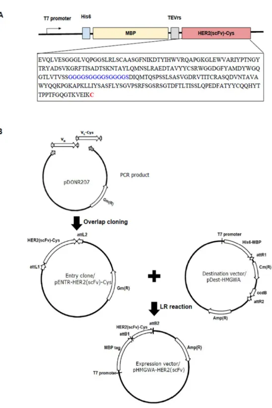
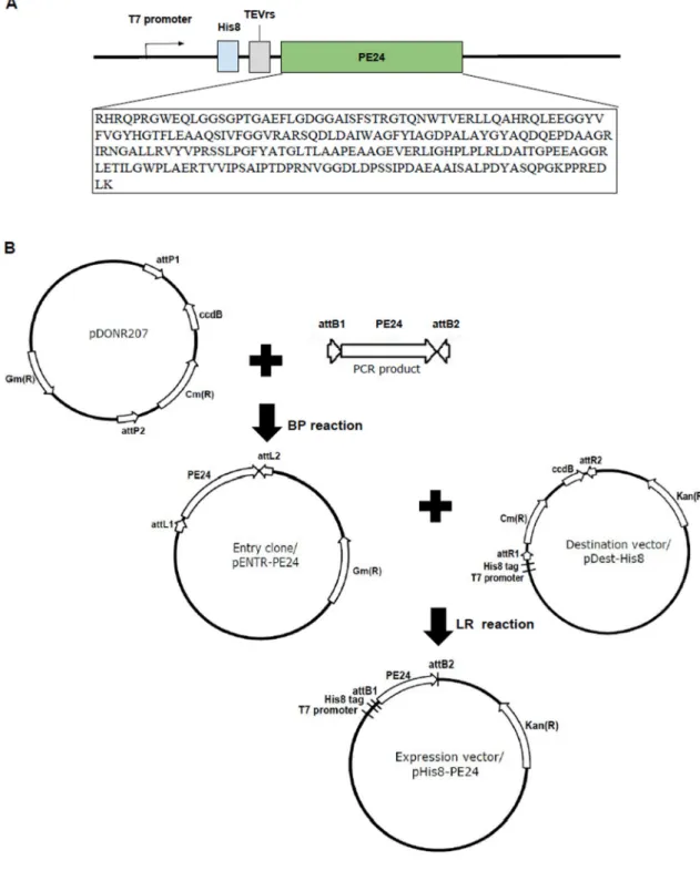
Construction of expression vector
Expression and solubility analysis of recombinant fusion protein in E. coli, BL21
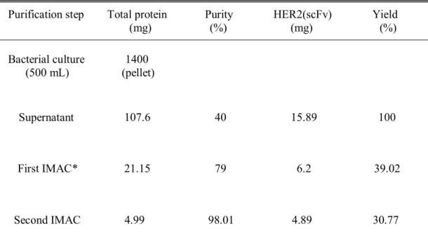
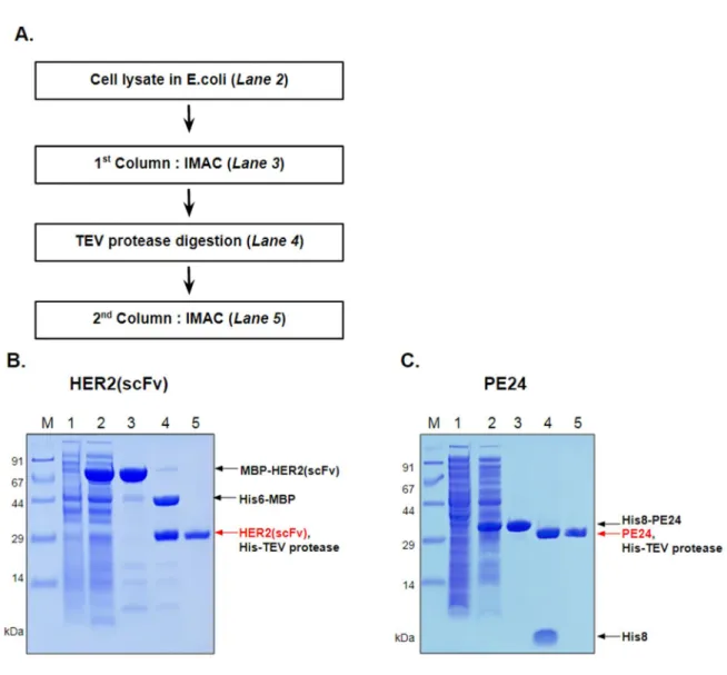
Purification of HER2(scFv)-Cys and PE24
After centrifugation, the supernatant was filtered with a 0.45 µm filter and applied to 10 mL HiTrap Ni HP which had already been equilibrated with buffer A. For purification of MBP–HER2(scFv), after application of the cell lysate, 30 mM imidazole was allowed to flow in the column for 5 CV to wash off unbound proteins. When 100 mM imidazole flowed into the column for 3-4 CV, MBP-HER2 fusion protein was eluted and collected.
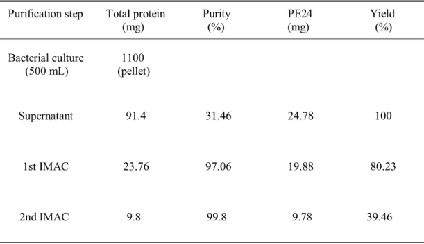
In the case of his8–PE24, unbound proteins were washed with 50 mM imidazole and the fusion protein was eluted with 500 mM imidazole. The TEV-treated mixture was dialyzed against buffer A and applied to 10 ml of HiTrap Ni HP equilibrated with the same buffer. His-MBP tag or His8 tag and His7-TEV protease were eluted at 500 mM imidazole.
Purified HER2(scFv) or PE24 was concentrated with Amicon Ultra from Merck Millipore by centrifugation at 3800 g and dialyzed against PBS at pH 7.4.
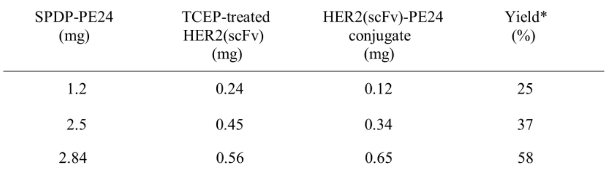
Crosslinking of HER2(scFv) and PE24
Purification of GFP and HER2(scFv)–GFP conjugate
Flow Cytometric Analysis
In vitro cytotoxicity assay
PEGylation of HER2(scFv)
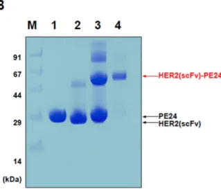
The expression vector for His8-PE24 was created by LR reaction with His8-tag containing pDEST-His8 and pENTR-PE24 (Figure 2A,B). TEV protease was treated with the eluted His8-PE24 at a ratio of 20:1 with 1 mM DTT addition. The purity of the purified HER2(scFv)-PE24 conjugate was verified by SDS-PAGE under non-reducing conditions (10% Tricine gel).
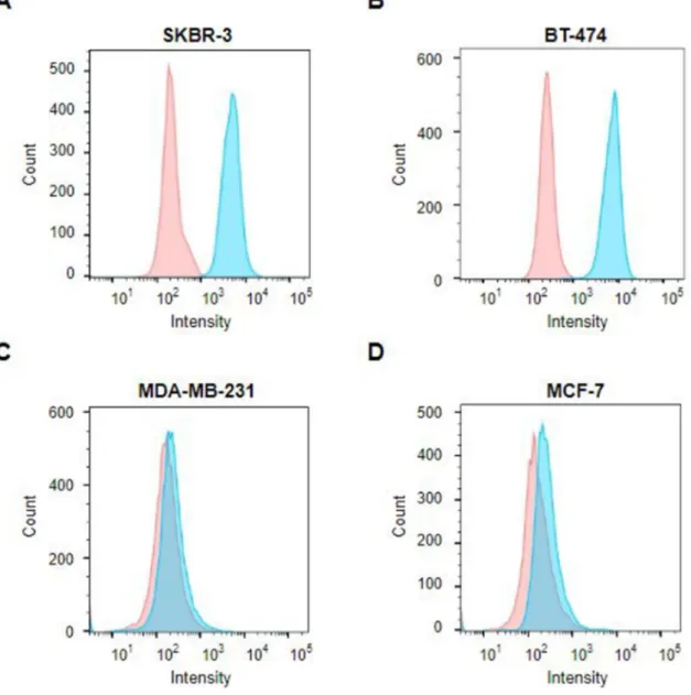
To evaluate the binding ability of the HER2(scFv)–PE24 conjugate, the HER2(scFv)-GFP conjugate was prepared and flow cytometry analysis was performed. After incubation with HER2(scFv)-GFP, a shift of the fluorescence histogram to the right was observed in the HER2-, SKBR3-, and BT-474-overexpressing cell lines ( Figure 6A, B ). From fluorescence-activated cell sorting (FACS) analysis data, we determined that the HER2(scFv)-PE24 conjugate binds strongly to HER2-expressing cells.
To determine the cytotoxicity of the HER2(scFv)–PE24 conjugate, HER2 over- and low-expressing cells were treated with HER2(scFv)–PE24, HER2(scFv) and PE24 at various concentrations. The HER2(scFv)-PE24 conjugate revealed high toxicity in HER2-overexpressing cell lines (SKBR-3 and BT-474) at the picomolar level. After adding SPDP-modified PE24 to PEGylated HER2, formation of PEGylated HER2(scFv)–PE24 was verified by SDS-PAGE under non-reducing condition.
And then formed PEGylated HER2(scFv)-PE24 was purified using cation exchange chromatography (Figure 8B, lane 3). MTT assay was performed to confirm that the cytotoxic effect of HER2(scFv)-PE24 remains unchanged after PEGylation. Four types of breast cancer cell lines were used to test the cytotoxicity of the chemically conjugated HER2(scFv)–PE24.
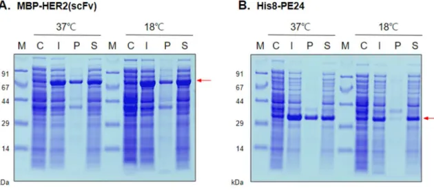
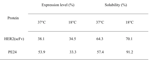
This cytotoxicity of the chemically conjugated HER2(scFv)-PE24 on the four breast cancer cells was comparable to that of the conventional HER2(scFv)-PE24 (unpublished result), demonstrating the feasibility of the chemically conjugated immunotoxin. Expression and solubility analysis of MBP–HER2 (scFv) and His8–PE24 in E.coli, BL21 strain. Expressions of (A) MBP–HER2 (scFv) and (B) His8–PE24 were induced at different induction temperatures of 18 °C or 37 °C.
A schematic overview and (B) SDS-PAGE analysis of the cross-linking process and purification of the conjugated HER2(scFv)–PE24. PEGylated HER2(scFv)–PE24 was formed; Lane 3, PEGylated HER2(scFv)–PE24 (72 kDa) conjugate purified by ion exchange chromatography. The cytotoxicity of the PEGylated HER2(scFv)–PE24 was determined on HER2 overexpressing cell line, BT-474 and HER2 low expressing cell line, MCF-7.
면역접합체 HER2(scFv)-PE24의 세포독성 효과.
Closslinking and purification of PEGylated HER2(scFv)-PE24 conjugate
In vitro cytotoxicity assay of PEGylated HER2(scFv)-PE24
Statistics
Cloning the constructs
Expression and solubility analysis of HER2(scFv) and PE24
However, when proteins were induced at 18°C, protein solubility increased compared to that at 37°C (Table 1).
Purification of HER2(scFv) and PE24
Chemical conjugation of HER2(scFv) and PE2
However, when the proteins were induced at 18°C, protein solubility was increased compared to that at 37°C (Table 1). 5B, lane 4).
HER2 expression of breast cancer cells
Cytotoxicity of HER2(scFv)–PE24 conjugate in vitro
PEGylation of HER2(scFv)–PE24 conjugate
Cytotoxicity of PEGylated HER2(scFv)–PE24 conjugate in vitro
Another advantage is that different combinations of immunotoxins can be made with less effort, because the chemical conjugation of the two components is simple. Instead, the chemical crosslinker in our experiment creates a disulfide-containing linkage between the HER2(scFv) and PE24, so that the linkage is cleaved within the cell due to the reducing intracellular environment. As expected, the cytotoxicity of the chemically conjugated immunotoxin was higher in SKBR-3 and BT-474 with IC50 of the picomolar range, while the other two cell lines showed IC50 of the nanomolar range (Figure 7).
ADA formation was delayed to allow multiple cycles, and 40% of patients showed dramatic tumor responses that significantly increased survival 26) . Construct design and cloning strategy for the MBP-HER2(scFv) expression vector. A) Designed constructs of MBP-anti-HER2(scFv). The TEV protease cleavage site was included at the N-terminus of HER2(scFv) for tag removal. expression vector was created by an overlapping cloning and a gateway cloning method.
Flow cytometry analysis of HER2-overexpressing cell and HER2-low-expressing cell after incubation with DAPI (red) or DAPI and HER2(scFv)-GFP conjugate (blue). Fluorescence histogram shows that HER2(scFv)-GFP binds strongly to HER2 receptors in (A) SKBR-3 and (B) BT-474 in contrast to low-expressing HER2 cell lines, (C) MDA-MB-231 and ( D) MCF-7. Importance of neonatal FcR in regulating the serum half-life of therapeutic proteins containing the Fc domain of human IgG1: a comparative affinity study of monoclonal antibodies and Fc fusion proteins to human neonatal FcR.
일반적으로 면역독소는 항체 단편과 독소 유전자를 융합시켜 단일 폴리펩티드로 생산된다. 그러나 본 연구에서는 독소 단백질과 항체 단편을 화학적으로 접합시켜 면역독소를 생산하였다. 이 방법의 장점은 일부 재조합 면역독소의 낮은 발현 및 수율 문제를 해결할 수 있다는 것입니다.
또 다른 장점은 클로닝, 발현, 정제 등에 비해 성분들의 화학적 결합이 상대적으로 간단하다는 점이다. 다양한 면역독소에 대해, 적은 노력으로 다양한 면역독소 조합을 생산할 수 있습니다. 개념 증명으로 트라스투주맙 단일 사슬 단편 항체(scFv)와 슈도모나스 PE24 외독소 단편이 대장균에서 생산되어 화학적으로 가교되었습니다. 제조된 면역독소를 서로 다른 4가지 인간상피성장인자수용체2(HER2) 발현 유방암 세포주에서 활성을 시험한 결과, 화학적 가교를 통해 생산된 면역독소는 HER2 발현량에 비례하는 세포독성을 나타냈다.
따라서 본 연구에서는 HER2 발현 유방암 세포의 생존율을 효과적으로 감소시키는 면역독소 구축을 위한 또 다른 대안을 제안하였다.