Background and Aims: Nasal floor tilt (NFT) is an incidental finding seen on an ostiomeatal computed tomography (OMU CT) image created by asymmetry in the levels of the two nasal floors. Despite its frequent occurrence, this finding has never been properly defined and its association with asymmetry of adjacent nasofacial structures has never been investigated. The purpose of this study was to reveal the incidence of tilting of the nasal floor in patients with sinonasal symptoms and to determine its relationship with the structures of the nasofacial skeleton. Materials and methods: From January 2008 to July 2017, patients who underwent OMU CT and facial photography before surgery were examined.
A random difference analysis of adjacent nasofacial asymmetry in the presence of NFT was performed. Perceived overall facial asymmetry was measured on the frontal facial photograph in addition to the anthropometric measurement of each upper, middle, and lower facial asymmetry. Conclusions: An inclination of the nasal floor can be observed in half of the patients complaining of nasal symptoms.
Key words: nasal floor, nasal floor tilt, nasal floor asymmetry, upper jaw, nasal septum deviation, facial asymmetry. Linear regression analysis of the degree of inclination of the nasal floor and the degree of asymmetry of bony nasofacial structures ··· 14. Anatomical relationship of the bony nasal septum and maxillary sinus in patients with inclination of the nasal floor ··· 16.
However, we often encounter unexpected anatomical features with unknown clinical significance, one of which is nasal floor tilt (NFT).
Study Design and Patient Selection
Acquisition of CT Image and Facial Photograph
The image was reconstructed with a slice thickness of 2 mm, both in the coronal and axial planes. The CT window was set to a window width of 2000 and a window level of 200, giving the best visibility of the osteomeatal unit (OMU) and nasofacial structures. All facial photographs were taken by a single, trained professional using standardized clinical photography protocol.
The patients were instructed to sit on a stool with facial muscles relaxed, in front of a blue-colored fabric. Using the Canon EOS 700 camera with EF24-105L lens, a full frontal view of the face was achieved. The distance between the patients and the camera lens was set so that the frontal face would occupy 80% of the field of view and the image would be focused on the center of the face.
The camera was installed with a built-in dot cross, and the center of the cross was focused on the vertical midpoint between both pupils. camera shutter speed remained constant for all patients.
Measurement of the Nasal Floor Tilting (NFT) and the Nasofacial Skeleton
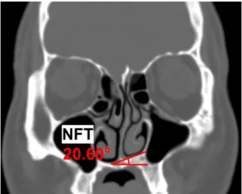
First, the most inferior point of the bony nasal floor in each nasal cavity was marked. The degree of angle between two lines was measured and represented as the 'NFT angle'. In patients who showed no level difference of the nasal floor (NFT angle . = 0°), they were categorized as the 'non-tilted group', while patients with NFT angle above 0° were classified as the 'tilted group' .
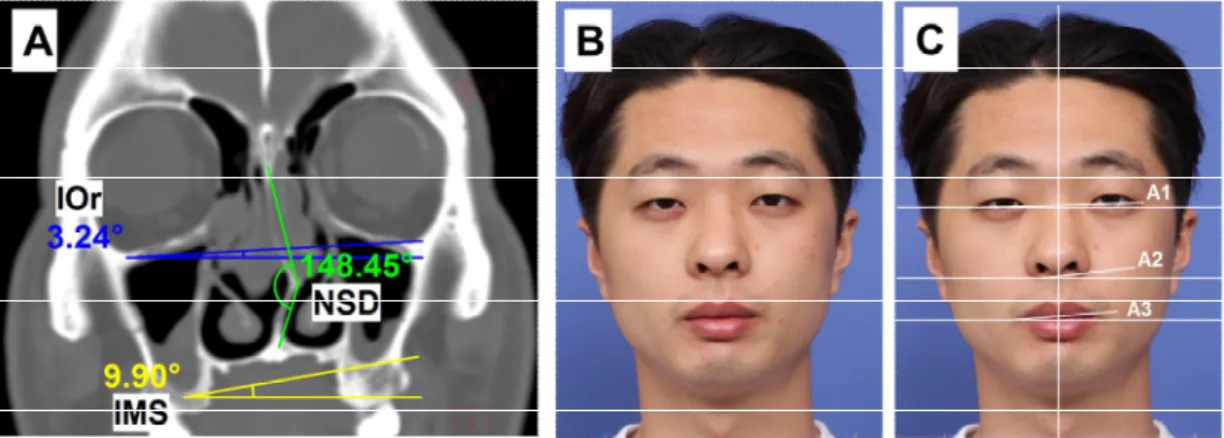
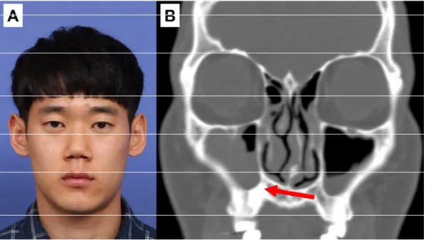
In the same coronal OMU CT image where the NFT was measured, the most inferior point of the orbit (IOr) and the most inferior point of the maxillary sinus (IMS) were marked on both sides (Figure 2, A). Similar to the NFT measurement, a higher side for both IOr and IMS was examined. A horizontal line was drawn from each IOr and IMS on each side, and a line connecting each marked points.
An angle was measured between the two lines, shown as IOr angle and IMS angle. To measure the nasal septal deviation (NSD) angle, the most protruding point of the bony nasal septum was marked, and the angle formed by the lines from the marked point to the center of the cribriform plate and the maxillary apex was measured. In patients with a deviated deformity of the bony nasal septum, the side of the more protruding bony septum was considered to be the side of NSD.
To examine the presence of level asymmetry in each facial subunit, the lateral canthus, the lowest point of the alar base, and the lateral angular margin were marked on both sides (Figure 2, C). Parallel to the NFT angle measurement, a horizontal line was drawn at one point and a line connecting both landmarks was drawn. The angles between two lines were measured and defined as A1, A2, and A3, which represent the upper face, the middle face, and the lower face, respectively.
Statistical Analysis
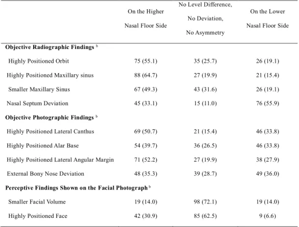
On a coronal image of the OMU CT image, the lowest point of the bony nasal floor in each nasal cavity was marked. The inferior point of the orbit (IOr) and the lowest point of the maxillary sinus (IMS) were marked on both sides. The degree of correspondence of each parameter, along with the direction of the NFT in 136 patients who had a tilted nasal floor, is shown in Table 3.
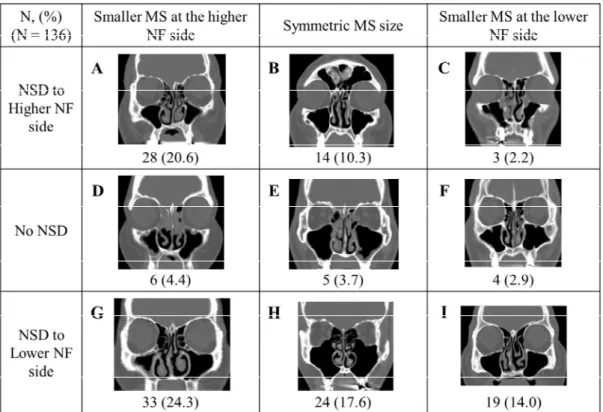
The direction of external deviation of the bony nose showed no difference according to the presence and side of the superior nasal floor. Linear regression analysis of the degree of inclination of the nasal floor and the degree of asymmetry of bony nasofacial structures (N = 265). Anatomical relationship of bony nasal septum and maxillary sinus in patients with nasal floor inclination (N = 136).
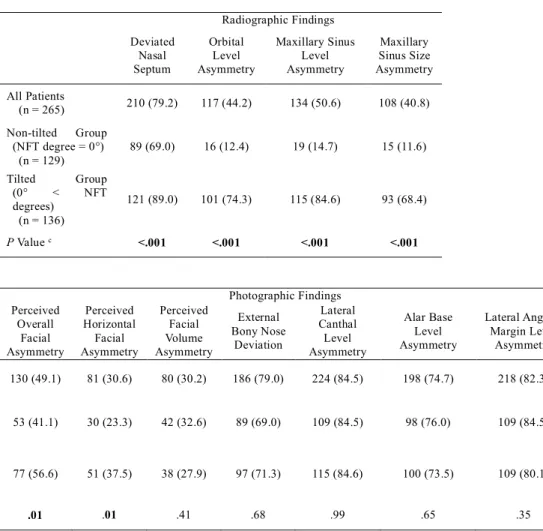
A–I, The incidence of each possible combination on the anatomical relationship between the bony nasal septum and the maxillary sinus is described. The slope of the nasal floor was observed in half of the patients who complained of nasal symptoms. In addition, the side of the higher nasofacial structures corresponded to the side of the higher nasal floor.
In the past, the irregular shape of the jaw bone and its connection with the deformed nasal septum were investigated by only a few scientists1-3). In addition, Gray revealed that the palatal edge of the nasal septum tends to luxate toward the inferior nasal floor side. In 1987, Mladina classified the types of deformities of the nasal septum according to the presence of jaw bone asymmetry2).
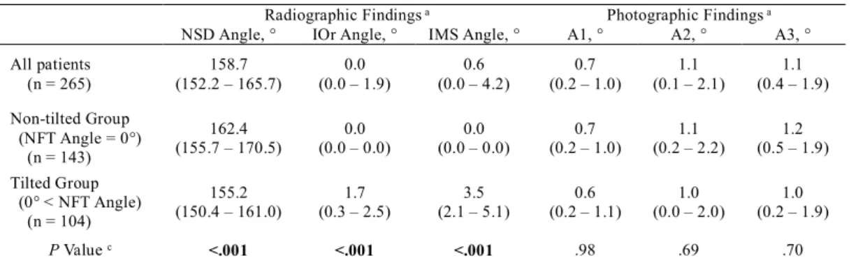
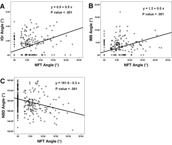
However, contrary to previous reports, we revealed the spatial relationship between the bony nasal septum and maxillary sinus, along with the asymmetry of the nasal floor levels. As shown in Table 3, it is noticeable that the bony components of the maxilla (orbit and maxillary sinus) were higher on the higher side of the nasal floor, showing a similarity of asymmetry. In addition, we found that the size of the maxillary sinus was probably smaller on the higher nasal floor side.
This finding was further supported by the smaller average IOr angle (1.7°) than the average IMS angle (3.5°), indicating a more prominent asymmetry in the middle part of the maxilla (represented as IMS) than the upper part (represented as IOr), resulting in smaller size maxillary sinus on the side of higher nasal floor. In previous studies, information regarding the structural relationship between the nasal septum and the palatal aspect of the maxilla was limited. The most rapid inferior expansion of the maxillary sinus occurs at age 7 to 1211).
We aimed to find out whether the presence of NFT was associated with facial asymmetry.