이경이의 이학석사학위 한국을 인준함. Cigarette smoke (CS) is considered the main causative factor for the development of emphysematous change in chronic obstructive airways disease. The aim of this study is to assess whether there is a regulatory role of PGRN in human alveolar epithelial cells exposed to cigarette smoke extract (CSE).
ER, endoplasmic reticulum CSE, cigarette smoke extract PERK, pancreatic ER kinase IRE-1, inositol-requiring protein ATF6, activating transcription factor 6 GRP78, glucose-regulated protein 78 IL-8, interleukin-8.
Introduction
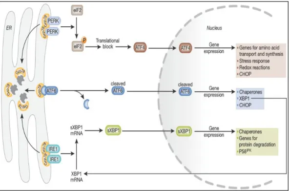
For maintenance of ER homeostasis, ER stress response typically activates 3 signaling pathways, including ER membrane, including pancreatic ER kinase (PERK), inositol-requiring protein (IRE-1) and activating transcription factor 6 (ATF6) (Fig. 3) . Recently, it has been reported that cigarette smoke extract (CSE) induced ER stress-mediated apoptosis of lung epithelial cells 3. However, whether PGRN directly regulates the ER stress response pathway and ER stress-mediated apoptosis has not yet been elucidated.
In addition, it is suggested that the enhanced expression of PGRN induced by CS exposure is critically linked to the regulation of ER stress response and protection from ER stress-mediated cellular apoptosis.
Materials & methods
Cell cultures and experimental design
Preparation of cigarette smoke extract
Generation of PGRN knock down or overexpression cells
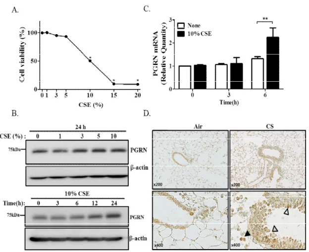
Next, to evaluate the expression of PGRN in the lungs of rats with CS-induced emphysema, we performed immunohistochemical staining of lung sections. For the overexpression of human PGRN in A549 cells, a pCMV3-SP-N-flag-hPGRN and pCMV-SP-N-flag as a negative control vector were purchased from Sino Biological Inc. Beijing, China) and were induced in these cells using ViaFect Transfection Reagent (Promega, Madison, WI, USA) with 1 μg of the indicated plasmids at a density of 3 x 10⁵ cells/well for 48 h.
Cell viability assay
Western blot analysis
Membranes were reblotted with anti-b-actin (1:5000; GeneTex Inc.) for normalization, and densitometry analysis was performed using the ImageJ software (NIH).
RT-PCR and quantitative real-time PCR
The reaction was performed in 20 µl SYBR Green mix containing 1 µl of each 10 µM forward and reverse primer and in a LightCycler 480 system (Roche Applied Science, Penzberg, Germany) using the following PCR conditions: initial incubation step of 2 min at 50 °C, reverse transcription of 60 min at 60 °C and 95 °C for 2 min, followed by 40 cycles of 15 sec at 95 °C for denaturation and 1 min at 60 °C for annealing and extension. The presence of a single specific PCR product was verified by melting curve analysis and confirmed on a 1.5% agarose gel. RT-PCR analysis of total RNA was performed to simultaneously detect both unspliced (XBP1u) and spliced (XBP1s) XBP1 mRNA and GAPDH was used for normalization of target gene expression data.
Briefly, 1 μg of total RNA was reverse transcribed with cDNA synthesis kit (Roche) as described above and amplified with AccuPower PCR Premix (Bioneer) using the following primer pair: 5'-TCTGCTTGATGTGTGTGTCCTCTT-3' and 5'-GTCCTACTCTGT 3' for XBP1, PCR product size is 289 bp amplicon was generated from unspliced XBP1, a 263 bp amplicon was generated from spliced XBP1. The reaction was performed in 20 µl of premix containing 1 ul of each 10 µM forward and reverse primer and in an S1000 Thermal Cycler (Bio-Rad Laboratories Inc.).
Immunohistochemistry for PGRN
Slides were washed three times in PBS, blocked with 0.25% casein in PBS (DaKo Protein Block Serum-Free) for 15 min at room temperature in a humidified chamber, and then incubated with rabbit polyclonal anti-PGRN antibody (Santa Cruz Biotechnology; 1:100 ) overnight in the dark at 4°C. After washing three times in PBS, the slides were incubated with polyclonal HRP-conjugated anti-rabbit immunoglobulin G (DakoCytomation) for 1 h at room temperature in the dark, and detection was by the DaKo EnVision HRP/DAB system (Dako, Carpinteria, CA, USA). . The slides were counterstained for 5~15 seconds in hematoxylin (Sigma) and then dehydrated and mounted using Permount (Thermo Fisher Scientific Inc. Rockford, IL, USA) with a coverslip.
Enzyme-linked immunosorbent (ELISA) assay
Flow cytometry
Results
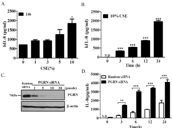
CSE induces PGRN expression in airway epithelial cells
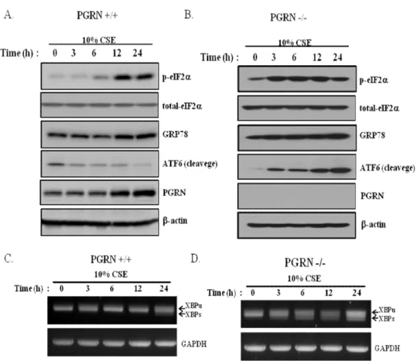
Induction of PGRN in airway epithelial cells by cigarette smoke exposure (CSE). (A) A549 cells were exposed to different concentrations of CSE for 24 h, and the cell viability of A549 decreased at high concentrations of CSE. Increased levels of PGRN expression were associated with CSE-induced ER stress via PERK or IRE1 signaling in airway epithelial cells.
Increased levels of PGRN expression were associated with CSE-induced ER stress via PERK or IRE1 signaling in airway epithelial cells
Next, for the evaluation of the IRE1 pathway, the XBP1 cleavage assay was performed since XBP1 is a known molecule to be bound by IRE-1 and leads to its translation into protein and further regulation of chaperones in an attempt to prevent overload of ER misfolded proteins. RNA samples from CSE-exposed cells were collected at several time points and analyzed by semi-quantitative RT-PCR with primers enabling the detection of the spliced and intact form of XBP1. The intensity of spliced XBP1 mRNA (XBP1s) represented by ER stress in intact PGRN cells exposed to CSE increased slightly as exposure time progressed, whereas spliced XBP1 in PGRN knockdown cells increased significantly at both time points earliest (Fig. 5C and D) .
Taken together, PGRN expression was upregulated under CSE exposure conditions, and this response was closely related to CSE-induced ER stress responses mediated primarily by the PERK and IRE1 pathways in PGRN-intact alveolar epithelial cells. Meanwhile, all 3 ER stress pathways appear to be involved with the exaggerated ER stress response under PGRN-deficient alveolar epithelial cells. PGRN-intact A549 cells (A and C) or PGRN-deficient A549 cells (B and D) were exposed to 10% CSE for different time periods as indicated, and then the levels of p-eIF2a, GRP78 and ATF6 were determined separately (50 kDa). by Western blot (A and B).
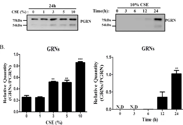
RNA was extracted from intact and PGRN-deficient A549 cells exposed to CSE at multiple time points, and XBP1 splicing was assessed by RT-PCR (C and D). Airway alveolar cells exposed to CSE-enhanced PGRN secretion and proteolytic cleavage by extracellular proteases.
Airway alveolar cells exposed to CSE augmented PGRN secretion and proteolytic cleavage by extracellular proteases
Increased proteolytic cleavage of secreted PGRN to GRN in culture supernatants from A549 cells exposed to CSE. (A) A549 cells were treated as described above for Fig.4B. The levels of secreted PGRN in the cell culture supernatant were detected by Western blot. Asterisks indicate statistical significance, **P<0.01, ***P<0.001 compared with no treatment group; ND, not detected.
CSE-induced PGRN modulates release of IL-8 from epithelial cells
After treatment with different concentrations of CSE for 24 h or (B) for different times with 10% CSE, cell-free supernatants from A549 cell cultures were collected and IL-8 release was measured by ELISA. Asterisks indicate statistical significance, **P<0.01, *** P<0.001 compared to no treatment group; ND, not detected. The efficiency of PGRN siRNA knockdown in cells and the measurement of IL-8 release in the culture medium were determined by Western blot and ELISA analysis.
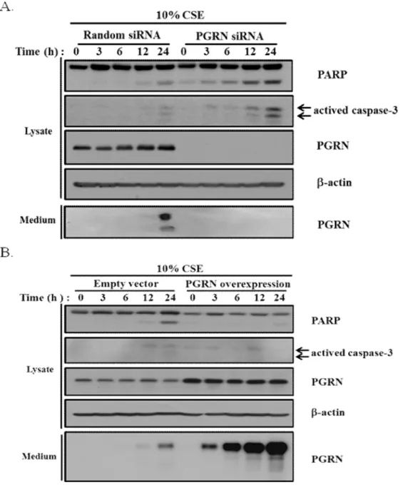
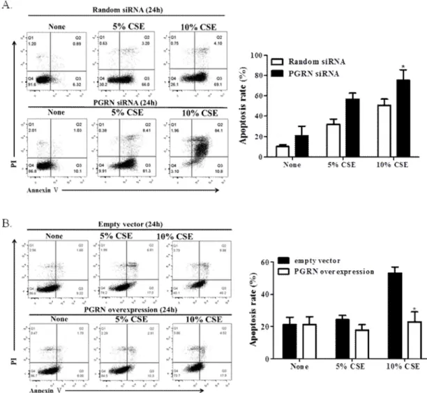
Intracellular PGRN prevents ER stress-induced cellular apoptosis under CSE treated conditions
The effect of PGRN on CSE-mediated apoptosis in epithelial cell using flow cytometry analysis. CSE for 24 h, representative images of cells obtained using flow cytometry analysis after Appendix V/propidium iodide (PI) staining. PGRN-depleted (A) and PGRN-overexpressed (B) A549 cells were exposed to 10% CSE for various time periods as indicated.
Apoptotic cells were determined by Western blot with either anti-cleaved caspase 3 or anti-PARP antibodies. b-actin was used as a loading control.
The regulatory role of PGRN in MAPK pathway and Akt signaling
Discussion
Moreover, the investigation of the relationship between GRP78 and PGRN in ER stress should be necessary. Considering that various cellular responses should be followed by ER stress, it is hypothesized that the increased production of intracellular PGRN may be closely related to the ER stress response to induce cellular homeostasis and protection against external stimuli. Although the precise relationship between IL-8 secretion and PGRN has not been elucidated in detail, several evidences were reported that ER stress-induced IL-8 production was enhanced by NF-kB activation 19 , 30 .
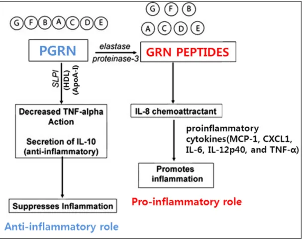
There have been other reports that CSE enhanced CCN1 expression and secretion via the induction of ER stress and the secreted CCN1-regulated increased IL-8 release through the activation of the Wnt pathway 31. It has also been reported that PGRN was involved in inflammatory response with its recruitment into sites of inflammation and competition between inflammatory mediators in acute skin injury. PGRN has been known to be a potent anti-inflammatory molecule5 based on the fact that PGRN can bind to the receptor for tumor necrosis factor-α (TNF-α), sortilin 34 as well as Toll-like receptor 9 (TLR9) 5, 25 and lead to inhibition of these mediators.
In relation to emphysema, it has been suggested that exposure of alveolar epithelial cells to CSE caused oxidative damage to respiratory epithelial cells followed by DNA damage and apoptotic cell death35. More attention has been paid to the mechanism of damage to the alveolar epithelium in addition to the destruction of the alveolar matrix. It was demonstrated in this study that PGRN-deficient cells became extremely susceptible to ER stress-induced apoptosis.
It has been reported that secreted PGRN binds to the receptor for TNF-α, which induces caspase-3 activation. The results of the current study showed that caspase-3 was activated in PGRN-deleted epithelial cells at high doses of CSE and PGRN may possibly has protective effect against CSE-induced apoptosis. Taken together, these results indicate that CSE-induced PGRN plays a role in ER stress response of the COPD airway that is reasonably associated with protection against epithelial apoptosis.
Conclusion
Progranulin does not bind tumor necrosis factor (TNF) receptors and is not a direct regulator of TNF-dependent signaling or bioactivity in immune or neuronal cells. Differential effects of cigarette smoke on oxidative stress and proinflammatory cytokine release in primary human airway epithelial cells and in a series of transformed alveolar epithelial cells. Novel mechanism of anti-apoptotic function of 78-kDa glucose-regulated protein (GRP78): endocrine resistance factor in breast cancer through release of B-cell lymphoma 2 (BCL-2) from BCL-2-interacting killer (BIK).
Progranulin is a substrate for neutrophil elastase and proteinase-3 in the airways, and its concentration correlates with mediators of airway inflammation in COPD. Progranulin growth factor binds to TNF receptors and is therapeutic against inflammatory arthritis in mice. Cigarette smoke induces apoptosis through caspase-3 activation in isolated type II fetal rat alveolar epithelial cells in vitro
Intraperitoneal injection of cigarette smoke extract caused emphysema and cardiac and skeletal muscle injury in BALB/C mice.