Other Biopotentials
EMG EEG
Visually Evoked Potentials Auditorily Evoked Potentials
Other Biopotentials
EMG EEG
Visually Evoked Potentials
Auditorily Evoked Potentials
Voltage and Frequency Ranges for Some Important Parameters That Are Measured in the Human Body
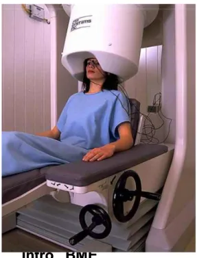
Intro. BME Intro. BME
Intro. BME Intro. BME
http://www.youtub
e.com/watch?v=k0
uSpYd_Ics
http://www.youtube.com/watch?v=3eZTAA It3QU
http://www.youtube.com/watch?v=M9XVm -ks1ME
http://www.youtube.com/watch?v=C4H- 0eLVZAk
Intro. BME Intro. BME
http://www.youtube.com/watch?v=3eZTAA It3QU
http://www.youtube.com/watch?v=M9XVm -ks1ME
http://www.youtube.com/watch?v=C4H-
0eLVZAk
EEG
• EEG configuration
21 electrodes, z~10kΩ max Top view
각 전극은 1cm 직경 정도의 면적을 봄.
신호강도 a ∝1/r 2
... ... ...
코20
20 10
• EEG configuration
21 electrodes, z~10kΩ max Top view
각 전극은 1cm 직경 정도의 면적을 봄.
신호강도 a ∝1/r 2
... ... ...
2032 channel
EEG
EEG
Dead Epilepsy 50uV
100ms
Wave d q a b g
0 ~ 4 ~ 8 ~ 13 ~ 22 ~ 30
Intro. BME Intro. BME
Awake
깊은잠 얕은 잠
REM sleep(Dream)
EEG source
Scalp EEG measures summated activity of post-synaptic currents.
While it is post-synaptic potentials that generate the EEG signal, it is not possible to determine the activity within a single dendrite or neuron from the scalp EEG. Rather, surface EEG is the summation of the synchronous activity of thousands of neurons that have similar spatial orientation, radial to the scalp. Currents that are tangential to the scalp are not picked up by the EEG.
The EEG therefore benefits from the parallel, radial arrangement of apical
dendrites in the cortex. Because voltage fields fall off with the fourth power of the radius, activity from deep sources is more difficult to detect than currents near the skull.
Scalp EEG activity is composed of multiple oscillations. These have different characteristic frequencies, spatial distributions and associations with different states of brain functioning (such as awake vs. asleep). These oscillations represent synchronized activity over a network of neurons. The neuronal network underlying some of these oscillations are understood (such as the thalomocortical resonance underlying sleep spindles), while many others are not (e.g., the system that generates the posterior basic rhythm still defies Scalp EEG measures summated activity of post-synaptic currents.
While it is post-synaptic potentials that generate the EEG signal, it is not possible to determine the activity within a single dendrite or neuron from the scalp EEG. Rather, surface EEG is the summation of the synchronous activity of thousands of neurons that have similar spatial orientation, radial to the scalp. Currents that are tangential to the scalp are not picked up by the EEG.
The EEG therefore benefits from the parallel, radial arrangement of apical
dendrites in the cortex. Because voltage fields fall off with the fourth power of the radius, activity from deep sources is more difficult to detect than currents near the skull.
Scalp EEG activity is composed of multiple oscillations. These have different
characteristic frequencies, spatial distributions and associations with different
states of brain functioning (such as awake vs. asleep). These oscillations
represent synchronized activity over a network of neurons. The neuronal
network underlying some of these oscillations are understood (such as the
thalomocortical resonance underlying sleep spindles), while many others are
not (e.g., the system that generates the posterior basic rhythm still defies
Electroencephalography (EEG) and magnetoencephalography (MEG) are non-invasive techniques for detecting and localizing electrical activities of the central nervous system. EEG systems measure the electric potentials induced on the surface of the scalp using electrodes (see Fig. 1). MEG systems measure the magnetic fields emanating from the brain with
SQUID biomagnetometers (SQUID is a Superconducting QUantum
Interference Device.)(see Fig. 2). Both EEG and MEG are non-invasive, have good temporal resolution, and directly yield information about
neurologic functions. Compared with EEG, MEG is more robust to
modeling inaccuracy, more comfortable, has a smaller procedural cost (shorter preparation time), but is also more expensive. EEG and MEG can be used in clinical applications such as seizure source localization in
epilepsy, fatal medicine, psychiatry, or in neuroscience to analyze sensorimotor or cognitive functions of the brain.
EEG and MEG
Intro. BME Intro. BME
Electroencephalography (EEG) and magnetoencephalography (MEG) are non-invasive techniques for detecting and localizing electrical activities of the central nervous system. EEG systems measure the electric potentials induced on the surface of the scalp using electrodes (see Fig. 1). MEG systems measure the magnetic fields emanating from the brain with
SQUID biomagnetometers (SQUID is a Superconducting QUantum
Interference Device.)(see Fig. 2). Both EEG and MEG are non-invasive, have good temporal resolution, and directly yield information about
neurologic functions. Compared with EEG, MEG is more robust to
modeling inaccuracy, more comfortable, has a smaller procedural cost (shorter preparation time), but is also more expensive. EEG and MEG can be used in clinical applications such as seizure source localization in
epilepsy, fatal medicine, psychiatry, or in neuroscience to analyze
sensorimotor or cognitive functions of the brain.
Fig. 1: 128 channel EEG system (courtesy of Electrical Geodesics).
Fig. 2: (a) 143 channel MEG system. (b) Illustration of the MEG sensor array. The contours correspond to the magnitude of the field induced by the source. (courtesy of VSM MedTech Ltd.)
Biopotentials related with Vision
• EOG : 안전도, eye battery 측정
일정한 거리의 두 점을 교대로 보게 하면서 뇌파 기록
• ERG : 망막전계, 광자극에 의한 망막의 전기적 반응 계측
• Visually Evoked Potential (VEP) and EEP (from retina prosthesis)
Intro. BME Intro. BME
• EOG : 안전도, eye battery 측정
일정한 거리의 두 점을 교대로 보게 하면서 뇌파 기록
• ERG : 망막전계, 광자극에 의한 망막의 전기적 반응 계측
• Visually Evoked Potential (VEP) and
EEP (from retina prosthesis)
Electro-Oculogram
Surface electrode on the temple Surface electrode
on the nose
Surface electrode on the temple
EOG Amplifier+filter
Intro. BME Intro. BME
http://www.adinstruments.com/e
ducation/experiments/applicatio
ns/Electrooculogram--EOG--
Recordings/
Electroretinogram & Visual Evoked Potential
Electroretinogram & Visual
Evoked Potential
The Electrical Basis of ERG Recordings
Intro. BME Intro. BME
Current pathway following light stimulation An equivalent electrical circuit of the eye
IaR1=Ib(R2+R3+R4+R5+R6)
ERG Parameters and Major Components
b
C d
a
The ERG of a cat in response to a 2 sec light stimulus.
Retina layer
The ERG Parameters Measured in the Ophthalmic Clinic
Intro. BME Intro. BME
ERG Responses from different light Adaptation
The same light stimulus was used.
ERG Responses from Different Species
Intro. BME Intro. BME
Typical ERGs as Recorded with
Different Electrodes
Typical ERGs as Recorded with Different Electrodes
Intro. BME Intro. BME
Fundus Photo of Human
Retina
ERGs from Normal Subject and RP Patient
Intro. BME Intro. BME
ERG & EERG (Electrically Evoked ERG)
(a) (b) (c)
1.2J Flash
480uA, 1 segment 480uA, 2 segment
Lb: 48mS;
Vb:125 uV
Lb: 24mS;
Vb:25 uV
Lb: 24mS;
Vb:31.2 uV
Lb: 24mS;
Lb: 24mS;
20mS/DIV
(a) (b) (c)
1.2J Flash
480uA, 1 segment 480uA, 2 segment
(a) (b) (c)
1.2J Flash
480uA, 1 segment 480uA, 2 segment
Lb: 48mS;
Vb:125 uV
Lb: 24mS;
Vb:25 uV
Lb: 24mS;
Vb:31.2 uV
Lb: 24mS;
Lb: 24mS;
20mS/DIV
(b), (c): subretinal stimulation; (d), (e): epiretinal stimulation
(d) (e)
480uA, 1 segment 1800uA, 1 segment
Lb: 24mS;
Vb:156 uV Lb: 24mS;
Vb:68.7 uV 20mS/DIV
100uV/DIV High Cut: 30Hz Low Cut: 0.5Hz
Recording
Condition
(d) (e)
480uA, 1 segment 1800uA, 1 segment
(d) (e)
480uA, 1 segment 1800uA, 1 segment
Lb: 24mS;
Vb:156 uV Lb: 24mS;
Vb:68.7 uV 20mS/DIV
100uV/DIV High Cut: 30Hz Low Cut: 0.5Hz
Recording Condition
The Retina and Visual System
Intro. BME Intro. BME
VEP Recording
Electrode Location A normal flash VEP
A Pattern Reversal VEP
Intro. BME Intro. BME
Electrically Evoked Potentials
EEP, 1 mA EEP, 2.5mA
EEP, 1 mA EEP, 2.5mA
Subretinal Electrical
Stimulation
Biopotentials
related with Hearing
Intro. BME Intro. BME
Biopotentials
related with Hearing
Auditory Pathway
Comopound Action Potential (ECAP)
Electrically evoked potentials of auditory neurons.
- Very short latency ( 0.2 to 0.5msec)
-Artifact removal is the most important techniques for successful ECAP measure
Cochlear stimulating electrodes are used as recording electrodes
Every Cochlear Implant Manufacturers offer functions for ECAP masure/analysis
(1) Cochlear Corp.
– “NRT(Neural Response Telemetry)”
(2) Advanced Bionics
- “NRI (Neural Response Imaging)”
(3) Med-El
- “ART(Auditory nerve Response Telemetry)”
Intro. BME Intro. BME Electrically evoked potentials of auditory
neurons.
- Very short latency ( 0.2 to 0.5msec)
-Artifact removal is the most important techniques for successful ECAP measure
Cochlear stimulating electrodes are used as recording electrodes
Every Cochlear Implant Manufacturers offer functions for ECAP masure/analysis
(1) Cochlear Corp.
– “NRT(Neural Response Telemetry)”
(2) Advanced Bionics
- “NRI (Neural Response Imaging)”
(3) Med-El
- “ART(Auditory nerve Response Telemetry)”
Artifact Removal in ECAP measure
Alterating stimulus polarity (Brown et al.,1990)
- relatively simple approach (used by Advanced Bionics Corp.)
- underlying assumption : “the neural response is identical either anodic or cathodic leading pulses.”
( but not always true (Van den Honert and Stypulkowski, 1987, Miller et al., 1998)
Template subtraction (Miller et al., 1998)
- use subthreshold response as a template (very linear and acurate amplifier is needed) - can be used with wide range of stimulus duration.
Two-pulse subtraction (Brown et al., 1990, Abbas et al., 1999)
- the most commonly used (Ineraid àCochlear Corp., àAdvanced bionics) - uses forward masking paragigm (refractory characteristic of neurons) - need careful optimization of amplifier gain and another parameters
Auditory Evoked Potential (AEP)
ABR, AMLR, LLAEP, etc.
Tests are far field recordings of neurophysiological responses to auditory stimulation…in a bioelectric background!
Can be measured using acoustic sound or electrical stimulation (C.I.) Used to identify auditory dys-synchrony (auditory neuropathy), a dysfunction of neural pathways
Intro. BME Intro. BME
ABR AMLR LLAEP
AEP Measure
AEP can be measured non-invasively
- scalp electrode is used.
Setup for AEP measure
(1) Recording electrode – scalp electrode (2) Stimulator
- for acoustic stimulation : speaker
(click or tone-burst sound) - for electrical stimulation : C.I. or other stimulator (3) Acquisition hardware
- Amplification, filtering - data recording/analysis
+ -
GND
AEP
Auditory Brainstem Response (ABR)
Most well known AEP
- Primarily used to evaluate neurological disorders at level of auditory nerve and brainstem
ABR
(first described by Jewett and Williston, 1971) - Short latency (~10msec) evoked potential- ABR measure can access lower part of the auditory system - Amplitude ranges a few uV
ABR consists of 7 peaks
Wave I - compound action potential of cochlear nerve Wave II – proximal region of cochlear nerve
Wave III – cochlear nucleus
Wave IV – superior olivery complex Wave V – lateral lemniscus
Wave VI and VII – inferior colliculus
(Presence of Wave V found to be reliable estimate of hearing ability in 2K-4K Hz range)
Intro. BME Intro. BME
Most well known AEP
- Primarily used to evaluate neurological disorders at level of auditory nerve and brainstem
ABR
(first described by Jewett and Williston, 1971) - Short latency (~10msec) evoked potential- ABR measure can access lower part of the auditory system - Amplitude ranges a few uV
ABR consists of 7 peaks
Wave I - compound action potential of cochlear nerve Wave II – proximal region of cochlear nerve
Wave III – cochlear nucleus
Wave IV – superior olivery complex Wave V – lateral lemniscus
Wave VI and VII – inferior colliculus
(Presence of Wave V found to be reliable estimate of hearing ability in 2K-4K Hz range)
Typical ABR waveform
Sources of ABR
(lower part of auditory system)
ABR vs. EABR
EABR
(Electrically evoked ABR): ABR evoked by electrical stimulation (such as C.I.)
EABR has similar characteristic to ABR
- same auditory processes are used.Some importane differences
(1) WaveeI is usually obscured.- due to stimulation artifact (2) Shorter latency
- EABR arise 1.0~1.5msec earlier than ABR - Electrical stimulus bypasses the transmission process of sound.
Typical response wave forms for
Intensity series of ABR and EABR
Intro. BME Intro. BME
Other AEPs – AMLR, LLAEP, etc.
AMLR Auditory Middle Latency Response
LLAEP Long-Latency Auditory Evoked Potential - P300 Event Related Response
- On-going studies regarding clinical utility of these tests continue…
- Most recorded since 1960s
: Not in widespread use outside of research sites
AMLR Auditory Middle Latency Response
LLAEP Long-Latency Auditory Evoked Potential - P300 Event Related Response
- On-going studies regarding clinical utility of these tests continue…
- Most recorded since 1960s
: Not in widespread use outside of research sites
Auditory Middle Latency Response (AMLR)
AMLR
- AEP that occurs after the ABR
- Typical latency : 10msec ~ 100msec
AMLR contains larger and broader peaks than those of ABR
- Na,Pa, Nb, Pb(o P1) peaks
(This form of representation is introduced by Goldstein and Rodman, 1967)
- Pa is mostly used for checking auditory function (often compared to wave V of ABR)
- Pb is highly variable and may not appear in normal subjects
AMLR measure is helpful in studying central auditory function in patient with language, speech and learning
- Neural generator of AMLR
(1) subcortical portion of the auditory pathway that develops early
(2) cortical portion that developes later
Sources of AMLR
Intro. BME Intro. BME
AMLR
- AEP that occurs after the ABR
- Typical latency : 10msec ~ 100msec
AMLR contains larger and broader peaks than those of ABR
- Na,Pa, Nb, Pb(o P1) peaks
(This form of representation is introduced by Goldstein and Rodman, 1967)
- Pa is mostly used for checking auditory function (often compared to wave V of ABR)
- Pb is highly variable and may not appear in normal subjects
AMLR measure is helpful in studying central auditory function in patient with language, speech and learning
- Neural generator of AMLR
(1) subcortical portion of the auditory pathway that develops early
(2) cortical portion that developes later
Long Latency Auditory Evoked Potential (LLAEP)
LLAEP is results of cognitive processing
- related with cognitive function of brain rather ( than with physical sensory input)
-P300 event related potentialis mostly invested by researchers
P300 event-related potential (Sutton et al, 1965)
- Positive peak with latency around 300msec after stimulation
- late cognitive component
Clinical use of P300
- diagnosis of
(1) epilepsy, (2) Alzheimer disease, (3) obsessive-compulsive disorder, etc.