저작자표시-비영리-변경금지 2.0 대한민국 이용자는 아래의 조건을 따르는 경우에 한하여 자유롭게
l 이 저작물을 복제, 배포, 전송, 전시, 공연 및 방송할 수 있습니다. 다음과 같은 조건을 따라야 합니다:
l 귀하는, 이 저작물의 재이용이나 배포의 경우, 이 저작물에 적용된 이용허락조건 을 명확하게 나타내어야 합니다.
l 저작권자로부터 별도의 허가를 받으면 이러한 조건들은 적용되지 않습니다.
저작권법에 따른 이용자의 권리는 위의 내용에 의하여 영향을 받지 않습니다. 이것은 이용허락규약(Legal Code)을 이해하기 쉽게 요약한 것입니다.
Disclaimer
저작자표시. 귀하는 원저작자를 표시하여야 합니다.
비영리. 귀하는 이 저작물을 영리 목적으로 이용할 수 없습니다.
변경금지. 귀하는 이 저작물을 개작, 변형 또는 가공할 수 없습니다.
의학석사 학위논문
148명의 양성 담도 협착 환자에서 제거 가능한 피막형 스텐트(Retrievable Covered Stent)를 이
용한 경피 경간적 치료법의 장기 추적 결과
Percutaneous Transhepatic Treatment Using Retrievable Covered Stents in Patients with Benign Biliary Strictures: Long-term Outcomes of 148 Patients
울산대학교 대학원 의학과
임병수
148명의 양성 담도 협착 환자에서 제거 가능한 피막형 스텐트(Retrievable Covered Stent)를 이
용한 경피 경간적 치료법의 장기 추적 결과
지도 교수 권 동 일
이 논문을 의학 석사 학위 논문으로 제출함 2022년 2월
울산대학교 대 학 원 의학과
임병수
임병수의 의학 석사 학위 논문을 인준함
심사위원 고 기 영 (인) 심사위원 정 동 환 (인) 심사위원 권 동 일 (인)
울 산 대 학 교 대 학 원
2022 년 2 월
국문요약 연구목적
양성 담도 협착은 표준 치료 이후에도 잦은 재발로 인한 반복적인 담관염과 이로 인한 높은 재입원율을 보이며, 따라서 어떻게 치료 할 것 인지에 있어서 의사와 환자에게 모두 중요한 이슈이다. 수술적인 교정술이 90% 정도의 성공률을 보이는 가장 좋은 치료법이지만, 수술을 한 뒤 재발했을 경우 이를 관리하기 상당히 어렵고 수술을 반복할수록 수술적 복원의 성공률은 점차 감소한다. 이에 내시경적인 접근법이 수술 후 재발한 양성 담도 협착 환자에게서 일차적인 방법으로 선택되지만, 이미 수술적으로 담도 장관문합술을 받은 환자에게서 문합 부위로의 접근이 불가능한 경우가 많다.
따라서 경피 경간적인 접근법이 이를 대체할 수 있는 하나의 치료 방법으로 선택된다.
경피적인 풍선 확장술과 카테터 삽입술이 가장 널리 사용되었던 방법이지만, 이는 반복적인 시술이 필요하며, 카테터와 연관된 합병증과 이로 인한 삶의 질이 저하된다는 단점을 지니며, 이전의 몇몇 연구에서 10년간 카테터의 개통률 (patency)이 49%에서 72%정도로 만족스럽지 못한 결과를 보여주었다.
최근에는 제거 가능한 스텐트를 일시적으로 삽입하는 방법이 시도되고 있으며, 스텐트의 넓은 직경와 높은 팽창력이 담도를 넓히고 유지하는데 효과가 있음이 알려졌고, 기존의 풍선 확장술 및 카테터 삽입술과 비교해서 높은 성공률과 짧은 치료기간이 보고되고 있다. 하지만 이는 소수의 환자를 대상으로 한 소규모 연구들이며, 또한 장기간의 예후 및 결과에 대한 연구도 부족했다. 이에 본 연구에서 양성 담도 협착 환자에서 제거 가능한 피막형 스텐트 (retrievable covered stent)를 사용한 장기간의 추적결과에 대해서 알아보고자 한다.
연구방법
2007년 3월부터 2019년 8월까지 양성 담도 협착으로 제거 가능한 피막형 스텐트 (retrievable covered stent)를 삽입하고 제거하였던 148명의 환자 (84 남성, 64 여성, 평균 연령 57세; 11-92세)들을 후향적으로 분석하였다. 본 연구에 포함시킨 환자의 기준은 양성 담도 협착으로 확인된 환자 중 이전에 담도 장관
문합술(bilioenterostomy)을 시행받았거나 내시경적인 담도 삽관에 실패하였던 환자군으로 설정하였다. 92명의 환자는 이전에 중재적인 치료를 받은 적이 없는 반면, 나머지 56명의 환자들은 경피적인 시술에도 불구하고 협착이 재발하거나 불응성(refractory) 협착을 보였다.
모든 환자들은 배액용 카테터(drainage catheter)를 제거 한 뒤 1, 3, 6, 12개월째 재발 평가를 위해서 영상의학과 인터벤션 외래 진료를 보았고, 만약 기대하지 않던 증상이 발생한 경우 재발로 간주하였다. 간 수치 및 CT 검사를 통한 추적 검사는 배액용 카테터 제거 후 6 - 8개월째 시행되었고 이후 3년동안 1년간격으로 시행되었으며, 5년째 이후부터는 환자가 사망하거나 재발이 확인되기 전까지 전화 문진을 통해서 추적관찰이 이루어졌다.
본 연구의 일차 평가 변수는 일차 개존율 (primary patency)로, 이는 배액용 카테터를 제거하고 나서부터 협착이 재발하기까지의 기간으로 정의하였고, 기술적 성공 (technical success), 임상적 성공(clinical success) 및 시술과 관련된 합병증을 연구의 2차 평가 변수로 설정하였다. 스텐트의 이동(migration)의 경우 1달 이내에 발생한 경우 조기 이동(early migration), 1달 이후에 발생한 경우 후기(late migration)으로 정의하였고, 처음 스텐트 삽입 위치보다 10mm 이상 벗어났지만 협착부위에 스텐트의 일부가 거치되어있는 경우를 부분적인 이동(partial migration), 협착부위에서 완전히 벗어난 경우를 완전 이동(total migration)으로 정의하였다.
결과
총 148명의 환자에서 성공적으로 스텐트가 삽입되었다. 이 중 28명 (18.9%)의 환자에서 스텐트의 이동(migration)이 발생하였다. 스텐트와 배액 카테터의 평균 거치 기간은 각각 2.5개월 (1.2 - 8.1개월), 4개월 (1.6 – 67.4개월) 이였다.
총 합병증 발생율은 15.5% (148명의 환자중 23명)였으며, 각각 14명의 환자에서 주요 합병증 (major complication), 5명의 환자에서 가벼운 부작용 (minor complication)이 보고 되었다. 시술 자체와 연관된 사망은 없었다.
스텐트 삽입 후 추적관찰에서 제외된 9명을 제외하고 총 131명(94.2%) 의 환자에서
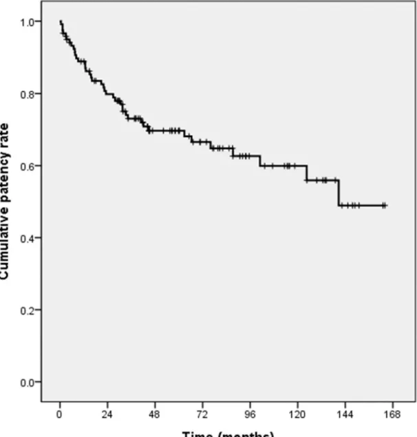
임상적인 성공을 보였다. 1, 3, 5, 7, 10년의 일차 개존율 (primary patency)은 각각 88.8%, 71.9%, 69%, 62.8%, 56.6%로 보고되었다.
스텐트 제거이후 추적관찰에서 제외된 20명의 환자를 제외하고, 평균 78.5개월 (3.1 - 164개월)의 추적관찰 기간 동안 119명의 환자 중 총 40명(33.6%)의 환자에서 임상적으로 유의한 협착이 재발했다.
Multivariate Cox’s proportional hazard regression 분석결과 협착 재발을 예측 할 만한 의미있는 예측 인자는 확인 되지 않았다.
결론
본 연구의 장기적인 추적 결과는 양성 담도 협착에서 제거 가능한 피막형 스텐트 (retrievable covered stent)를 사용한 경피 경간적 치료법이 임상적으로 효과적인 방법임을 시사한다.
차례
국문요약………
표차례………
그림차례………
서론………1
재료 및 방법………2
결과………6
고찰………9
결론………13
참고문헌………14
표………18
그림………20
영문요약………26
표 차례
Table 1. Demographics and characteristics of 148 patients with benign biliary strictures
Table 2. Results of Cox’s regression analysis for the evaluation of the factors associated with primary patency
그림 차례
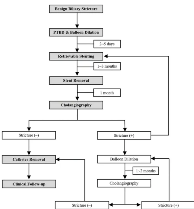
Figure 1. Schematic diagram of our treatment protocol
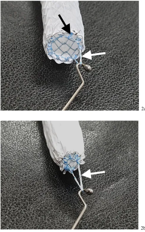
Figure 2. Retrievable covered stent and retrieval hook wire used in the procedures
Figure 3. A 60-year-old man with choledochojejunostomy stricture following pylorus-preserving pancreatoduodenectomy
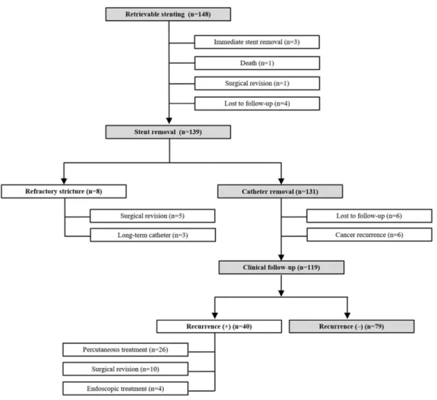
Figure 4. Flow chart of the study patients. n = number of patients Figure 5. Kaplan-Meier curve of the primary patency rate
Figure 6. Kaplan-Meier curves showing the primary patency rate according to the treatment period
Ⅰ. 서론 (INTRODUCTION)
Benign biliary strictures represent a major issue for patients and physicians, as the recurrence rate after standard treatment is quite high, with relevant problems for patients, such as recurrent cholangitis and the need for multiple readmissions to hospital (1). Surgical revision of a benign biliary stricture is still considered the best treatment, with a primary success rate of up to 90% (2). However, if the biliary strictures recur after surgery, it is quite difficult to manage them, and the success rate of surgical restoration decreases with each successive surgical intervention (3). Endoscopic treatment is the first- line management option for most patients with benign biliary strictures; however, endoscopic treatment is almost impossible in patients who have already undergone bilioenterostomy (4, 5). Therefore, percutaneous transhepatic approaches can be alternative treatment options (5-9).
Percutaneous balloon dilatation and large bore catheter placement are the most widely used alternatives to endoscopic treatment (5-11). However, this treatment requires multiple procedures and the prolonged use of indwelling percutaneous catheters that are often associated with catheter-related complications and a decreased quality of life. Limited data exist regarding the long-term outcomes of percutaneous treatment, but previous studies have reported 10-year patency rates between 49% and 72% (9-11).
Recently, several investigators have reported that percutaneous transhepatic placement of a temporary covered stent is feasible for the treatment of benign biliary strictures (12-18). The use of a temporary covered stent has been postulated to have a higher expansion force and a larger diameter compared to indwelling catheters and to lead to a sustained dilatation effect. Compared to percutaneous balloon dilatation and large-bore catheters, stents may increase the possibility of stricture resolution and shorten the treatment duration (17-19). Different types of temporary covered stents have been used to treat benign biliary strictures for varying durations and have achieved successful stricture resolution in 80%
to 90.6% of the patients (12-18). However, data about the percutaneous use of temporary covered stents during the treatment of benign biliary strictures are limited, and the reported series deal with relatively small groups of patients (16-19). Most patients with benign biliary strictures have a life expectancy greater than 5 years, and to our knowledge, no long-term patency data (>5 years) exist for radiologic management. Therefore, the purpose of this study was to investigate the long-term outcomes of using temporary placement of retrievable covered stents for the treatment of patients with benign biliary strictures.
Ⅱ. 재료 및 방법 (Materials and Methods)
Patient Population
This study was approved by the institutional review board, which waived the need for written informed consent because of the retrospective nature of this study. This study enrolled 148 patients (84 male, 64 female; mean age, 57 years; age range, 11‒92 years) who underwent percutaneous transhepatic placement and removal of a retrievable covered stent between March 2007 and August 2019 for the treatment of benign biliary strictures. Baseline characteristics of the 148 patients with benign biliary strictures are presented in Table 1. The initial diagnosis of a biliary stricture was based on the clinical symptoms or biochemical data of the patients, and the results of imaging with various modalities, including ultrasonography, computed tomography, magnetic resonance cholangiopancreatography, and percutaneous transhepatic cholangiography. Patients with benign biliary strictures were selected for inclusion in this study using the following inclusion criteria: (a) initially documented benign biliary strictures, (b) previously underwent bilioenterostomy, or (c) failed endoscopic cannulation of the intrahepatic or common bile duct. Among 148 patients with benign biliary strictures, 85 (57.4%) patients received a retrievable covered stent placement as the initial treatment of a new lesion and 63 (42.6%) patients had recurrent or refractory benign biliary strictures despite repeated balloon dilatation and prolonged catheter placement. Patients who had previously undergone liver transplantation were excluded from this study.
Technique
Our treatment protocol is summarized in Figure 1. All procedures, including percutaneous percutaneous transhepatic biliary drainage (PTBD) and placement and removal of the retrievable stent, were performed by two experienced radiologists (D.I.G. and G.Y.K.) with 20 and 26 years of experience, respectively. All procedures were performed under conscious sedation using intravenous pethidine hydrochloride (Demerol, Keukdong Pharmaceuticals, Seoul, Korea) and with local anesthesia using intramuscular lidocaine (Jeil Pharmaceuticals, Taegu, Korea). Broad-spectrum antibiotics were administered intravenously 2 h before the procedures and for at least 48 h afterward.
PTBD was performed under the guidance of fluoroscopy or ultrasound. A right or left approach was determined based on the stricture location. Before inserting a drainage catheter, dilation of the stricture
was performed using a balloon catheter 6–8 mm in diameter (Synergy or Mustang; Boston Scientific, Galway, Ireland). An internal-external 8.5-F drainage catheter (Cook, Bloomington, IN) was then placed across the stricture.
A retrievable expanded polytetrafluoroethylene (e-PTFE)-covered stent (Song retrievable stent;
TaeWoong Medical, Kimpo, Korea) with two drawstrings attached to the upper margin of the stent was used (Fig. 2a). Balloon dilation was performed using a balloon catheter with a diameter of 8–10 mm (Boston) just before and after stent placement. A stent diameter of 8–12 mm was chosen depending on the diameter of the bile duct and the bilioenterostomy site. In patients who had complex-type biliary strictures, two stents were deployed in a single session. To prevent distal migration of the stent after deployment, a 10–14-F multi-side-holes, pigtail-shaped drainage catheter was placed across the stent, and the catheter tip was placed beneath the distal margin of the stent. After placement, the internal drainage catheter was capped. The duration of stent indwelling was determined according to the severity of the biliary stricture. If the patients had initial benign biliary strictures, the indwelling period was one or two months; on the other hand, if the patients had recurrent or refractory strictures despite the previous percutaneous treatment, the indwelling period was three or four months. To evaluate stent migration, the patients returned for cholangiography via the drainage catheter in 1 month after the stent placement. If the stent had migrated, repositioning of the stent was performed using a retrieval hook wire or balloon catheter. If repositioning of the stent failed, another stent was inserted.
For each patient, the stent was electively removed. A safety wire was placed into the small bowel or the common bile duct in order to gain access. Then a 9-F sheath with a dilator was passed over the guide wire into the proximal lumen of the stent. After the dilator was removed from the sheath, a retrieval hook wire was introduced into the sheath to remove the stent, and the hook was pulled out of the stent so that the hook caught the extraction strings. Traction on any one drawstring caused the end of the stent to pull together and collapse into a funnel configuration (Fig. 2b). When this occurred, the retrieval hook wire was withdrawn through the sheath to collapse the stent. The stent and retrieval hook wire was then pulled out of the biliary duct through the sheath.
Cholangiography was obtained immediately after stent removal via the sheath to evaluate the stricture.
If the absence of a significant stricture was noted on cholangiography obtained immediately after stent removal, a 10–14-F external drainage catheter was placed in the intrahepatic bile duct with its tip clamped until 1 month follow-up cholangiography. If there was a persistent stricture, depending on the severity of the stricture, a retrievable covered stent was reinserted across the stricture site for an additional 1–3 months or one to two sessions of balloon dilatation with a 1 month interval was
recurrence of patient symptoms or changes in biochemical data, the drainage catheter was removed (Fig.
3).
Follow-up
All patients were evaluated clinically at the interventional radiology outpatient clinic at 1, 3, 6, and 12 months after drainage catheter removal to check for a recurrence, or if there were unexpected symptoms that suggested a recurrence. Liver enzyme analysis was also measured during this follow-up period and imaging studies, including CT, were performed at 6–8 months after drainage catheter removal. After that, the patients were followed at one-year intervals for 3 years. Telephone interviews were performed from 5 years after drainage catheter removal until the stricture recurred or the patient died. The patient was advised to visit our clinic if symptoms recur.
Study Endpoints, Definitions, and Statistical Analysis
The primary endpoint was primary patency, defined as the interval between drainage catheter removal and stricture recurrence. In patients with no recurrence, primary patency was assumed to be the interval between drainage catheter removal and the last follow-up. Secondary endpoints were technical success, clinical success, and procedure-related complications. Technical success was defined as successful stent deployment across the stricture with confirmation of patency using cholangiography and stent removal without bile duct injury. Clinical success was defined as the disappearance of the patient's symptoms and normalized biochemical data during the clamp test period until removal of the drainage catheter.
Complications were classified as major and minor according to the guidelines of the Society of Interventional Radiology Standards of Practice Committee (20). Based on the time frame, early migration was defined as stent migration being recognized within 1 month of stent placement, whereas late migration was detected 1 month after stent placement. Recurrence was defined as the development of a clinically significant recurrence of the stricture based on clinical manifestations, serum biochemical data, and imaging that required subsequent intervention.
Primary patency rates were calculated according to the Kaplan-Meier method, and differences between curves were analyzed by the log-rank test. The following variables were included in the analysis: age, gender, underlying disease, stricture site, stricture type, combined with biliary stones, a history of previous treatment, and treatment duration. Variables found to be significant by univariate analysis (p
< 0.05) were considered candidate variables for multiple analysis, which was performed using the Cox’s
proportional hazard model. Variables were selected in a stepwise forward selection manner. IBM SPSS Statistics for Window, version 23.0 (IBM Corp., Armonk), was used for the statistical analyses. A two- sided p value less than 0.05 was considered statistically significant.
Ⅲ. 결과 (Results)
Retrievable Stent Insertion
The results are summarized in Figure 4. Stent deployment was technically successful in all 148 patients.
Ten patients required two stent placements because of two separated bilioenterostomy strictures (n=6) and complex-type strictures (n=4); a single stent was sufficient to treat the biliary strictures in the remaining 138 patients.
Cholangiography immediately after stent placement showed ipsilateral or opposite intrahepatic bile ducts occlusion by the stents in 35 of 148 patients. In 32 patients, the stents had been placed in situ because their liver enzymes were within the normal range. In the remaining three patients, the stents were removed 1‒3 days after stent placement due to markedly elevated liver enzymes; the strictures were then managed using repeated balloon dilatation. Among the 145 patients, six patients were excluded before the 1-month follow-up cholangiography; four patients were lost to follow-up, one underwent a revisional operation, and one died. Therefore, 1 month follow-up cholangiography was performed on 139 patients.
One-month follow-up cholangiography after stent placement showed early stent migration in 14 patients.
Among these 14 patients, 12 patients experienced total downward stent migration into the jejunum;
therefore, an additional stent placement was performed. The other two patients showed upward stent migration; the stents were successfully repositioned using a balloon catheter.
Retrievable Stent and Catheter Removal
Stent removal was performed in 139 patients. The mean indwelling period of the stent was 2.5 months (range, 1.2‒7.7 months). In seven patients, the indwelling period of the stent was more than 5 months.
Cholangiography obtained at the time of stent removal revealed late stent migration in 14 of 139 patients.
In the 11 patients who showed distal stent migration, the stent had migrated distally and was evacuated from the bowel. In the three patients who showed proximal stent migration, the stent was successfully removed via the percutaneous route. Among the 14 patients with late stent migration, 12 patients had improved strictures whereas two required additional treatments due to persistent strictures. The overall stent migration (early migration [n=14], late migration [n=14]) rate was 18.9% (28 of 148 patients).
Except for 11 patients who showed distal stent migration, percutaneous stent removal was performed
in the remaining 128 patients. In 125 patients, after removal of the stents, the e-PTFE material was seen to be intact and the lumen of the stent was also visualized to be sure it was clear. In the remaining three patients with bilioenterostomy strictures, partial tearing of the e-PTFE membrane with partial luminal narrowing (n=2; indwelling period, 5.5 months and 6 months, respectively) or complete occlusion (n=1;
indwelling period, 7 months) due to sludge was seen. During the stent removal procedure in one patient, the drawstring became untied with the stent remaining in the peripheral intrahepatic bile duct, and the stent could not be removed under fluoroscopic guidance. The stent was removed surgically. Except for this one patient, percutaneous stent removal was successfully achieved in the remaining 124 patients.
Therefore, including 3 patients who showed proximal stent migration and had percutaneous removal, the technical success rate of stent removal was 99.2% (127 of 128 patients). Cholangiography immediate stent removal in 96 patients revealed a widened stricture with free passage of contrast media.
However, the remaining 32 patients showed a persistent stricture. In these 32 patients, 13 patients needed a second session of stent placement (n=11) or a third session of stent placement (n=2), and 19 patients needed 1‒5 sessions (mean, 2 sessions) of balloon dilatation.
Completion cholangiography obtained 1‒3 months after stent removal or additional balloon dilatation revealed a patent stricture with free passage of the contrast media in 131 patients. Their drainage catheters were then removed. Therefore, the mean treatment period from stent placement to catheter removal in 131 patients who successfully removed the drainage catheter was 8.1 months (range, 0.7‒
143.6 months). However, the remaining eight patients showed refractory strictures. These eight patients underwent surgical revision (n=5) or long-term large-bore catheter placement (n=3). After excluding the nine patients who dropped out after stent placement, clinical success was achieved in 131 (94.2%) of 139 patients.
Major complications included markedly elevated liver enzymes after stent placement in three patients, stent migration needing a second stent placement in 14 patients and the failure of percutaneous stent removal in one patient. Procedure-related minor complications occurred in five patients. In these five patients, self-limiting hemobilia was noted after balloon dilatation that was completely resolved 1-2 days later. Therefore, the overall complication rate was 15.5% (23 of 148 patients). There was no procedure-related mortality.
Follow-up and Primary Patency
Among 131 patients with clinical success, six patients were lost to follow-up and six patients had cancer recurrence at the bilioenterostomy site at a mean of 16.4 months (range, 1.8‒37.8 months). Therefore,
clinical follow-up data until the end of this study could be obtained for 119 patients and the cutoff date for the analysis was August 31, 2020. During the mean follow-up period of 78.5 months (range, 3.1‒
164 months), 20 patients died, but their causes of death were not related to the biliary obstruction. The remaining 99 patients are still alive.
The primary patency rates at 1, 3, 5, 7, and 10 years were 88.8%, 71.9%, 69%, 62.8%, and 56.6%, respectively (Fig. 5). During the follow-up period, 40 (33.6 %) of 119 patients had a recurrence of clinically significant strictures at a mean of 31 months (range, 0.5–140.7 months). For ten patients, the decision was made to resort to surgery; four patients underwent endoscopic treatments; and in 26 patients, eight patients received a retrievable covered stent and 18 patients were treated with repeat balloon dilatation with a large-bore catheter.
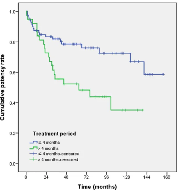
On multivariate Cox’s proportional hazard regression analysis, the treatment period (the time from stent placement to catheter removal > 4 months) (adjusted odds ratio, 2.201; 95% confidence interval, 1.140 4.246; p = 0.019) was identified as an independent risk factor for stricture recurrence (Table 2) (Fig. 6).
Other variables, including age, gender, underlying disease (benign versus malignant), stricture site (anastomotic versus nonanastomotic), stricture type (simple versus complex), combined with biliary stones, and a history of previous treatment (initial versus recurrent or refractory) did not show statistically significant associations. Among the 64 patients who underwent retrievable covered stent placement for the initial strictures, 46 patients (71.9%) showed improvement of the strictures by the end of the study period (a mean of 73.1 months after stent removal). Among 55 patients who underwent retrievable covered stent placement for recurrent or refractory strictures after balloon dilatation and long-term catheter placement, 33 patients (60%) showed improvement of the strictures at the end of the study period (mean of 68 months after stent removal).
Ⅳ. 고찰 (Discussion)
In the present study, the primary patency rates at 1, 3, 5, 7, and 10 years were 88.8%, 71.9%, 69%, 62.8%, and 56.6%, respectively. We determined the probability of a patient not having a clinically significant recurrence over the long-term (as long as 13.7 years) after the retrievable stent treatment. In the previous studies, the primary patency rates at 1 and 3 years were 82.6‒91% and 69.3‒87%, respectively (16-19). Although it is difficult to compare out results with the prior findings, the short- term primary patency rates we observed were similar to those in previous studies using percutaneous placement of temporary covered stents. These long-term primary patency rate showed comparable or superior to balloon dilatation (7, 9, 11), but stent placement maintained a slightly longer primary patency period (27, 31). With regard to complications, the overall complication rate of retrievable stent in the present study did not seem to differ significantly from that of balloon dilatation, which was reported in 8% to 11% of previous studies (7, 11). But considering that balloon dilatation required repeated procedures, retrievable stent also superior in terms of the patients’ quality of life (8, 10, 11, 27).
In most of the previous studies, only patients who underwent temporary covered stent placement for initial biliary stricture were included. In the present study, we also evaluated the efficacy of retrievable stent placement for recurrent or refractory benign biliary strictures in a relatively large number of subjects with long follow-up periods. It is noteworthy that nearly half of patients in the present study had a recurrent or refractory stricture. There was no significant difference between the initial group and the refractory group in terms of the patency rate. We also found that among the 55 patients who underwent retrievable stent placement and removal for recurrent or refractory strictures after repeated balloon dilatation, 33 patients (60%) showed improvement of the strictures until the end of the study period (mean of 68 months after catheter removal). Therefore, in addition to the primary use of temporary covered stents in patients with benign biliary strictures, retrievable stents appeared to provide sufficient relief of strictures even in cases recurrent or refractory to previous treatments, including repeated balloon dilatation or large-bore catheters.
Ideally, covered stents for benign biliary strictures should demonstrate a persistent dilatation effect and be easily removable. From this perspective, biodegradable stents made of polydioxanone material may be an ideal choice, providing advantrages of stenting without the need for removal. However, the limitation of biomaterials regarding expansile force and degradation profiles are still remain a problem, and data about effectiveness of these stents are limited not yet (28-30). Therefore, fully covered stents are closest to the ideal for now. Among several available covered metallic stents, the e-PTFE-covered
stent is considered to be the most effective choice for benign biliary strictures (12-14, 24) because the completely e-PTFE-covered material serves as an effective mechanical barrier to tissue ingrowth and as a relatively friction-free surface, which is particularly suitable for removal without becoming incorporated into the bile duct wall (21, 22). And also, since a nitinol stent is able to recompress to a small volume, this facilitates its removal through a relatively narrow channel without requiring an increase in the diameter of the tract (14).
There are two concerns associated with the placement of a covered stent. First, placement of covered stents across the bilioenterostomy anastomosis may cause cholangitis in the ipsilateral or contralateral bile duct. In the present study, cholangiographic occlusions of the ipsilateral or opposite intrahepatic bile duct by the stents were observed in 35 of 148 patients. Except for three patients who had the stent removed 1‒3 days after stent placement due to markedly elevated liver enzymes, the stents had been placed in situ in the remaining 33 patients because their liver enzymes were within the normal range despite cholangiographic occlusion. Gwon et al. suggested that cholangiographic occlusion of bile ducts by the e-PTFE-covered retrievable stent did not represent functional occlusion (16). Peterson et al. also observed a flow of contrast material between the bile duct wall and the covering of the PTFE-covered stent for as long as 4‒6 months following stent placement (12). Therefore, we suggest that blockage of the intrahepatic bile ducts caused by an e-PTFE-covered retrievable stent may not result in major complications, although close follow-up is necessary in such cases.
Second, migration and dislocation during or after stent placement is still a major concern. Previous studies have reported 6‒21% migration rates following percutaneous transhepatic placement of retrievable covered stents (12, 14, 16, 19). In the present study, the stent migration rate was 18.9% (28 of 148 patients). Positioning of the pigtail-shaped tip of the catheter beneath the distal margin of the stent did not completely prevent stent migration. In order to prevent stent migration, a few anti- migration designs have been tested, including stents with anti-migration fins and stents with percutaneous fixation strings. Peng et al. reported a transhepatic fixation string could prevent distal stent migration in 25 of 26 patients (18). Wang et al. used a covered stent with anchoring fins; however, they found mucosal ulceration of the bile duct due to anchoring fins of the stents (23). Therefore, innovative stent design modifications are required to ensure the ability to remove the stents and to prevent their migration and dislocation.
It remains to be determined how long temporary stents should be left in place. A short stent indwelling period might result in insufficient resolution of the strictures, whereas a longer indwelling period may not be desirable due to its association with stent-related complications such as stent occlusion or
In the previous studies, the mean indwelling period of retrievable stents was 2.5‒4 months (15, 16, 18, 19). Because of the long-term period of the present study, the target duration of stent placement varied a little, but the mean duration was close to that of the previous studies. We also found that complete stent occlusion due to sludge was observed in one patient with a stent indwelling period of 7 months.
Unfortunately, no current materials have 100% bioresistance in the hostile environment of the liver and bowel. The membrane is exposed to bile and potentially gastric acid, which results in degradation of the membrane. Previous investigators reported that the polyurethane and silicone membrane may be degraded by bile, pancreatic juice or gastric juice (21, 22). Moreover, the e-PTFE membrane is more resistant to acids and alkalis. However, Bang et al. reported that e-PTFE showed stronger tensile and tear strengths and tended to form biofilms more frequently than polyurethane and silicone during bile exposure (24). Gwon et al. reported that intact e-PTFE coverings with clean stent lumens as well as intact drawstrings and nylon loops were observed in the removed stents after up to 64 days (25).
Petersen et al. reported that moderate luminal narrowing (50‒60%) was observed at 5-6 months and one stent was occluded at 9 months (12). Tomishima et al. reported stent fracture occurred in two cases (7.7%) in which the retrievable covered stents were placed for more than 6 months (26). According to these previous studies and our outcomes, leaving the covered stents in situ for 6 months seems to be acceptable for benign biliary diseases in terms of stent durability and safety, although there is anecdotal evidence that some stents remain intact for a significantly longer period.
In the present study, there were no statistically significant predictive factors for recurrence. Although it seemed to be slight high recurrence rate in patients with a longer treatment period of 4 months or longer, it was not statistically significant (p=0.019). In patients with long treatment periods, the high recurrence rate is thought to be due to additional stent placement or balloon dilatation for persistent or recurrence stricture immediately after removal of the stents initially inserted, but the treatment period itself does not appear to be a predictor of recurrence.
The present study had several limitations. First, this study is retrospective and was conducted at a single center with a nonrandomized design, which may have decreased its statistical strength. Some unpredictable drop-outs were due to unsuspected diagnoses of anastomotic recurrence (4.1%) during follow-up, but only 2.7% of patients were lost to follow-up during the stenting period, while 4.1% of the patients were lost during follow-up after catheter removal. However, the patients were consecutively enrolled and long-term follow-up data were obtained from most of them. Nevertheless, it remains possible that the sequential nature of the study may have introduced some bias. Second, the study patients did not undergo homogenous treatments. Prior to retrievable stent treatment, 63 of 148 patients with recurrent or refractory benign biliary strictures despite prolonged interposition of the catheter after
balloon dilatation were included in this study. However, because the patients had different causes, locations, and types of stricture, it was not possible to apply a homogeneous treatment regimen.
Ⅴ. 결론 (Conclusion)
In conclusion, the long-term outcomes suggest that percutaneous treatment of benign biliary strictures using a retrievable covered stent is a clinically effective method.
Ⅵ. 참고문헌 (References)
1. Judah JR, Draganov PV. Endoscopic therapy of benign biliary strictures. World J Gastroenterol 2007;13(26):3531-3539. doi: 10.3748/wjg.v13.i26.3531
2. Bolton JS, Braasch JW, Rossi RL. Management of benign biliary stricture. Surg Clin North Am 1980;60(2):313-332. doi: 10.1016/s0039-6109(16)42083-9
3. Mueller PR, vanSonnenberg E, Ferrucci JT, Jr., Weyman PJ, Butch RJ, Malt RA, Burhenne HJ.
Biliary stricture dilatation: multicenter review of clinical management in 73 patients. Radiology 1986;160(1):17-22. doi: 10.1148/radiology.160.1.3715030
4. Visrodia KH, Tabibian JH, Baron TH. Endoscopic management of benign biliary strictures. World J Gastrointest Endosc 2015;7(11):1003-1013. doi: 10.4253/wjge.v7.i11.1003
5. Köcher M, Cerná M, Havlík R, Král V, Gryga A, Duda M. Percutaneous treatment of benign bile duct strictures. Eur J Radiol 2007;62(2):170-174. doi: 10.1016/j.ejrad.2007.01.032
6. Weber A, Rosca B, Neu B, Rösch T, Frimberger E, Born P, Schmid RM, Prinz C. Long-term follow- up of percutaneous transhepatic biliary drainage (PTBD) in patients with benign bilioenterostomy stricture. Endoscopy 2009;41(4):323-328. doi: 10.1055/s-0029-1214507
7. Glas L, Courbière M, Ficarelli S, Milot L, Mennesson N, Pilleul F. Long-term outcome of percutaneous transhepatic therapy for benign bilioenteric anastomotic strictures. J Vasc Interv Radiol 2008;19(9):1336-1343. doi: 10.1016/j.jvir.2008.05.019
8. Bonnel DH, Fingerhut AL. Percutaneous transhepatic balloon dilatation of benign bilioenteric strictures: long-term results in 110 patients. Am J Surg 2012;203(6):675-683. doi:
10.1016/j.amjsurg.2012.02.001
9. Cantwell CP, Pena CS, Gervais DA, Hahn PF, Dawson SL, Mueller PR. Thirty years' experience with balloon dilation of benign postoperative biliary strictures: long-term outcomes. Radiology 2008;249(3):1050-1057. doi: 10.1148/radiol.2491080050
10. DePietro DM, Shlansky-Goldberg RD, Soulen MC, Stavropoulos SW, Mondschein JI, Dagli MS, Itkin M, Clark TW, Trerotola SO. Long-term outcomes of a benign biliary stricture protocol. J Vasc Interv Radiol 2015;26(7):1032-1039. doi: 10.1016/j.jvir.2015.03.002
11. Janssen JJ, van Delden OM, van Lienden KP, Rauws EA, Busch OR, van Gulik TM, Gouma DJ, Laméris JS. Percutaneous balloon dilatation and long-term drainage as treatment of anastomotic and
nonanastomotic benign biliary strictures. Cardiovasc Intervent Radiol 2014;37(6):1559-1567. doi:
10.1007/s00270-014-0836-y
12. Petersen BD, Timmermans HA, Uchida BT, Rabkin JM, Keller FS. Treatment of refractory benign biliary stenoses in liver transplant patients by placement and retrieval of a temporary stent-graft: work in progress. J Vasc Interv Radiol 2000;11(7):919-929. doi: 10.1016/s1051-0443(07)61812-0
13. Kuo MD, Lopresti DC, Gover DD, Hall LD, Ferrara SL. Intentional retrieval of viabil stent-grafts from the biliary system. J Vasc Interv Radiol 2006;17(2 Pt 1):389-397. doi:
10.1097/01.Rvi.0000194867.86371.0b
14. Gwon DI, Shim HJ, Kwak BK. Retrievable biliary stent-graft in the treatment of benign biliary strictures. J Vasc Interv Radiol 2008;19(9):1328-1335. doi: 10.1016/j.jvir.2008.05.017
15. Kim J, Ko GY, Sung KB, Gwon DI, Lee SG, Kim KM, Kim KA, Yoon HK. Percutaneously placed covered retrievable stents for the treatment of biliary anastomotic strictures following living donor liver transplantation. Liver Transpl 2010;16(12):1410-1420. doi: 10.1002/lt.22173
16. Gwon DI, Ko GY, Ko HK, Yoon HK, Sung KB. Percutaneous transhepatic treatment using retrievable covered stents in patients with benign biliary strictures: mid-term outcomes in 68 patients.
Dig Dis Sci 2013;58(11):3270-3279. doi: 10.1007/s10620-013-2784-9
17. Yun G, Yoon CJ, Seong NJ. Percutaneous treatment of benign bilioenteric anastomotic strictures:
temporary covered stent placement versus balloon dilatation. Eur Radiol 2019;29(5):2690-2697. doi:
10.1007/s00330-018-5776-5
18. Ye P, Zeng Q, Miao H, Pang H, Chen Y. Percutaneous Treatment of Benign Biliary Anastomotic Strictures: Retrievable Covered Self-Expandable Metal Stent with Fixation String Versus Large-Bore Catheters. J Vasc Interv Radiol 2021;32(1):113-120. doi: 10.1016/j.jvir.2020.01.034
19. Kim JH, Gwon DI, Ko GY, Sung KB, Lee SK, Yoon HK, Shin JH, Song HY. Temporary placement of retrievable fully covered metallic stents versus percutaneous balloon dilation in the treatment of benign biliary strictures. J Vasc Interv Radiol 2011;22(6):893-899. doi: 10.1016/j.jvir.2011.02.009 20. Sacks D, McClenny TE, Cardella JF, Lewis CA. Society of Interventional Radiology clinical practice guidelines. J Vasc Interv Radiol 2003;14(9 Pt 2):S199-202. doi:
10.1097/01.rvi.0000094584.83406.3e
21. Schoder M, Rossi P, Uflacker R, Bezzi M, Stadler A, Funovics MA, Cejna M, Lammer J. Malignant biliary obstruction: treatment with ePTFE-FEP- covered endoprostheses initial technical and clinical
experiences in a multicenter trial. Radiology 2002;225(1):35-42. doi: 10.1148/radiol.2251011744 22. Bezzi M, Zolovkins A, Cantisani V, Salvatori FM, Rossi M, Fanelli F, Rossi P. New ePTFE/FEP- covered stent in the palliative treatment of malignant biliary obstruction. J Vasc Interv Radiol 2002;13(6):581-589. doi: 10.1016/s1051-0443(07)61651-0
23. Wang AY, Ellen K, Berg CL, Schmitt TM, Kahaleh M. Fully covered self-expandable metallic stents in the management of complex biliary leaks: preliminary data - a case series. Endoscopy 2009;41(9):781-786. doi: 10.1055/s-0029-1215050
24. Bang BW, Jeong S, Lee DH, Lee JI, Lee SC, Kang SG. The biodurability of covering materials for metallic stents in a bile flow phantom. Dig Dis Sci 2012;57(4):1056-1063. doi: 10.1007/s10620-011- 1958-6
25. Gwon DI, Ko GY, Sung KB, Kim JH, Yoon HK. Percutaneous transhepatic treatment of postoperative bile leaks: prospective evaluation of retrievable covered stent. J Vasc Interv Radiol 2011;22(1):75-83. doi: 10.1016/j.jvir.2010.10.004
26. Tomishima K, Ishii S, Fujisawa T, Ikemura M, Ushio M, Takahashi S, Yamagata W, Takasaki Y, Suzuki A, Ito K, Haga K, Ochiai K, Nomura O, Saito H, Shibuya T, Nagahara A, Isayama H. Evaluation of the Feasibility and Effectiveness of Placement of Fully Covered Self-Expandable Metallic Stents via Various Insertion Routes for Benign Biliary Strictures. J Clin Med 2021;10(11). doi:
10.3390/jcm10112397
27. Chinmay Bhimaji K, Sreekumar Karumathil P, Srikanth M, Nirmal Kumar P, Puthukudiyil Kader N, and Ramiah Rajesh K. Percutaneous transhepatic balloon dilatation of benign bilioenteric strictures:
Analysis of technique and long-term outcome. Int J Gastrointest Interv 2015;4(2):112-119. doi:
10.18528/gii150001
28. Gwon DI, Laasch H-U. Radiological approach to benign biliary strictures. Gastrointestinal Intervention 2015;4(1):9-14. doi: https://doi.org/10.1016/j.gii.2015.01.001
29. Kapoor BS, Mauri G, Lorenz JM. Management of Biliary Strictures: State-of-the-Art Review.
Radiology 2018;289(3):590-603. doi: 10.1148/radiol.2018172424
30. Mauri G, Michelozzi C, Melchiorre F, Poretti D, Pedicini V, Salvetti M, Criado E, Falcò Fages J, De Gregorio M, Laborda A, Sonfienza LM, Cornalba G, Monfardini L, Panek J, Andrasina T, Gimenez M. Benign biliary strictures refractory to standard bilioplasty treated using polydoxanone biodegradable
biliary stents: retrospective multicentric data analysis on 107 patients. Eur Radiol 2016;26(11):4057- 4063. doi: 10.1007/s00330-016-4278-6
31. Park SJ, Chung HH, Lee SH, Cho SB, Kim YH, Seo TS, Song MG. Long-term balloon indwelling technique for the treatment of single benign biliary stricture. Diagn Interv Radiol 2019;25(1):90-94. doi:
10.5152/dir.2018.18225
표 (Tables)
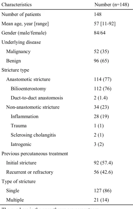
Table 1: Demographics and characteristics of 148 patients with benign biliary strictures
Characteristics Number (n=148)
Number of patients 148
Mean age, year [range] 57 [11-92]
Gender (male/female) 84/64
Underlying disease
Malignancy 52 (35)
Benign 96 (65)
Stricture type
Anastomotic stricture 114 (77)
Bilioenterostomy 112 (76)
Duct-to-duct anastomosis 2 (1.4) Non-anastomotic stricture 34 (23)
Inflammation 28 (19)
Trauma 1 (1)
Sclerosing cholangitis 2 (1)
Iatrogenic 3 (2)
Previous percutaneous treatment
Initial stricture 92 (57.4)
Recurrent or refractory 56 (42.6)
Type of stricture
Single 127 (86)
Multiple 21 (14)
The numbers in the parentheses are percentages
Table 2: Results of Cox’s regression analysis for the evaluation of the factors associated with primary patency
Factors Hazard ratio 95% CI P value
Female 1.505 0.806 – 2.810 0.199
Age 0.599 0.321 – 1.119 0.108
Underlying disease (malignancy) 0.518 0.258 – 1.038 0.064
Postoperative stricture 1.130 0.573 – 2.229 0.725
Complex type 1.858 0.723 – 4.772 0.198
Biliary stone 1.384 0.714 – 2.684 0.336
Recurrent stricture 1.440 0.772 – 2.689 0.252
Stent length 0.561 0.245 – 1.284 0.171
Treatment duration > 4 months 2.501 1.331 – 4.700 0.004
Note.— CI = confidence interval
그림 (Figures)
Figure 1: Schematic diagram of our treatment protocol.
2a
2b
Figure 2: Retrievable covered stent and retrieval hook wire used in the procedures. (a) “End on” view of the proximal end of the retrievable covered stent. A 2-mm-diameter nylon loop (black arrow) is hooked inside each bend of the proximal end of the stent and attached to the upper inner margin of the stent. Two drawstrings are passed through each of the nylon loops. Note the two drawstrings (white arrow) caught by the retrieval hook wire. (b) Traction on the drawstrings (white arrow) causes the end of the stent to be pulled together.
3a 3b 3c
3d 3e 3f
Figure 3: A 60-year-old man with choledochojejunostomy stricture following pylorus-preserving pancreatoduodenectomy. (a) Cholangiography shows severe stricture at the choledochojejunostomy anastomosis. (b) A 10-mm-diameter and 80-mm-long retrievable covered stent (white arrows) was inserted across the stricture, and a 10-F pigtail catheter was inserted across the stent for follow-up cholangiography and stent removal. To prevent stent migration, the pigtail-shaped drainage catheter tip (black arrow) was placed just beneath the distal stent margin. (c) A fluoroscopic image obtained 2 months after stent insertion shows a good stent position without migration. A retrieval hook (white arrow) for stent removal was inserted through a 9-F sheath (black arrow). The stent was successfully removed through the 9-F sheath. (d) A cholangiography obtained immediately after the stent removal shows the anastomosis is patent. (e) A follow-up cholangiography obtained 1 month after stent removal shows the anastomosis to be patent without recurrence. The drainage catheter was removed immediately after the follow-up cholangiography. (f) Contrast-enhanced coronal CT image obtained 74 months after the drainage catheter removal shows patent anastomosis (white arrow). The patient remained healthy
Figure 4: Flow chart of the study patients. n = number of patients
Figure 5: Kaplan-Meier curve of the primary patency rate
Figure 6: Kaplan-Meier curves showing the primary patency rate according to the treatment period.
영문요약
Purpose: To investigate the long-term outcomes of a retrievable covered stent for the treatment of benign biliary strictures.
Materials and Methods: We retrospectively assessed 148 patients (84 men, 64 women; mean age, 57 years; range, 11‒92 years) who underwent percutaneous transhepatic placement and removal of a retrievable covered stent between March 2007 and August 2019 for the treatment of benign biliary strictures. Ninety-two patients had not previously undergone interventional treatment, whereas 56 had recurrent or refractory strictures despite previous percutaneous procedures.
Results: Placement of the stents was technically successful in all 148 patients. Stent migration occurred in 28 (18.9%) patients. The mean indwelling period of the stent and drainage catheter was 2.5 months (range, 1.2‒8.1 months) and 4 months (range, 1.6‒67.4 months), respectively. Excluding the nine patients who dropped out after stent placement, clinical success was achieved in 131 (94.2%) of 139 patients. The overall complication rate was 15.5 % (23 of 148 patients; 14 patients in major complications, 5 patients in minor complications), and there was no procedure-related mortality. The primary patency rates at 1, 3, 5, 7, and 10 years were 88.8%, 71.9%, 69%, 62.8%, and 56.6%, respectively. During the mean follow-up of 78.5 months (range, 3.1‒164 months), excluding the 20 patients who dropped out after stent removal, 40 (33.6%) of 119 patients had a recurrence of clinically significant strictures. In multivariate Cox’s proportional hazard regression analysis, there were no statistically significant predictive factors for recurrence.
Conclusion: The long-term outcomes suggested that percutaneous treatment of benign biliary strictures using a retrievable covered stent was a clinically effective method.