Engineered Carbon-Nanomaterial-Based Electrochemical Sensors for Biomolecules
Jitendra N. Tiwari,* Varun Vij, K. Christian Kemp, and Kwang S. Kim*
Center for Superfunctional Materials, Department of Chemistry, Ulsan National Institute of Science and Technology (UNIST), Ulsan 689-798, Korea
ABSTRACT: The study of electrochemical behavior of bioactive molecules has become one of the most rapidly developing scientificfields. Biotechnology and biomedical engineeringfields have a vested interest in constructing more precise and accurate voltammetric/amperometric biosensors. One rapidly growing area of biosensor design involves incorporation of carbon-based nanomaterials in working electrodes, such as one-dimensional carbon nanotubes, two-dimensional graphene, and graphene oxide. In this review article, we give a brief overview describing the voltammetric techniques and how these techniques are applied in biosensing, as well as the details surrounding important biosensing concepts of sensitivity and limits of
detection. Building on these important concepts, we show how the sensitivity and limit of detection can be tuned by including carbon-based nanomaterials in the fabrication of biosensors. The sensing of biomolecules including glucose, dopamine, proteins, enzymes, uric acid, DNA, RNA, and H2O2traditionally employs enzymes in detection; however, these enzymes denature easily, and as such, enzymeless methods are highly desired. Here we draw an important distinction between enzymeless and enzyme-containing carbon-nanomaterial-based biosensors. The review ends with an outlook of future concepts that can be employed in biosensor fabrication, as well as limitations of already proposed materials and how such sensing can be enhanced. As such, this review can act as a roadmap to guide researchers toward concepts that can be employed in the design of next generation biosensors, while also highlighting the current advancements in thefield.
KEYWORDS: carbon nanotubes, graphene, glucose, dopamine, proteins, uric acid, DNA, RNA, H2O2, biosensors
T
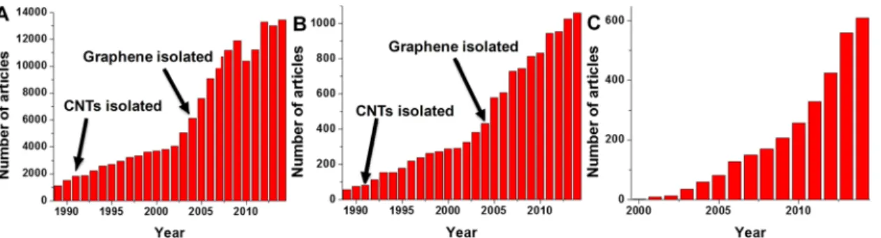
he use of femto-, pico-, and nanosensitive biosensors is of critical importance not only in obvious medical applications,1such as glucose monitoring in diabetics,2 but also in environmental monitoring, water remediation, molecular imaging, and noxious gas sensing, among other applications.3−6In fact, thisfield is so important that there are multiple dedicated journals focused on either biosensors and/or sensors, with multiple reviews published on a wide range of highly specific sensor-related topics.4−8 Additionally, the amount of papers published with sensor in the title has increased dramatically from 1131 articles in 1989 to 13461 in 2014 (Figure 1A). In the same way, the amount of papers published with biosensor in the title has also increased exponentially (Figure 1B). What is interesting to note in these graphs is that with the isolation of carbon nanotubes (CNTs) in 1991 and the isolation of graphene in 2004 there has been a rapid rise in the amount of articles published.9−12 Figure 1C is a graph indicating the rise in the number of publications that contain the title words: sensor, graphene, biosensor, and carbon nanotube. These statistical results are indicative of the importance and application of these carbon nanomaterials in sensors and biosensors.To date, reviews focused on the electrochemical properties of carbon nanomaterials9,10or a single carbon nanomaterial acting as sensors have appeared in the literature.11,12However, there are no definitive reviews that focus on the application of the various carbon nanomaterials in the important fields of biosensing and single-molecule biosensing. It is for this and other reasons that this review focuses on the experimental and theoretical use of carbon nanomaterials in biosensors.13,14 Thus, the main objective of this review is to present a comprehensive overview of the fundamental principles for carbon-based material voltammetric biosensor design, fabrica- tion, and operation mechanisms, as well as to provide insight into their rapidly growing future potential in the fields of biomedical and biological engineering. We also summarize and discuss the most recent developments in voltammetric biosensing, while addressing key challenges and opportunities for next generation biosensing.
The carbon-based electrode was introduced by Adams in 1958.15Besides renewability by simple polishing of the working
Received: September 9, 2015 Accepted: November 18, 2015 Published: November 18, 2015
Review
www.acsnano.org copying and redistribution of the article or any adaptations for non-commercial purposes.
electrode, the use of carbon or carbon-based materials offers many advantages including easy preparation, uniform distribu- tion of the catalyst, reproducibility, stability, low ohmic resistance, and robustness in aqueous solutions. In the beginning, progress in the development of ordinary carbon- based voltammetric biosensors was limited with respect to detection limits. Basically, these biosensors suffered from a lack of surface architectures, allowing high sensitivity with the desired biochemical/physiological events that characterize the detection of biomolecules responses. As such, there has been a call for more research on the use of graphene and CNTs to reduce the dimensions of voltammetric biosensors elements to sizes which can increase the signal-to-noise ratio for the processes occurring at the interface of the device, as well as methods which utilize multiple enzymatic labels to enhance the signal per event. The use of one-dimensional (1D) and two- dimensional (2D) carbon-based nanomaterials integrated with nanoparticles (NPs), ionic liquids, polymers, enzymes, and DNA offers multiple routes toward constructing voltammetric biosensors which utilize diverse sensing methods.
One-dimensional CNTs (single-walled carbon nanotubes (SWCNTs) and multiwalled carbon nanotubes (MWCNTs)) and 2D graphene (graphene nanoribbon (GNR), graphene oxide (GO), and reduced graphene oxide (rGO)) possess unique mechanical, electrical, and optical properties that present multiple new avenues for utilization in biosen- sors.9−12,14−22 The use of graphene/CNTs has been vastly extended by tuning the physicochemical properties through modification of their surface. Although other carbon nanoma- terials such as the zero-dimensional fullerenes and carbon quantum dots have been applied as biosensors,23−25they are not as widely applied as the 1D and 2D carbon nanomaterials in voltammetric biosensing applications. For this reason, this review will deal specifically with the application of 1D and 2D carbon nanomaterials in voltammetric biosensors.
The 2D graphene (a flat monolayer of sp2-bonded carbon atoms tightly packed into a honeycomb lattice, with a thickness of 0.34 nm) has attracted enormous attention due to its mechanical strength, tunable optical properties, surface area (theoretical surface area ∼2630 m2/g), and electrical conductivity.26−34 Graphene is produced by either top-down (e.g., chemical/mechanical exfoliation of graphite) or bottom- up (e.g., chemical vapor deposition) methods.33,34The family of 2D graphene-related materials include structural or chemical derivatives of graphene such as pristine graphene, few-layered graphene (flake-like stacks of less than 10 graphene layers), GNRs, GO (highly oxygenated form of graphene synthesized by harsh oxidation of graphite), and rGO (the product of thermal/chemical GO reduction).35−42 Depending on the reduction method employed to produce rGO, the functional groups and conductivity of the material can vary dramati- cally.35,38,41−43For this reason, rGO and GO are ideal graphene analogues to be employed in biosensor fabrication as they afford multiple sites for easy surface modification with tunable conductivity.44−46
The 1D CNTs can be considered as either a rolled-up GNR sheet (SWNT with a diameter of 0.4−2 nm) or multiple rolled- up GNR sheets (MWCNT with a diameter of 2−100 nm).47−49 As with graphene, CNTs can be chemically modified, and these modifications have a direct impact on the physicochemical properties of the CNTs such as conductance, mechanical strength, optical properties, etc.50,51 These changes in the CNTs’ intrinsic properties can lead to enhanced catalytic or electrical properties in materials which contain these carbon nanomaterials.52−54
Since the development of thefirst O2biosensor, biosensors have been employed in a wide range of applications such as analyses of food, beverages, environments, and agriculture, as well as clinical applications.55−60 In essence, a voltammetric biosensor is an analytical device which converts a biological response into a current signal which is then measured.Figure Figure 1. Number of articles over the time period of 1989 to 2014 with the keyword sensor in the title (A) and biosensor in the title (B), and number of articles, over the time period of 2000 to 2014, with the keywords sensor or biosensor in combination with graphene or carbon nanotube in the title (C). All statistics were obtained using Thomson Reuters Web of Science.
Figure 2. (A) Schematic of a typical carbon nanomaterial voltammetric biosensor made up of a reference electrode (RE), carbon-based working electrode (WE), and counter electrode (CE). (B) Basic principle of a voltammetric biosensor.
ACS Nano
2A shows the components of a typical voltammetric biosensor, which consists of a working electrode (e.g., platinum, gold, or several forms of carbon, including carbonfiber, epoxy graphite, graphene, glassy carbon, commercial screen-printed electrode, etc.), counter electrode, and reference electrode. What makes voltammetric methods ideal for biosensing is that they offer high sensitivity, convenience, good selectivity, simple designs, low costs, and fast analysis.
In this review, the working principles of voltammetric biosensors will be elaborated on in the section“Voltammetric Biosensors”. Additionally, the detection limits of electroactive carbon-based materials will be reviewed.“Overview of Carbon- Based Voltammetric Bisosensors”details the various biosensing techniques that rely on the use of a graphene- or CNT-based material working electrodes. This section is subdivided into subsections dealing with the type of analytes being sensed, that is, hydrogen peroxide, DNA, single molecules, etc. Finally,
“Outlook: Future Prospects, Problems, and Challenges” highlights the outlook, potential future applications, and challenges of carbon-based nanomaterials for detecting biomolecules using the voltammetric method.
VOLTAMMETRIC BIOSENSORS
Principles. Voltammetric biosensors are based on electro- analytical chemistry techniques in which quantitative analyte sensing is made by varying the potential and measuring the resulting current as an analyte reacts electrochemically with the working electrodes surface (seeFigure 2B). There are multiple techniques whereby the potential can be varied in a voltammetric sensing technique, such as linear sweep, differ- ential staircase, normal pulse, reverse pulse, and differential pulse. The most commonly applied technique for determi- nation of redox potential and electrochemical reaction rates of analyte solutions is linear sweep cyclic voltammetry (CV).61 The general shape of a CV is determined by a number of factors such as analyte reduction potential, sweep rate, electrolyte, analyte isomerization, electrode surface, stability of reduced/oxidized analyte, electrochemical reversibility of the analyte,etc. The advantages of the CV approach are (i) high sensitivities and low detection limits, (ii) quantitative analysis of processes, and (iii) fast and clear characterization of the processes that take place on the surface of the sensing electrode.61
Sensitivity and Limit of Detection.In analytical electro- chemistry, a calibration curve is used to determine the sensitivity and limit of detection (LOD) of a sensor. While an extremely high sensitivity may seem advantageous, in application, the LOD is of far greater importance toward reproducibility. To calculate the LOD and sensitivity, calibration curves are constructed by plotting measured current (I) versus concentration (C) of the analyte. Importantly, only the linear portions of these graphs are used to calculate sensitivity and LOD.12,19,62,63
The sensitivity (S) of a sensor is defined as the gradient of the linear portion of the calibration curve, as shown ineq 1.
= Δ Δ
S I/ C (1)
The LOD is defined as the lowest concentration of analyte that can be reproducibly determined, with a specified level of confidence, in a sample compared to a blank measurement. The LOD can be calculated usingeq 2.
= k×σ S
LOD / (2)
whereσ is the standard deviation of the blank measurements (i.e., noise level),Sis the sensitivity calculated usingeq 1, andk is the confidence level parameter (k= 1, 2, and 3 correspond to a statistical confidence of 68.2, 95.4, and 99.6%, respectively).
The confidence level parameter is set as k = 3 in the determination of the LOD and is reported as such in this review.63,64Alternatively, the confidence level parameter is set as k = 10 in the determination of the level of quantification (LOQ).
Tuning the Sensitivity and Limit of Detection in Carbon-Based Biosensors. If the current in the working electrode of the sensor can be tuned in a way that less noise is observed (i.e., smallerσ), then the LOD can be increased. In the same way, the sensitivity of a biosensor can be increased if a larger response is measured when the analyte interacts with the working electrode. Carbon nanomaterials with their large surface areas and high conductivity can be both advantageous and disadvantageous in tuning the LOD and sensitivity in biosensors.
In general, a larger electrode surface area affords an increased total current response for an analyte in solution due to more reactive sites, which is beneficial for sensing small analyte concentrations. However, this increased current response is offset by an increase in the background current which limits the sensitivity and in turn raises the LOD.9,12,26,65 This effect is noted to a large degree in pure carbon nanomaterial sensors, which require active (nonreduced carbon or oxygenated) sites for sensing. As such, extensive reduction of the carbon surface leads to an increased background current. This problem can be magnified by active carbon sites being reduced electrochemi- cally during the sensing process. For this reason, it is usually necessary to modify the carbon surface, that is, functionalization of the carbon surface and/or attachments of metal NPs, biomolecules,etc., so that reproducible trace detection can be attained.
By these modifications of the carbon electrode surface, one is able to improve the sensitivity and LOD by improving electron transfer between analyte and electrode. For example, when we attach an enzyme directly to the carbon-based sensor, we are able to enhance direct electron transfer between enzyme and electrode without use of a chemical mediator which would be necessary under normal circumstances. It should be noted that an additional benefit of modifying the carbon electrode with biomolecules is that a highly selective signal is produced, provided that their three-dimensional shape is not distorted on attachment.
Another method which can be employed to enhance the sensitivity and LOD in biosensors is to use layer-by-layer self- assembly. Layer-by-layer self-assembly is an uncomplicated and inexpensive method for assembling ultrathin films of organic and inorganic compounds. The benefit of this technique is that it is very easy to control the thickness of the thinfilm layer.
Additionally, research shows that the layer-by-layer method provides not only a direct electron transfer between redox sites and the working electrode but also a satisfactory microenviron- ment for enzymes.66,67 For these reasons, layer-by-layer self- assembly techniques have been used for the design and construction of enzyme-containing, as well as enzymeless, biosensors.68,69
ACS Nano
OVERVIEW OF CARBON-BASED VOLTAMMETRIC BIOSENSORS
One of the primary advantages of using carbon-based electrodes is that they usually are easily renewable when fouled by analyte solutions by simple polishing. Besides renewability, the use of carbon or carbon-based materials offers many advantages including easy preparation, uniform distribution of the catalyst into the paste, reproducibility, stability, and low ohmic resistance. It should also be mentioned here that screen- printed carbon electrodes (SPCE) offer a number advantages over rod tube electrodes (i.e., glassy carbon electrode (GCE)), as they are suitable for working with microscale amounts and
for decentralized assaying. This allows the development of mass-produced portable, accurate, and reproducible sensors.70
CNTs have been widely used as an ideal material to fabricate linear sweep voltammetric biosensors, due to their free-standing structure, low background noise, high tensile strength, low cost, wide potential window, high stability, and impressive electrical conductivity. Interestingly, the electrical conductivity of CNTs facilitates rapid electron transfer, but this is not sufficient to explain the major effect that occurs when CNTs are incorporated in biosensors.
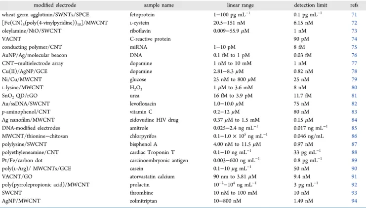
The enhanced effect of CNT inclusion in modified voltammetric biosensors (e.g., enzyme-based biosensors, Table 1. Recent High-Performance Reports Utilizing CNT-Based Biosensors for the Detection of Various Enzymatic or Nonenzymatic Biomolecules
modified electrode sample name linear range detection limit refs
wheat germ agglutinin/SWNTs/SPCE fetoprotein 1−100 pg mL−1 0.1 pg mL−1 71
[Fe(CN)5(poly(4-vinylpyridine))10]/MWCNT L-cystein 20.5−151 nM 6.15 nM 72
oleylamine/NiO/SWCNT riboflavin 0.009−55.9μM 1 nM 73
VACNT C-reactive protein 90 pM 74
conducting polymer/CNT miRNA 1−10 pM 8 fM 75
AuNP/Ag/molecular beacon DNA 0.1 fM to 1 pM 0.03 fM 76
CNT−multielectrode array dopamine 1 nM to 10 mM 1 nM 77
Cu(II)/AgNP/GCE dopamine 2.81−8.3μM 0.82 nM 78
Ni/Cu/MWCNT glucose 25 nM to 800μM 25 nM 79
L-lysine/MWCNT H2O2 1μM to 3.6 mM 8 nM 80
SnO2QD/rGO urea 16 fM to 3.9 pM 11.7 fM 81
Au/ssDNA/SWCNT levofloxacin 1.0−10.0μM 75 nM 82
p-aminophenol/CNT vitamin C 0.2−12μM 80 nM 83
Ag nanofilm/MWCNT zidovudine HIV drug 0.37μM to 1.5 mM 0.15μM 84
DNA-modified electrodes amitrole 0.025−2.4 ng mL−1 0.017 ng mL−1 85
MWCNT/thionine−chitosan chlorpyrifos 0.1−1.0×10⁵ng mL−1 0.046 ng/mL 86
polylysine/SWCNT bisphenol A 4.00 nM to 11.5μM 0.97 nM 87
polyethyleneamine/CNT cardiac Troponin T 0.1−10 ng mL−1 33 pg mL−1 88
Pt/Fe/carbon dot carcinoembryonic antigen 0.003−600 ng mL−1 0.8 pg mL−1 89
poly(L-Arg)/ MWCNTs/GCE casein 0.1−10μg mL−1 50 nM 90
VACNT/GO atorvastatin calcium 90 nm to 3.81μM 9.4 nM 91
poly(pyrrolepropionic acid)/MWCNT prolactin 10−2−104ng mL−1 3 pg mL−1 92
SWCNT thrombine 10 nM to 100 mM 10 nM 93
AgNP/MWCNT zolmitriptan 10−800 nM 1.49 nM 94
Table 2. Recent High-Performance Reports Utilizing Graphene-Based Biosensors for the Detection of Various Enzymatic or Nonenzymatic Biomolecules
modified electrode sample name linear range detection limit refs
CuO NP−graphene glucose 0.5−2000μM 0.09μM 95
3D graphene dopamine 0.1−200μM 19.4 nM 96
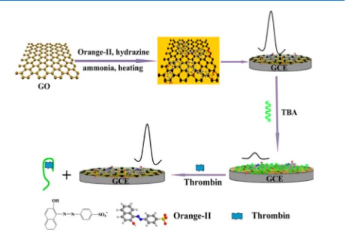
graphene−Orange II thrombin 1.0−400 pM 0.35 pm 97
graphene−Orange II lysozyme 5.0−700 pM 1.0 pm 97
graphene−Au cysteine 0.1−24μM 20.5 nM 98
Au-polydopamine−thionine−GO fetoprotein 0.1−150 ng mL−1 30 pg mL−1 99
graphene ovalbumin 1 pg mL−1to 0.5μg mL−1 0.83 pg mL−1 100
pErGO uric acid 0.1−10μM 50 nM 101
cobalt oxide/rGO hydrogen peroxide 5μM to 1 mM 0.2μM 102
Au/GO DNA 1.0 fM to 0.1μM 0.35 fM 103
Gr/CuPc/PANI vitamin C 0.5−12μM 63 nM 104
carbon/Co2+-Y vitamin B2 1.7−34μM 0.71μM 105
carbon/ZrO2/ionic liquids vitamin B6 0.8−550μM 0.1μM 106
graphene−hydroxyapatite luteolin 0.02−10μM 10 nM 107
TrGO baicalein 10 nM to 10μM 6.0 nM 108
rGO−tetraethylene pentamine carcinoembryonic antigen 0.05−20 ng mL−1 13 pg mL−1 109 rGO−tetraethylene pentamine squamous cell carcinoma antigen 0.03−20 ng mL−1 10 pg mL−1 109
ACS Nano
immunosensors, and nucleic acid biosensors) is believed to be due to the strong π−π interactions between CNT and the immobilized host. This immobilization has the added benefit of inhibiting CNT aggregation in solution, which is a common problem encountered when working with CNTs. Additionally, noncovalent modification does not disturb the electronic structure of CNTs, and as such, the CNT maintains its electrical conductivity. These two factors mentioned above contribute to the low detection limits that are found when utilizing CNT-based biosensors. Lastly, CNT hybrid materials (i.e., CNTs combined with conducting polymers, redox mediators, or metal NPs) have been receiving increased attention in biosensors. This is due to the various synergistic effects observed when these hybrid materials, such as the enzymeless biosensors which afford greater durability and are less expensive, are used.
Another very common carbon material recently utilized in biosensors is graphene in its various forms (i.e., rGO, GO, CVD graphene, etc.). As for CNTs, pristine graphene can be noncovalently functionalized through strong π−π interactions between its surface and the immobilized host. Additionally, rGO offers additional covalent functionalization options due to multiple oxygenated functional groups. Although rGO is less conductive than pristine graphene, it maintains a large degree of conductivity, compared to the insulating GO, as large domains on its basal plane are reduced, thereby forming a π-electron- conjugated system similar to graphene. For these reasons, graphene and chemical derivatives of graphene are emerging as a preferred choice for the fabrication of various biosensors.
Furthermore, various polymers, noble metals, metal oxides, spinels, and metal complexes have been used as effective modifiers in conjunction with graphene-based materials for sensing biomolecules. Tables 1 and 2 list recent results for CNT- and graphene-based enzymatic and nonenzymatic biosensors.
In the following sections, the CNT- and graphene-based applications for electrochemical biosensors are reviewed.
Glucose Sensing. Diabetes is a chronic condition which leads to elevated blood glucose levels, which if left untreated can lead to major health problems and/or death. For this reason, reliable and accurate glucose detection in blood is of critical importance. Additionally, glucose sensing is important in food and textile industries, as well as environmental monitoring.110,111 Usually, glucose sensing relies on the use of glucose oxidase (GOx), which catalyzes glucose in the presence of O2 to afford gluconic acid and H2O2.112,113 The concentration of glucose is then determined by monitoring the number of electrons flowing through the enzyme, or the concentration of the as formed H2O2 is determined quantitatively as an indicator of the amount of glucose present in the solution.
As enzymes such as GOx lose activity with changes in temperature or pH, there is a lot of research interest in the development of a cheap, sensitive, and interference-free sensors for nonenzymatic glucose detection.114−116 In this vein, Mohamedi and colleagues showed that gold nanostructured layers deposited by pulsed laser onto CNTs could be used as a nonenzymatic electrochemical glucose biosensor, LOD 0.1 mM with a sensitivity of 25μA cm−2mM−1.115They showed it by adjusting the deposition vacuum level and the number of pulses that the electroactive surface area (max 6.55 cm2for 10000 laser pulses) and roughness factor could be effectively controlled.
Additionally, they found that the as-formed gold nanostructures
contributed toward a lower onset potential for glucose oxidation.
It is evident from this study, among many others, that the CNTs afforded the sensors with unique electrochemical behavior by changing the morphology of the deposited NPs.
This method of depositing metal particles as isolated NPs or nanostructures with different morphologies onto CNTs offers one of best strategies to develop efficient voltammetric biosensors. Similarly, graphene-based voltammetric biosensors have also been applied in glucose sensing as graphene materials provide a large surface area and unique electrochemical behavior.117
Glucose Oxidase−Carbon-Nanotube-Based Sensors. Ra- mesh and colleagues have shown that the sensitivity and LOD of a glassy carbon electrode (GCE) toward glucose can be improved by modifying the GCE with SWNTs dispersed in a polymer matrix (polyethylenimine, polyethylene glycol, or polypyrrole) and then a layer of GOx.118These polymer layers are beneficial to the enzyme as they provide not only binding places but also stability. Importantly, the authors showed that the high purity and large surface area of the SWNTs was of critical importance to the response of the electrode. The use of high-purity SWNTs resulted in a high conductivity, enzyme stability, and fast electron transfer rate. The response time of the electrodes was shown to be less than 5 s with a LOD of 0.2633 μM. You and co-workers have shown that N-doped carbon nanofibers (NCNF) prepared from electrospun polyacrylonitrile fibers have a large electrocatalytic activity toward the oxygen reduction reaction (ORR).119This activity is believed to be due to the presence of abundant defective sites and high pyrrolic-N content in the NCNF. These NCNFfilms coated with GOx and Nafion showed high sensitivity (LOD of 0.6 mM), stability, and selectivity toward glucose. Additionally, this GOx/NCNF/GCE electrode exhibited successful detection of glucose in the presence of commonly existing interfering species such as ascorbic acid (AA), uric acid (UA), and dopamine (1 mM). This work shows that the doping of SWNTs and other carbon nanomaterials with N can be beneficial in increasing the selectivity and sensitivity of glucose biosensors.
Layer-by-layer self-assembly methods have been applied in the fabrication of GOx-containing glucose biosensors, as these layers provide enzyme stability. Importantly, when these self- assembly layers contain either MWNTs or SWNTs, the current response and electrical conductivity between the electrode and the GOx is improved.66,120As such, this allows for an increased sensitivity and stability of the GOx. For example, Wanget al.
showed that the glucose oxidation current of a gold electrode modified by layer-by-layer self-assembly of (poly- [(vinylpyridine)Os(bipyridyl)2Cl2+/3+] and GOx SWNTs in- creases 17 times.120
In another study, Wu and colleagues showed that layered multilayers of poly(allylamine),N-hydroxysuccinimide-oxidized MWCNTs, cysteamine, gold NPs, and GOx on a platinum electrode could be employed as an amperometric biosensor.69 The authors showed that the biosensor exhibited a large linear range for glucose detection (0.1−10 mM) and a LOD of 6.7 μM. Additionally, the sensor displayed long-term stability (92%
current response retention after 1 month) and was not influenced by the addition of AA, UA, and acetaminophen.
Porterfield et al.have shown that GOx and SWNTs can be assembled in a layer-by-layer method by first dispersing the SWNTs in single-stranded DNA (Figure 3A).121 In this way, ACS Nano
self-assembly can proceed by electrostatic interactions between the negatively charged DNA-coated SWNTs, which was then coated with a thin layer of cationic poly(ethylenimine) and finally negatively charged GOx. This material showed an impressive linear range up to∼9.4 mM, with minimal deviation at low concentrations (Figure 3B−D). This biosensors stability and enhanced sensitivity is believed to be due to the electrostatic interactions between the positive and negative layers, as well as the large amount of GOx included in the sensor. Additionally, it can be seen in Figure 3B−D that this biosensor response was not affected by spiking with either phosphate-buffered saline solution (PBS), AA, UA, and acetaminophen.
Recently, Kwon et al. immobilized GOx on CNTs (GOx/
CNT) to sense glucose, with the authors showing that a larger CNT content contributes to an improved amperometric response.122This modified sensor shows increased sensitivity (53.5 μA mM−1 cm−2), glucose activity (86% activity maintained after 2 weeks), and large electron transfer rate constant (1.14 s−1). Interestingly, the Wei group has confirmed an earlier study that CNTs simultaneously modified with GOx and its cofactor, flavine adenine dinucleotide (FAD) show electron transfer kinetics similar to those observed in isolated FAD.123 This implies that the FAD is directly wired to the CNT, and as such, electron transfer is unimpeded between the cofactor and support structure, allowing for increased sensitivity. Importantly, as the FAD is itself embedded in the GOx, this in turn allows rapid electron transfer.123
Enzymeless Carbon-Nanotube-Based Sensors.Enzymeless glucose biosensors which rely on NPs for the detection of glucose can similarly be enhanced through the introduction of CNTs.124−129For example, Lin and co-workers79have shown that the sequential electrodeposition of Ni and CuNPs on a MWCNT-modified GCE (Ni/Cu/MWCNT/GCE) can in- crease the amperometric response of the biosensor by 2.5−20 times compared to either Ni/GCE, Cu/GCE, Ni/MWCNT/
GCE, or Cu/MWCNT/GCE. This sensor exhibits two linear response ranges both in the low (25 nM to 0.8 mM) and high
(2−8 mM) concentration regions, with an impressive LOD of 25 nM. Importantly, when this sensor was applied to glucose detection in human blood serum, it exhibited a recovery rate of
≥95%. This result indicates that the sensor is able to avoid interference from other molecules and could be applied in practical applications.
Choiet al.synthesized a CNT−Ni hybrid using atomic layer and chemical vapor deposition of Ni on oxygen- or bromine- functionalized CNT surfaces.130 The as-fabricated sensor exhibited a wide linear response window (5 μM to 2 mM), short response time (3 s), small LOD (2μM), high sensitivity (1384.1μA mM−1cm−2), good selectivity, and reproducibility in alkaline media. Alizadehaet al.131reported the fabrication of a highly sensitive enzyme-free amperometric glucose sensor by codeposition of copper oxide NPs/multiwalled CNTs onto the surface of a GCE. At the optimized potential, the LOD, linear range, and sensitivity of the sensor were calculated as 0.07 (±0.03)μmol L−1, 0.5−2000.0μmol L−1, and 3968.42 (±0.84) μA L mmol−1 cm−2, respectively. The optimized sensor was used to detect glucose in blood samples, with results that are comparable to that of a commercial enzymatic sensor.
It has been reported that a MWCNT and GO hybrid material deposited on a GCE can be used as a scaffold to electrodeposit Ni(OH)2 NPs which act as a glucose biosensor.132 Before the deposition of the NiOH2 NPs, the GO is electrochemically reduced (ErGO). This electro- reduction leads to an increase in conductivity in the sensor, while the MWNTs act as conducting bridges to enhance electron transfer between independent ErGO sheets and the GCE. While this sensor exhibits a LOD of 2.7 μM, it is important to note that it exhibits a selective response to glucose in the analysis of a human urine sample.
Glucose Oxidase−Graphene-Based Sensors. GOx cova- lently attached to either ErGO or GO deposited on a GCE has been demonstrated as a biosensor for the detection of glucose in PBS.133,134The advantage of using graphene-based materials such as GO is that GO has been shown to be biocompatible while providing carboxylic oxygen functionalities for the GOx Figure 3. (A) Schematic diagram of layer-by-layer electrostatic self-assembly of single-stranded DNA (ssDNA)−SWNT, polyethylenimine (PEI), and GOx on a Pt/Ir electrode (inset: structural closeup of ssDNA−SWNT). (B) Amperometric response of the GOx/
polyethylenimine/single-stranded DNA−SWNT/Pt biosensor at +500 mV (vsAg/AgCl) upon successive addition of glucose solution to 20 mL of pH 7.0 PBS stirred at 350 rpm. (C) Calibration curve of amperometric response toward glucose concentration variation. (D) Amperometric responses to PBS, ascorbic acid (0.1 mM AA), uric acid (0.1 mM UA), acetaminophen (0.1 mM AP), and 1 mM glucose.
Adapted from ref121. Copyright 2014 American Chemical Society.
ACS Nano
amino groups to covalently attach.134,135 On the other hand, the use of rGO allows for an enhanced conductivity while still providing attachment sites for the GOx amino groups due to the partially reduced nature of GO.136,137
To improve the GOx biosensing ability, Teymourian et al.
have shown that GOx can be immobilized on a rGO/Fe3O4 magnetic NP-modified GCE.138The authors showed that this biosensor has a LOD of 0.05 mM, a linear sensing range of 0.5−12 mM, and a response time of ∼6 s. Importantly, the authors demonstrated that the rGO/Fe3O4-modified GCE readily immobilizes biomolecules, such as human immunoglo- bulin E, making this a readily applicable biosensing material.
The use of graphene/NP composite materials in glucose biosensors has been further demonstrated by Bai and Shiu, who showed that a rGO/AuNP-modified GCE can be further modified to detect glucose when a layer of chitosan/GOx is coated onto the rGO/Au surface.139 The fabricated sensor exhibits an impressive LOD of 76 μM, with a retention of sensitivity for 0.1 mM of glucose after 36 days of >70% of the original sensitivity.
The carboxylic acid groups on the edges of GO and the amino groups of GOx can covalently link through peptide bonding. Utilizing this phenomenon, Hasanet al.showed that a borosilicate glass capillary coated with GO/GOx can be inserted into the intracellular environment of a single human cell to detect glucose. This sensor exhibited a linear electrochemical potential difference over a glucose concen- tration range of 10−1000 mM.140Chiaet al.reported that an electrochemical glucose biosensor can be fabricated by immobilizing GOx on exfoliated pristine graphene through noncovalentπ−πinteractions. As for CNTs,123this interaction results in enhanced electron transfer kinetics between the FAD redox sites of GOx at the modified electrode surface.141 As such, the modified nonfunctionalized pristine graphene- containing biosensor resulted in enhanced stability, reproduci- bility, and selectivity for glucose detection in comparison to unmodified electrodes. These examples show that covalent bonding is not essential for protein adhesion to electrode surfaces.
Chitosan (CS), a polysaccharide with plentiful amino groups, has a pKa value of approximately 6.3 and displays pH- dependent solubility. As such, CS provides a matrix for immobilization of enzymes and nanomaterials, which make CS a promising material to modify electrochemical sensors.
Keeping this in mind, Fang et al. immobilized GOx onto a biocompatible CS−rGO−AuNP hybrid-modified Pt elec- trode.142 The sensor showed electrochemical detection of glucose over a wide linear range of 15μM to 2.13 mM, with a sensitivity of 102.4 μA mM−1 cm−2 and LOD of 1.7 μm.
Similarly, Yeet al. reported that immobilized GOx on a CS/
rGO/Au hybrid displays fast electron transfer at a working voltage of−0.45 V (vsAg/AgCl).143The sensor gives a linear response to glucose in the 0.05 to 1.2 mM concentration range, with a sensitivity of 13.58μA mM−1cm−2and a 0.52μM LOD.
Enzymeless Graphene-Based Sensors. An enzymeless glucose biosensor based on Ni−Co nanostructures electro- deposited by dynamic potential scanning on a rGO-modified GCE has been prepared.144 The sensor exhibited a LOD of 3.79μM and a linear glucose detection range of 10μM to 2.65 mM. With respect to selectivity, this sensor exhibited no noticeable amperometric change on the addition of simple cations and anions such as Fe3+, Fe2+, SO42−, BrO3−, IO3−, NO2−, NO3−, and Cl−. In contrast, the addition of biomolecules
such as fructose, D-galactose, and AA resulted in a slight amperometric response (<10%), while the addition of UA showed no interference. This amperometric response, while small, is an issue that needs to be addressed for successful application of these types of nanostructures in biosensors. In a similar manner, Szuneritset al.showed that electrodeposition of Ni(OH)2 on a rGO-modified GCE can be used as a glucose biosensor.145 The authors showed that this electrode exhibits negligible amperometric response on the addition of UA, AA, and dopamine, as well as a LOD of 15μM. This result is similar to that obtained using the enzymeless Ni(OH)2/MWCNT biosensor,146which would indicate that Ni(OH)2is a material that warrants further investigation in the development of enzymeless glucose biosensors. The authors of this study also noted that the electrofabrication method leads to high reproducibility and stability of the biosensor compared to drop-casting methods. Utilizing a galvanic replacement fabrication method, Chen and co-workers were able to prepare a hollow Pt−Ni-rGO hybrid-functionalized GCE for glucose detection, which displayed enhanced selectivity (no response to AA, UA, 3,4-dihydroxyphenylacetic acid), sensitivity (LOD of
∼2μM), and stability (93% response after 1 month).146This material was also applied in the determination of glucose in human blood serum, with an accuracy that is comparable to that of commercially available sensors.
In multiple biosensors, the active sites are adhered to the working electrode through the use of a conductive polymeric binder, which is not active for glucose sensing. However, with the use of graphene-based materials, this can be avoided, as has been demonstrated by Alizadehet al., who showed that a rGO/
CuO NP-modified GCE electrode can be used as a glucose biosensor without adding Nafion as a binder.95This electrode showed good selectivity for glucose over other sugars as well as long-term stability (95% response after 30 days).
Tianet al.reported the microwave-assisted synthesis of CuO NPs on sulfur-doped graphene (SG) as an electrode material for nonenzymatic glucose detection with a LOD of 80 nM.147 The improved GOx sensing performance of the CuO NPs deposited on SG compared to rGO is attributed to conductivity of SG, enhanced electron transfer between the Cu−S interaction, and increased surface area. Other biosensors based on graphene, functionalized with metal oxides like Mn3O4and Co3O4, have been employed in the sensitive and selective detection of glucose.148,149
Zhang et al. have shown that the bimetal oxide CuNiO decorated on graphene sheets can be utilized in the highly stable and sensitive enzymeless sensing of glucose with a LOD of 16 μM.150 In another example of bimetal oxide-based catalysts, Dhara et al. used PdCuO deposited on rGO as a nonenzymatic glucose sensor with a LOD of 30 nM.151 Although, metal NPs are research targets for enzymeless glucose sensing due to their high specific surface area, excellent conductivity, and catalytic activity, it should be noted that these particles are highly prone toward agglomeration, which results in a decrease in catalytic activity. Taking this fact into consideration, many groups have dispersed NPs over graphene to reduce the NP aggregation. This has allowed nonenzymatic glucose sensors to possess rapid response, good stability, selectivity, and low LODs.152−154
In most reported cases of enzymeless glucose biosensing mentioned above, a NaOH solution (pH 13) is required to catalyze the oxidation of the metal center which then catalyzes the reduction of the glucose to gluconolactone.95,144As such, ACS Nano
when human samples, such as blood or urine, are tested using these biosensors, the biological samples have been diluted in a NaOH solution.
Dopamine, Uric Acid, and Ascorbic Acid Sensing.The quantification of dopamine and its metabolic derivatives is important because of the role played by these substances in mammalian central nervous systems. For example, low concentrations and inactivity of dopamine functioning in the central nervous system may lead to Parkinson’s disease, while on the other hand, an elevated dopamine level has been linked to schizophrenia.155
Various sensing methods, mainly based on the chemical modification of working electrode materials, have been developed for detection of dopamine. In this regard, CNTs and graphene nanosheets have been widely used to modify the working electrode surfaces for the analysis of dopamine. The use of these carbon materials is highly desired, as they provide enhanced electrical conductivity, large specific surface area, and chemical stability.
UA (2,6,8-trihydroxypurine) is the end product of purine (an essential component of DNA and RNA) metabolism in humans. An abnormally high UA level has been associated with multiple diseases such as gout, hyperuricemia, leukemia, pneumonia, Lesch-Nyhan syndrome, etc.156 For this reason, monitoring the levels of UA is of great importance. Generally, UA biosensors are based on the oxidation of UA by uricase in the presence of O2to produce H2O2, allantonin, and CO2.157 This chemical reaction can be expressed as
+ + ⎯⎯⎯⎯⎯→ + +
uric acid H O2 O2 uricase allantonin H O2 2 CO2 According to Noroozifar, the two most promising UA detection techniques, that is, oxidation of UA using phosphortungstate or uricase, have the disadvantage of long reaction times, large LODs, and high operational cost.158Therefore, it is clinically important to develop sensitive and effective sensing techniques for analysis of UA. UA is a voltammetrically active chemical, and as such, voltammetric biosensors based on uricase enzyme- modified electrodes are an extremely attractive means of detecting UA.159Additionally, the use of enzymeless electrodes, such as NP-modified electrodes, for the detection of UA is highly desired, as these methods hold many advantages, such as simplicity, ease of miniaturization, high selectivity, and low cost.
Considerable attention has been devoted in recent years to the voltammetric determination of UA by CNT and graphene- modified electrodes.158−163 In comparison to CNT-modified electrodes, graphene, GO, and rGO have been much more successfully explored as biosensors. This is believed to be due to graphene’s 2D π-conjugated structure, which makes its electronic structure very sensitive to the local chemical environment.
Carbon Nanotubes. Ardakani et al. have shown that a CNT/graphite paste electrode modified with [1,1′-binaphtha- lene]-4,4′-diol can be employed in the sensitive and simultaneous detection of dopamine, folic acid (FA), and UA.164The addition of the [1,1′-binaphthalene]-4,4′-diol into the paste electrode increases the sensitivity of the electrode and allows for LODs of 0.49, 4.28, and 7.69 mM for dopamine, UA, and FA, respectively. The electrode exhibited negligible amperometric response toward 100 mM (Na+, Cl−, K+), 80 mM (Mg2+, Ca2+), and 6 mM (L-lysine, glucose, glutamic acid, glycine, L-cystine, L-cysteine, acetaminophen, nicotinamide adenine dinucleotide (NADH)) spiking into a 0.1 mM
dopamine solution. The precursor and metabolites of dopamine (i.e., levodopa, epinephrine, and norepinephrine) interfered with the detection of dopamine, showing the general applicability of this sensor toward dopamine and its metabolites. In another study, this same group showed that in a similar way the CNT/graphite paste can be modified with t h e S c h iff b a s e , 2 , 2′- [ 1 , 4 - p h e n y l e n e d i y l b i s - (nitrilomethylidene)]bis(4-hydroxyphenol) to afford similar simultaneous and sensitive results for dopamine, FA, and UA.165 Interestingly, this electrode could also be used to simultaneously determine dopamine and acetaminophen, which differs from the previous result and shows the applicability of modification with the Schiffbase.
The selective detection of dopamine is challenging in biological samples, as its oxidation potential range is very close to those of the commonly interfering analytes AA and UA.166,167Utilizing a GCE modified with ferrocene (Fc)-filled double-walled CNTs (Fc@DWNTs), Liet al.have shown that dopamine sensing can be achieved.168In this study, the authors noted, however, that the sensitivity and the LOD (0.3μM) was not as high as that reported by other groups. However, by building on their previous study, this group has shown that a Fc-filled SWCNT (Fc@SWNTs)-modified GCE was far more responsive and sensitive (LOD, 0.1μM).169This is believed to be due to the mechanism of electro-oxidation and reduction, which is bidirectional (dopamine oxidation and reduction) for the Fc@SWNTs, while it is unidirectional (only dopamine oxidation) for the Fc@DWNTs. This difference in electro- activity is believed to be due to the dopamine not entering the DWNTs, while in contrast, it can enter the SWNTs, which is only one carbon layer thick and contains more defects.
The Vasantha group have reported on the sonochemical synthesis of a nickel tetrasulfonated phthalocyanine (NiTsPc)- functionalized multiwalled CNT hybrid, where the NiTsPc acts as a dispersing agent for the MWCNTs.170The authors showed that a GCE coated with this MWCNT−NiTsPc hybrid can be used for monitoring dopamine in the presence of UA and AA.
This sensor displayed a linear response range from 20 nM to 1.384 mM with a LOD of 1 nM.
By electrodepositing NiO NPs on a CNT/dihexadecylphos- phatefilm-modified GCE, Filho and colleagues have shown that it is possible to simultaneously determine dopamine and epinephrine.171The authors found this simultaneous detection to be possible due to the difference between the two analyte reduction peaks of ∼360 mV, which is in contrast to what is normally found in the detection of dopamine and its metabolites.164The LOD for dopamine and epinephrine were determined to be 50 and 82 nM, respectively. It should also be noted that this biosensor was successfully applied in the detection of dopamine and epinephrine in cerebrospinalfluid, human blood serum, and lungfluid.
The Anandan et al. group reported on the simultaneous detection of dopamine and UA by modification of a GCE with a silicate (N-[3(trimethoxysilyl)propyl]ethylenediamine;
EDAS) network which interlinks gold NPs (3−8 nm) and MWCNTs.172This sensor exhibited a wide linear response for AA and dopamine over the concentration range of 1×10⁻⁷to 9×10⁻⁶M and 1×10⁻⁷to 8×10⁻⁶M with LODs of 0.07 and 0.08 μM, respectively. Tsierkezos et al. reported the simultaneous detection of dopamine, UA, and AA by modifying a GCE with boron-doped MWCNTs (B-MWCNTs). This sensor exhibited dopamine, UA, and AA detection at working ACS Nano
potentials of ∼0.267, ∼ 0.412, and ∼0.127 V with LODs of 0.11, 0.65, and 1.21μM, respectively.173
A multielectrode array (MEA) dopamine sensing chip has been created by electroplating CNTs onto an indium−tin oxide MEA.77This sensor was used to sense dopamine (LOD, 1 nM) release from mouse striatal brain slices (coronal and sagittal slices), and these results corresponded with the expected results found in other studies using carbon fiber electrodes (Figure 4).174,175This MEA was further applied to determine real-time
sensing in hippocampal neuronal cultures and hippocampal slices by monitoring the action potentials andfield postsynaptic potentials. The results from these experiments show that this MEA can be applied in real-time sensing, and that the probe is noninvasive as neuronal cells cultured on these MEAs survived for more than 1 month.
Ionic liquids have been shown to be biocompatible toward biomolecules, as well as capable of enhancing their bioactivity.176,177 In this vain, Noroozifar and colleagues showed that by utilizing the ionic liquid 3-hydroxypropanami- nium acetate together with a nanosized zeolite and MWCNT paste the simultaneous detection of UA and dopamine could be achieved.178Utilizing differential pulse voltammetry (DPV), the authors showed that the linear ranges of detection were 0.812 to 301 μM (LOD, 0.116 μM) and 0.931 to 336 μM (LOD, 0.133 μM) for dopamine and UA, respectively. These biosensors were used to analyze human urine and blood serum samples, with recovery rates of ∼100% across all samples, indicating the potential use of this material in commercial applications.
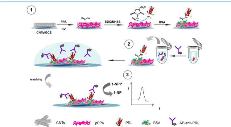
Besides the enzymeless detection of dopamine detailed above, the use of enzymes in conjunction with CNTs has been employed in the determination of dopamine. In this manner, Pereiraet al.immobilized horseradish peroxidase from zucchini (Cucurbita pepo L.) directly onto MWCNTs using covalent bonding.78These functionalized MWCNTs were then mixed with graphite and paraffin oil to create a dopamine biosensor.
The biosensor was successfully applied in the detection of dopamine in pharmaceutical formulations with an accuracy equivalent to high-performance liquid chromatography
(HPLC). Additionally, it was shown that this biosensor performance is not compromised in the presence of AA and UA.
Noroozifar et al. fabricated a enzymeless holmium fluoride (HoF3) NP/MWCNT-functionalized GCE for the sensitive determination of UA in the presence of interfering analytes AA and dopamine.158Comparison of the electrocatalytic activity of the GCE/MWCNT/HoF3 NPs and GCE/MWCNT sensors showed that HoF3NPs are the dominating participator in the electrocatalytic activity of UA in the presence of AA and dopamine. The fabricated sensor exhibited a linear response range from 0.2 to 500μM with a LOD of 0.16μM. To confirm the practical application of the sensor, the electrode was successfully employed in the detection of UA in human serum and urine samples with recovery rates of 95.5 and 102.2%, respectively.
Liuet al.reported the nonenzymatic sensing of UA using a CNT ionic liquid paste electrode that was modifiedin situwith electropolymerized poly(β-cyclodextrin) (β-CD).179 The as- fabricated working electrode shows a linear response range from 0.6 to 400μM with a LOD of 0.3μM in the presence of ascorbic acid, with the selective response of this sensor being due to host−guest recognition between β-CD and UA.
Recently, the Mascarenhas group reported the selective detection of UA in the presence of ascorbic acid, dopamine, andL-tyrosine by utilizing FeNPs coated on CNTs.180Under optimized experimental conditions, the differential pulse voltammetry curve displayed two linear concentration ranges for UA of 7.0×10⁻⁸to 1.0×10⁻⁶M and 2.0×10⁻⁶to 1.0× 10⁻⁵ M with a LOD of (4.80 ± 0.35) × 10−8 M. To demonstrate the practical applicability, the electrode was successfully applied to the determination of UA in (spiked) human urine samples with good recovery rates.
Numnuamet al.fabricated an amperometric UA biosensor by immobilizing uricase on an electrospun nanocomposite of a chitosan−CNT nanofiber covering an electrodeposited layer of AgNPs on a Au electrode.160 The basis of the UA detection method was to monitor the change in the reduction current for the dissolved O2, which forms during oxidation of uric acid by the immobilized uricase. This biosensor’s ability to detect UA (LOD, 1 μM) was not affected by the introduction of AA, glucose, and lactic acid. This biosensor was also successfully applied in the determination of UA in human serum samples, with a sensitivity equivalent to that obtained using enzymatic colorimetric detection.
Graphene/Carbon Nanotube Composites. One of the problems associated with the application of CNTs in sensing is the fouling that takes place. With the accumulation of oxidation products on the surface of the CNTs (fouling), the sensitivity and LOD of the biosensor can be altered negatively.
For this reason, CNTs are often incorporated with other nanomaterials to prevent fouling.181−183This section deals with the use of CNT/graphene materials in dopamine, UA, and AA biosensors,184which have the added benefit of antifouling on the CNTs through incorporation of the graphene.
Chen and colleagues have shown that dopamine and acetaminophen can be simultaneously determined using a MWCNT/GO composite-modified GCE.185 The composite material is formed simply through π−π interactions between the GO and the MWCNT by sonication of the two components together. The LODs for the biosensor were 22 and 47 mM for dopamine and acetaminophen, respectively.
Importantly, the authors showed that the composite material Figure 4. Real-time measurements of dopamine release from mouse
striatal brain slices. (A) Mouse striatal slice cut in the coronal plane and mounted on a 4 × 104 μm2 CNT plated indium−tin oxide MEA. (B) Amperometric responses to dopamine discharge from a coronal striatal slice at +0.3 V (black) or 0 V (gray). (C) Superimposed waveforms of dopamine responses from coronal (red) and a sagittal section (blue). (D) Mean half-decay time (t1/2) for coronal and sagittal sections (n= 6). Adapted with permission from ref77. Copyright 2013 Elsevier B.V.
ACS Nano
out-performed both the GO-modified GCE and the MWCNT- modified GCE, while interference studies demonstrated that UA, AA, and NADH have negligible effect on the simultaneous sensing ability of the biosensor. Additionally, the biosensor proved to be stable over 1 week (91.3% signal retention) as well as successive measurements (∼3% standard deviation). In a related study, it has been shown that a MWCNT-bridged mesocellular graphene foam, nanocomposite-modified GCE can be used to simultaneously determine AA, dopamine, UA, and tryptophan.186The authors of this study showed that this biosensor exhibited a far greater electrochemical response in terms of selectivity and catalytic activity compared to GCEs modified with mesocellular graphene foam, MWNTs, or MWNT/GS.
To further enhance the sensing ability of a GCE modified with a GO/MWCNT composite, Yang et al.have shown that including cetyltrimethylammonium bromide (CTAB) in the composite allows for the selective and sensitive simultaneous detection of dopamine (LOD, 1.0μM), AA (LOD, 1.5 μM), UA (LOD, 1.0μM), and nitrite (LOD, 1.5 μM).187 Perhaps, this enhanced electrochemical activity can be attributed to the highly porous 3D nanohybrid structure that the CTAB−GO/
MWNT composite forms, which offers more active sites for dopamine oxidation, compared to the smooth packed surface of CTAB−GO/GCE.
Using a PtNP-coated graphene−CNT (Pt−G−CNT) hybrid, Ramakrishnanet al.demonstrated a high electrocatalytic activity toward the oxidation of AA, dopamine, and UA in 0.1 M phosphate buffer solution (pH 7.0).188 Under optimal conditions, the authors reported simultaneous detection of AA, dopamine, and UA, in the linear concentration ranges of 200− 900, 0.2−30, and 0.1−50 mM, respectively, with LODs of 0.186 (AA), 9.199 (dopamine), and 9.386 mA mM−1 cm−2 (UA).
The fabricated sensor was also used in the simultaneous detection of the three biomolecules in a vitamin C tablet solution, human serum, and urine. In another study, the Yuan group reported the simultaneous determination of dopamine, AA, and UA using a MWCNT/rGO hybrid functionalized with PAMAM and AuNPs (rGO−PAMAM−MWCNT−AuNPs).189 This hybrid material exhibited large electrocatalytic activities, which translated into linear response ranges for the determination of AA, DA, and UA in mixed analytes systems of 20 to 1.8 mM (LOD 6.7 mM), 10 to 0.32 mM (LOD 3.3 mM), and 1 to 0.114 mM (LOD 0.33 mM), respectively.
Graphene. The use of graphene-based electrodes in the detection of dopamine has been widely reported.190−194 Depending on the type of graphene material employed, the electroactivity is enhanced in differing ways. For example, it is believed that in graphene/rGO-based materials the electro- activity toward dopamine is enhanced by the large surface area and superior conductivity.
Two separate groups have shown that a GCE190or a carbon fiber electrode191can be modified through the electrochemical reduction of GO (ErGO) onto their surface. These electrodes were then used in the simultaneous detection of AU, UA, and dopamine. In both of these studies, the ErGO-modified electrode showed a separation of three analyte peaks, while a broad merged peak with a lower response was observed for the unmodified surface. The LODs for the ErGO/carbon fiber electrode were determined to be 4.5 (AA), 0.77 (dopamine), and 2.23 μM (UA), while for the ErGO/GCE, they were 0.3 (AA), 0.5 (dopamine), and 0.5 μM (UA). In a related study, Nancyet al.showed that solar reduced GO could be used to modify a GCE without the addition of a binder.192As with the previously mentioned studies, this electrode could be used in the simultaneous oxidation and detection of AU, UA, and dopamine; however, the authors only reported the LOD for dopamine (2.8μM). It is important to note that although this biosensor is believed to be stable under electrochemical detection conditions, no long-term stability studies were conducted.
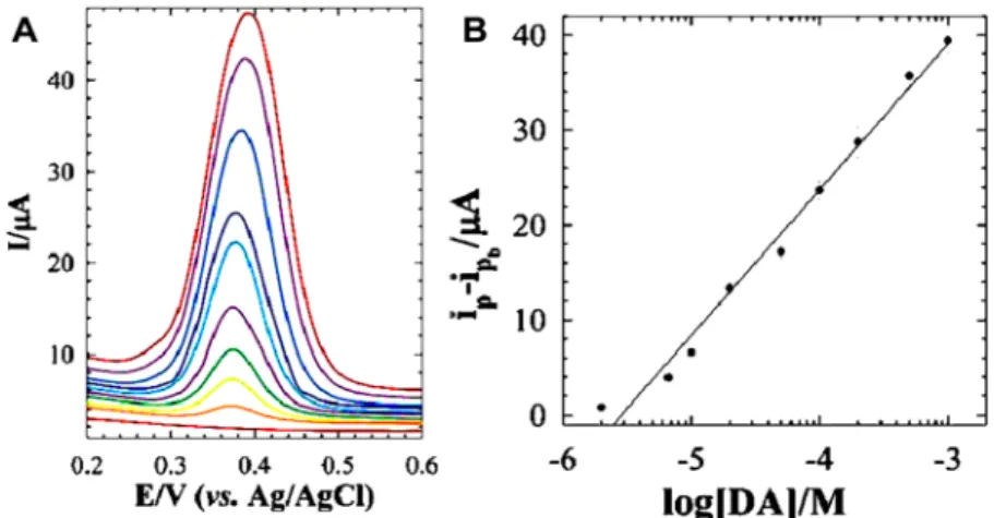
In a similar way, Gao and co-workers have shown that a GO- modified GCE can be used in the highly sensitive and selective detection of dopamine in the presence of AA.193The dopamine LOD for the electrode was determined to be 0.27 μM, while the addition of AA does not affect the signal. It is believed that electrostatic repulsion between the GO and AA makes oxidation of the AA at the electrode surface impossible. In contrast, theπ−πstacking and electrostatic attraction between the dopamine and GO allows for facile oxidation to take place.
The different electrochemical responses of dopamine and ascorbic acid on the working electrode are shown inFigure 5.
To overcome the selectivity problems occurring with the introduction of analytes which interfere with the dopamine signal, Bagherzadeh and Heydari showed that by modifying a carbon paste electrode with graphene nanosheets dopamine could be preconcentrated on the electrodes surface.194 This preconcentration method allows for the sensitive (LOD, 8.5 Figure 5. Difference in the electrochemistry of dopamine (adsorbed) and ascorbic acid (repelled) on a GO/GCE. Adapted with permission from ref193. Copyright 2013 Elsevier B.V.
ACS Nano
μM) and selective determination of dopamine in the presence of interfering analytes. This method employs a dopamine preconcentration step followed by stripping the electrodes in a dopamine-free solution, as such any possibility of interference from other interfering analytes is removed (Figure 6). Similar to the GO case, it is believed that the residual carboxylic acid groups in the graphene nanosheets attract the dopamine electrostatically, which increases the overall π−π stacking between dopamine and graphene.
Qiet al.have prepared pristine graphene (PG) using organic salt-assisted exfoliation; this PG was then applied in the simultaneous electrochemical determination of AA, DA, and UA with LODs of 6.45, 2.00, and 4.82μM.195The reason the authors used PG over chemically converted graphene (CCG) is that some oxygen-containing functional groups on the CCG surface are negatively charged and provide an electrostatic repulsion to the also negatively charged AA. As such, CCG- based sensors are much less sensitive than PGs toward the detection of AA.
As has been noted so far, one way to enhance the sensitivity of a biosensor is to increase the surface area. In this way, 3D graphene synthesized by chemical vapor deposition, freeze- casting, and electrochemical polymerization techniques (poly- pyrrole-coated 3D graphene) has been explored for dopamine sensing.96,196 These materials exhibited remarkable sensitivity and LODs, due to the high conductivity and large specific surface area of the graphene.
Chemical vapor deposition 3D graphene foam electrodes have been manufactured and applied in dopamine detection (LOD, 25 nM) in the presence of UA.196To improve the LOD, selectivity, and sensitivity of 3D foams, Liu et al. coated a graphene foam electrode with polypyrrole which binds favorably with dopamine.96 The 3D polypyrrole-coated foam electrode displayed predicted selectivity, sensitivity, wide linear response range (0.1−200 μM), and slightly improved LOD (19.4 nM). It should be noted, however, that this coated material was shown to be selective for dopamine detection in the presence of both UA and AA.
Graphene-based electrodes have additionally been modified with various polymers, noble metals, metal oxides, spinels, and metal complexes to produce either doped graphene or other graphene composite materials. Some of these materials can be applied in biosensors for the simultaneous sensing of dopamine, AA, and/or UA.197−203
A screen-printed carbon electrode modified with N-doped graphene material, synthesized through thermal expansion/
reduction of GO and melamine, has been used in the simultaneous sensing of dopamine, AA, and UA (Figure 7).197While the authors believe the shift in the peak oxidation
potentials, which allows simultaneous detection, arises from enhanced oxidation of the analytes at the pyrrolic-N groups, they do point out that the observed effect is due to the total N- doping of the material. The LOD (0.93μM) of this material is similar to that of undoped graphene; however, its simultaneous sensing abilities should allow its further application.
Utilizing the enhanced nitrogen activity toward analytes, various groups have modified graphene using nitrogen- containing polymers to achieve simultaneous sensing. By modifying a GCE with a polyaniline-coated GO material, Viswanathan and co-workers showed that UA (LOD, 0.2μM), AA (LOD, 20 μM), and dopamine (LOD, 0.5 μM) can be simultaneously determined in solution.198 The enhanced oxidation peak potential observed is attributed to electrostatic andπ−πinteractions between the analytes and the polyaniline/
Figure 6. (A) DPV curves recorded on the carbon-paste-electrode/graphene nanosheet in dopamine-free PBS, after 25 min preconcentration in PBS containing different concentrations of dopamine. (B) Calibration curve obtained from changes in the DPV anodic peak currentvs dopamine concentration. Adapted with permission from ref194. Copyright 2013 Royal Society of Chemistry.
Figure 7. Linear sweep voltammetry plots using a screen-printed carbon electrode modified with N-doped graphene 0.1 M PBS containing varying concentrations of AA, dopamine (DA), and UA at a sweep rate of 20 mV/s. From bottom to top, the concentrations are 0.6, 0.7, 0.8, 1, 1.2 mM for AA, 0.12, 0.14, 0.16, 0.19, and 0.22 mM for DA, and 0.1, 0.15, 0.17, 0.2, and 0.25 mM for UA, respectively. Adapted with permission from ref 197. Copyright 2013 Elsevier B.V.
ACS Nano