Item Type Preprint
Authors Alowaysi, Maryam; Lehmann, Robert; Al-Shehri, Mohammad;
Baadheim, Moayad; AlZahrani, Hajar; Aboalola, Doaa; Zia, Asima;
Malibari, Dalal; Daghestani, Mustafa; Alghamdi, Khalid; Haneef, Ali; Jawdat, Dunia; Hakami, Fahad; Gomez-Cabrero, David;
Tegner, Jesper; Alsayegh, Khaled
Citation Alowaysi, M., Lehmann, R., Al-Shehri, M., Baadheim, M.,
Alzahrani, H., Aboalola, D., Zia, A., Malibari, D., Daghestani, M., Alghamdi, K., Haneef, A., Jawdat, D., Hakami, F., Cabrero, D.
G., Tegner, J., & Alsayegh, K. (2023). HLA-Based Banking of Human Induced Pluripotent Stem Cells in Saudi Arabia. https://
doi.org/10.1101/2023.09.16.557826 Eprint version Pre-print
DOI 10.1101/2023.09.16.557826
Publisher Cold Spring Harbor Laboratory
Rights This is a preprint version of a paper and has not been peer
reviewed. Archived with thanks to Cold Spring Harbor Laboratory.
The copyright holder for this preprint is the author/funder, who has granted bioRxiv a license to display the preprint in perpetuity.
It is made available under a CC-BY-NC-ND 4.0 International license. http://creativecommons.org/licenses/by-nc-nd/4.0/
Download date 23/09/2023 06:39:06
Item License http://creativecommons.org/licenses/by-nc-nd/4.0/
Title
:HLA-Based Banking of Human Induced Pluripotent Stem Cells in Saudi
1
Arabia
2
Authors:
3
Maryam Alowaysi a, Robert Lehmann b, Mohammad Al-Shehri a, Moayad Baadheim a, Hajar 4
Alzahrani a, Doaa Aboalola a, Asima Zia b, Dalal Malibari a, Mustafa Daghestani c, Khaled 5
Alghamdi d, Ali Haneef a, Dunia Jawdat a, Fahad Hakami c, David Gomez-Cabrero b, e, Jesper 6
Tegner b, e, Khaled Alsayegh a. 7
a King Abdullah International Medical Research Center (KAIMRC), King Saud bin Abdulaziz University for Health Sciences, King Abdulaziz Medical
8
City, Jeddah, Saudi Arabia
9
b Biological and Environmental Science and Engineering Division, King Abdullah University of Science and Technology (KAUST), Thuwal, Saudi
10
Arabia
11
c Molecular Medicine Section, Department of Pathology and Laboratory Medicine, Ministry of the National Guard - Health Affairs, Jeddah, Saudi
12
Arabia
13
d Forensic Laboratories, Criminal Evidence Department, Jeddah, Saudi Arabia
14
e Computer, Electrical and Mathematical Sciences and Engineering Division, King Abdullah University of Science and Technology (KAUST),
15
Thuwal, Saudi Arabia
16
Abstract:
17
Human iPSCs' derivation and use in clinical studies are transforming medicine. Yet, there is a high 18
cost and long waiting time for autologous iPS-based cellular therapy, and the genetic engineering 19
of hypo-immunogenic iPS cell lines is hampered with numerous hurdles. Therefore, it is 20
increasingly interesting to create cell stocks based on HLA haplotype distribution in a given 21
population. In this study, we assessed the potential of HLA-based iPS banking for the Saudi 22
population. First, we analyzed the HLA database of the Saudi Stem Cell Donor Registry (SSCDR), 23
which contains high-resolution HLA genotype data of 64,315 registered Saudi donors at the time 24
of analysis. We found that only 13 iPS lines would be required to cover 30% of the Saudi 25
population, 39 iPS lines would offer 50% coverage and 596 for more than 90% coverage.
26 27
Next, As a proof-of-concept, we launched the first HLA-based banking of iPSCs in Saudi Arabia.
28
Using clinically relevant methods, we generated the first iPSC line from a homozygous donor for 29
the most common HLA haplotype in Saudi. The two generated clones expressed pluripotency 30
markers, could be differentiated into all three germ layers, beating cardiomyocytes and neuronal 31
progenitors. To ensure that our reprogramming method generates genetically stable iPSCs, we 32
assessed the mutational burden in the generated clones and the original blood sample from 33
which the iPSCs were derived using whole-genome sequencing. All detected variants were found 34
in the original donor sample and were classified as benign according to current guidelines of the 35
American College of Medical Genetics and Genomics (ACMG).
36
This study sets a road map for introducing iPS-based cell therapy in the Kingdom of Saudi Arabia.
37 38
Introduction
39
Induced pluripotent stem cells (iPSCs) are a type of stem cell that can be generated from adult 40
somatic cells by reprogramming them to a pluripotent state (Takahashi et al., 2007). Human iPSCs 41
can indefinitely proliferate in the lab and be directed to differentiate into derivatives of all three 42
germ layers (Takahashi et al., 2007; Yu et al., 2007). These two characteristics make iPSCs an 43
attractive source of cells for cell therapy (Singh et al., 2015; Mandai et al., 2017). Upon their 44
discovery, iPSCs were hailed as a promising alternative to human embryonic stem cells (hESCs), 45
as they overcome the ethical problems associated with hESCs derivation and alleviate the risk of 46
immunological rejection (Takahashi et al., 2007; Taylor et al., 2012). However, it became evident 47
that developing autologous iPS-based cell therapy products for every patient is a laborious 48
process that is currently prohibitively expensive and time-consuming (Habka, Mann et al. 2015;
49
Huang, Liu et al., 2019; Bravery et al., 2015).
50
Alternatively, Human Leukocyte Antigen (HLA)-based banking of iPSCs for allogeneic cell therapy 51
became a more attractive option (Taylor et al., 2005). HLA-matched cell therapy has been widely 52
employed for hematopoietic stem cell transplantation for patients with blood cancers and other 53
hematological disorders (Gragert et al., 2014; Park et al., 2012). However, HLA loci are highly 54
polymorphic; therefore, generating thousands of iPS lines would be impractical. To mitigate this, 55
it has been previously proposed that the generation of iPS cell stocks from carefully selected 56
donors who are homozygous for the most common HLA haplotypes found in a given population, 57
could offer coverage for every patient in need and could allow for the development of off-the- 58
shelf cell therapy products (Taylor et al., 1993; Opelz and Dohler, 2007; Johnson et al., 2010;
59
Opelz and Dohler 2010; Lee et al., 2018; Álvarez-Palomo, et al., 2021).
60
To evaluate the feasibility of HLA-based banking of iPSCs in Saudi Arabia, we analyzed the 61
database of the Saudi Stem Cell Donor Registry (SSCDR), which is a registry established to 62
facilitate patient-donor matching for hematopoietic stem cell transplantation. The SSCDR 63
database contained 64,315 high-resolution HLA genotypes of registered Saudi citizens at the time 64
of our analysis. We found that HLA-based banking of iPSCs may be a suitable strategy for pilot 65
implementation and introduction of iPS-based cell therapy in Saudi Arabia. Additionally, we 66
herein describe the establishment of the first two iPS lines from a Saudi donor who is 67
homozygous for the HLA haplotype with the highest frequency in the population and provide 68
maximal coverage. We describe the donor recruitment process, the reprogramming method to 69
be used, and quality control tests that will be employed in the establishment of the HLA 70
haplobank of iPSCs in Saudi Arabia.
71 72
Results
73
Identification of HLA Homozygous Donors in the Saudi Population. 74
To identify potential homozygous donors, we examined the SSCDR HLA database for haplotype 75
frequencies for HLA-A, HLA-B, and HLA-DRB1. Matching for these loci reduces the allograft 76
rejection and diminishes the use of immunosuppressive drugs (Opelz and Dohler 2010). Our 77
analysis showed that generating iPS lines from homozygous donors for the ten most frequent 78
haplotypes can be expected to offer haplotype matching for 12.94% of the Saudi population 79
(Table 1). We also performed a 5-locus based analysis of the SSCDR database and compared our 80
results with those described by Alfraih et al., 2021,which yielded a good correspondence 81
(Supplementary figure 1)( Table 1).
82
Haplotype Cumulative
Frequency
Population Frequency
1 A*02:01g~B*50:01g~DRB1*07:01 1.94% 1.94%
2 A*02:05g~B*50:01g~DRB1*07:01 3.75% 1.81%
3 A*23:01g~B*50:01g~DRB1*07:01g 5.15% 1.40%
4 A*26:01g~B*08:01g~DRB1*03:01g 6.54% 1.39%
5 A*31:01g~B*51:01g~DRB1*13:01g 7.85% 1.31%
6 A*02:01g~B*07:02g~DRB1*15:01g 9.08% 1.23%
7 A*02:01g~B*51:01g~DRB1*04:02g 10.29% 1.22%
8 A*01:01g~B*41:01g~DRB1*07:01g 11.20% 0.91%
9 A*24:02g~B*08:01g~DRB1*03:01g 12.08% 0.87%
10 A*68:01g~B*08:01g~DRB1*03:01g 12.94% 0.87%
83
Table 1: Frequencies and cumulative frequencies of the 10 most common HLA-A, HLA-B, and HLA-DRB1 84
in the Saudi Arabian population.
85
Linkage disequilibrium (LD) scores between alleles of some of the most frequent haplotype in the 86
Saudi population were shown to be low in some cases, suggesting a considerable possibility of 87
recombination (Jawdat et al., 2020; Alfraih et al., 2021). We therefore modified the matching 88
procedure by splitting haplotypes into individual loci and performing matching per locus, where 89
each of the two possible alleles can be counted as potential match. Iterative selection and 90
removal of the haplotype matching the most individuals (see Materials and Methods for details) 91
yields a very similar order of the top ten haplotypes (Table 2), including only one new haplotype 92
A*02:01g~B*51:01g~DRB1*03:01g. The fraction of HLA matches offered by these ten haplotypes, 93
however, increases significantly to 26.9% (Table 2).
94
Haplotype Cumulative
Coverage
Coverage Number of Homozygous Donors
1 A*02:01g~B*50:01g~DRB1*07:01 6.10% 6.08% 72
2 A*02:05g~B*50:01g~DRB1*07:01 9.40% 3.72% 64
3 A*26:01g~B*08:01g~DRB1*03:01 12.30% 3.05% 39 4 A*31:01g~B*51:01g~DRB1*13:01 14.90% 2.77% 57 5 A*02:01g~B*07:02g~DRB1*15:01 17.30% 2.81% 27 6 A*02:01g~B*51:01g~DRB1*04:02g 19.60% 2.71% 44 7 A*23:01g~B*50:01g~DRB1*07:01g 21.80% 3.24% 56 8 A*02:01g~B*51:01g~DRB1*03:01g 23.70% 2.51% 8 9 A*24:02g~B*08:01g~DRB1*03:01g 25.40% 2.24% 21 10 A*01:01g~B*41:01g~DRB1*07:01g 26.90% 1.90% 35
Table 2: Top 10 haplotypes that maximize the coverage across the population, using allele-wise 95
matching across both haplotypes 96
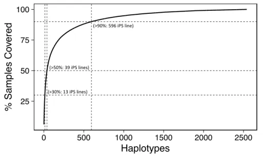
In extension, when using the maximized coverage approach, we found that a total of 13 97
haplotypes are estimated to have a match of 30% of the Saudi population (Figure 1), versus 51 98
lines, when selecting by maximum population frequency and using haplotype-wise matching. The 99
number of required haplotypes covering 50% of the population increases to 39 and 220 for the 100
maximum-coverage allele-wise, and maximum-frequency haplotype-wise approach, 101
respectively. Since the generation of 39 iPS lines to cover >50% of the Saudi population is feasible, 102
HLA-based banking of iPSCs may be a suitable strategy for the pilot implementation and 103
introduction of iPS-based cell therapy in Saudi Arabia.
104
105
Figure 1: Estimated numbers of iPSC lines homozygous for HLA-A, HLA-B, and HLA-DRB1 106
(haplolines) and their coverage percentages for the Saudi population. The dotted lines mark 30, 107
50, and 90% coverage, for 13, 39 and 596 iPS lines, respectively.
108
Donor recruitment and Derivation of HLA-haplobank iPS lines 109
In collaboration with the SSCDR, we identified a registered donor who is homozygous for the 110
most common HLA haplotype (Table 1). This donor’s iPSCs would offer 6.1% coverage. The donor 111
was initially contacted through the phone and upon approval, in-person interview was scheduled.
112
After signing the informed consent, 10 ml peripheral blood sample was collected from the donor 113
and erythroid progenitor cells (EPCs) were isolated expanded in culture for eight days. EPCs were 114
chosen as the starting cell population for reprogramming due to their lack of DNA alterations and 115
genomic structural variation including the absence of TCR/BCR genes recombination found in T- 116
cells (Chou et al., 2011; Perriot et al., 2018; Araki et al., 2020).
117
Flow cytometry analysis showed that >69% of cells stained positive for the erythroid markers 118
CD71 and CD235a after EPCs expansion (Figure 2A, 2B). On day 8 of expansion, reprogramming 119
was initiated by electroporating EPCs with non-integrating episomal plasmids encoding OCT4, 120
SOX2, KLF4, L-MYC, LIN28A, dominant-negative form of TP53, and EBNA1. At day 25 post 121
transfection, numerous embryonic stem cell (ESC)-like colonies were identified with typical ESC 122
morphological characteristics (i.e., distinct borders, bright centers, tight-packed cells, and a high 123
nucleus-to-cytoplasm ratio) (Figure 2C). Such colonies were manually picked, expanded, and 124
cryopreserved. Based on their ideal ESC-like morphology, two clones were chosen to be passaged 125
and subjected to downstream pluripotency validation. The derived lines were registered in the 126
Human Pluripotent Stem Cell Registry (hPSCreg)
127
(https://hpscreg.eu/user/cellline/edit/KAIMRCi002-A, 128
https://hpscreg.eu/user/cellline/edit/KAIMRCi002-B) 129
To assess the genomic integrity of HLA-iPSC#1 and #2, high-resolution G banding was performed 130
after 12 passages in culture. More than 25 prometaphase spreads per clone were analyzed and 131
showed normal female chromosomal number and structure (Figure 2C). Short tandem repeats 132
(STR) assay confirmed the matching identity of the isolated iPS lines and the donor EPCs 133
(Supplementary Figure 2A). Moreover, PCR analysis showed that the episomal plasmids were 134
undetected in HLA-iPSC#1&2 after 12 passages (Supplementary Figure 2B). Additionally, 135
mycoplasma testing showed that the generated iPSC lines are mycoplasma-free (Supplementary 136
Figure 2C).
137
138
Figure 2: Generation and characterization of HLA-universal iPSCs. (A) Schematic representation 139
of ReproTeSRTM and episomal reprogramming method. (B) Flow cytometry histogram for 140
erythroid markers CD71 and CD235a EPCs culture on day 8 shows that ~70% of cells express the 141
erythroid cell markers. Phase-contrast images of mesenchymal-to-epithelial transition and 142
colonies appearance during reprogramming (days 11 to 28). (C) Top: representative images of 143
HLA-universal iPS cell colonies generated from Erythroid Progenitor Cells exhibit more defined 144
borders and compact morphology. Bottom: representative G-banded karyotype analysis for HLA- 145
universal iPSCs shows normal karyotypes 46, XX. (D) Flow cytometry histograms of OCT4, NANOG, 146
and SOX2 in HLA-universal iPSCs and H1 hESCs positive control. (E) Graph showing mRNA 147
expression levels of pluripotency markers for the indicated iPSC lines presented as fold change 148
relative to H1 hESC. Bars are median std of 3 biological replicates for each sample. (F) 149
immunofluorescence staining of the pluripotency markers OCT4 (green), NANOG (red), and SOX2 150
(yellow), Nuclei were stained with DAPI (blue). Scale bar = 200 μm.
151
Validation of iPSCs’ self-renewal and pluripotency properties 152
Pluripotency markers OCT4, NANOG, and SOX2 were detected at the mRNA and protein levels in 153
both clones. Flow cytometry histograms demonstrated that >98% of cells stained positively for 154
OCT4, >96% for NANOG, and >94% for SOX2 (Figure 2D). Moreover, the derived iPSC lines 155
displayed positive expression of OCT4, NANOG, SOX2, and LIN28 by RT-qPCR (Figure 2E) and 156
OCT4, NANOG, and SOX2 by immunofluorescence (Figure 2F). Direct in-vitro differentiation to 157
the three germ layers, mesoderm, definitive endoderm, and ectoderm was used to demonstrate 158
the tri-lineage differentiation capacity. We observed a down-regulation of OCT4 and NANOG and 159
an upregulation of germ layer-specific markers by RT-qPCR (Figure 3B). Immunostainings for the 160
neural progenitor marker (NESTIN) indicated ectodermal differentiation. The positive expression 161
of Brachyury, a member of the Tbox family, showed an early determination of mesoderm. We 162
further assessed the presence of the endodermal marker SRY-Box Transcription Factor 17 163
(SOX17) (Figure 3A). We, therefore, proved that the constructed HLA-universal iPSC lines possess 164
bona fide characteristics of pluripotent stem cells. All performed quality control tests are 165
summarized in Table 3.
166
Classification Test Results Data
Morphology Phase-contrast imaging Typical primed pluripotent human stem cell morphology
Figure 2C, Top
Karyotype G-banding 46, XX Figure 2C, Bottom
Pluripotency status
Quantitative analysis (Flow cytometry, RT- qPCR)
Flow Cytometry: 98% OCT4, 96% NANOG, 99% SOX2.
RT-qPCR: positive expression for OCT4, NANOG, SOX2, LIN28.
Figure 2D, 2E
Qualitative analysis (Immunocytochemistry)
Positive for the pluripotency markers:
OCT4, NANOG, and SOX2.
Figure 2F
Genetic identity STR profiling 24 loci tested, all matched between donor EPCs and derived iPSC lines
Supplementary Figure 2A Verification of
the absence of episomal vectors
PCR analysis The five episomal DNA plasmids were undetected after 12 passages
Supplementary Figure 2B
Mycoplasma testing
RT-qPCR Negative Supplementary
Figure 2C Multilineage
differentiation potential
Directed differentiation RT-qPCR measurement of expression levels of ectoderm (PAX6 and NESTIN),
mesoderm (BRACHYURY and CDX2), and endoderm (SOX17 and GATA4).
Figure 3A, 3B
Immunocytochemistry: Positive for NESTIN, BRACHYURY, and SOX17.
HLA typing WGS Homozygous at class I loci A, B, C, and class II loci DQB1 and DRB1, with only DPB1 being heterozygous
Figure 1
SV analysis WGS;
Manta+Delly+SURVIVOR
No SVs in iPSC#2; one tandem duplication in iPSC#1
Supplementary Figure 3 SNP analysis WGS; GATK, Mutect2 No high-impact SNP acquired during cell
line establishment; SNPs in genetic background of the donor classified as benign
Supplementary table 3.
Table 4.
Table 3: Summary of characterization tests performed on HLA-iPSC lines#1 and #2.
167 168
Furthermore, the differentiation potential of the iPSC lines toward central nervous system (CNS)- 169
type neural progenitor cells (NPCs) and beating cardiomyocyte was tested. CNS-type NPC 170
differentiation induced a marked increase in key neuronal markers such as SOX1, PAX6 and 171
TUBB3 (Figure 3C, 3D).
172
The HLA-iPSC clones were also differentiated into cardiomyocytes (CMs) through a step-wise 173
protocol. By day 15, the iPSC-derived CMs displayed spontaneous contractions, a unique 174
functional property of pulsating heart muscles (Supplemental file# movie). Flow cytometry 175
histograms showed that >94% of cells stained positively for Cardiac Troponin I (cTnI) (Figure 3E).
176
Moreover, RT-qPCR demonstrated that iPSC-derived CMs expressed the Cardiac Muscle Troponin 177
T (TNNT2), myocardial markers NK2 Homeobox 5 (NKX2.5), and GATA family of zinc-finger 178
transcription factor (GATA4) at mRNA levels (Figure 3F).
179
180
Figure 3: Differentiation potential of HLA-universal iPSCs. (A) immunofluorescence staining of specific 181
markers for the three germ layers Ectoderm (NESTIN), Mesoderm (Brachyury), Endoderm 182
(SOX17), Nuclei were stained with DAPI (blue). Scale bar = 200 μm.(B) Graphes showing mRNA 183
expression levels of the lineage-specific markers for the three germ layers Mesoderm (CDX2 and 184
Brachyury), Endoderm (GATA4 and SOX17), and Ectoderm (NESTIN and PAX6) presented as fold 185
change relative to undifferentiated cells. Bars are median std of 3 biological replicates for each 186
sample. Student’s t-tests, *p < 0.05. (C) Phase-contrast images of CNS-type NPC differentiation 187
display typical NPC morphology 34 days post differentiation. Scale bar = 200 μm. (D) Graph 188
showing mRNA high expression levels of CNS-type NPCs markers SOX1, PAX6, and a low or 189
negative expression of β-tubulin III presented as fold change relative to undifferentiated cells.
190
Bars are median std of 2 biological replicates for each sample. Student’s t-tests, *p < 0.05. (E) 191
Flow cytometry histograms display positive cTnI expression in iPSC-derived CM. (F) Graphes 192
showing mRNA expression levels of cardiac markers TNNT2, NKX2.4, and GATA4. Bars are median 193
std of 2 biological replicates for each sample. Student’s t-tests, *p < 0.05.
194 195
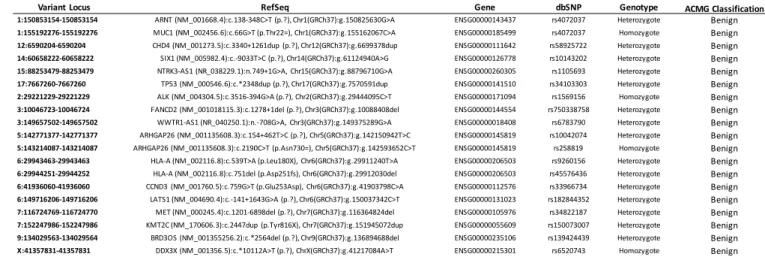
Whole-Genome Sequencing of Generated iPS Lines 196
To ascertain the genotype and whether the genomic integrity of the constructed iPSC lines was 197
maintained during reprogramming and prolonged cultivation, we sequenced the genomes of the 198
parental blood sample and the progenies iPSC#1 and iPSC#2 at passage 13. Genotyping of the 199
HLA loci using the 23x-26x coverage read datasets confirmed homozygous status at class I loci A, 200
B, C, and class II loci DQB1 and DRB1, with only DPB1 being heterozygous 201
(A*02:01~A*02:01~B*50:01~B*50:01~C*06:02~C*06:02~DPB1*02:01~DPB1*04:01~DQB1*02:0 202
2~DQB1*02:02~DRB1*07:01~DRB1*07:01). Genomic variants were called in parental and 203
progeny samples using GATK. This yielded a total of 5.4 million polymorphic sites with a mean 204
genotype call rate of 99.2% and a heterozygosity ratio of 1.7. Out of the 4.3 million single 205
nucleotide polymorphisms each sample had about 3.9 million polymorphic loci (Supplementary 206
Table 2). We first focused on SNPs that were found polymorphic in all three samples to generate 207
a high confidence variant set for the genetic background of the donor. We then examined any 208
variants that might affect cancer-related genes based on the COSMIC Cancer Gene Census 209
database and Shibata list as described previously (Yoshida et al., 2022) which involved 15 210
heterozygous SNVs and 4 homozygous SNVs in (see Materials and Methods for details). However, 211
in the categories of sequence variants developed by the American College of Medical Genetics 212
and Genomics (ACMG), we found that the 19 variants are almost certainly benign (Table 4). Thus, 213
the direct link between these mutations and tumorigenicity was eliminated since the HLA 214
universal donor was healthy at the time of iPSC generation.
215
216
Variant Locus RefSeq Gene dbSNP Genotype ACMG Classification
1:150853154-150853154 ARNT (NM_001668.4):c.138-348C>T (p.?), Chr1(GRCh37):g.150825630G>A ENSG00000143437 rs4072037 Heterozygote Benign 1:155192276-155192276 MUC1 (NM_002456.6):c.66G>T (p.Thr22=), Chr1(GRCh37):g.155162067C>A ENSG00000185499 rs4072037 Homozygote Benign 12:6590204-6590204 CHD4 (NM_001273.5):c.3340+1261dup (p.?), Chr12(GRCh37):g.6699378dup ENSG00000111642 rs58925722 Heterozygote Benign 14:60658222-60658222 SIX1 (NM_005982.4):c.-9033T>C (p.?), Chr14(GRCh37):g.61124940A>G ENSG00000126778 rs10143202 Heterozygote Benign 15:88253479-88253479 NTRK3-AS1 (NR_038229.1):n.749+1G>A, Chr15(GRCh37):g.88796710G>A ENSG00000260305 rs1105693 Heterozygote Benign 17:7667260-7667260 TP53 (NM_000546.6):c.*2348dup (p.?), Chr17(GRCh37):g.7570591dup ENSG00000141510 rs34103303 Heterozygote Benign 2:29221229-29221229 ALK (NM_004304.5):c.3516-394G>A (p.?), Chr2(GRCh37):g.29444095C>T ENSG00000171094 rs1569156 Homozygote Benign 3:10046723-10046724 FANCD2 (NM_001018115.3):c.1278+1del (p.?), Chr3(GRCh37):g.10088408del ENSG00000144554 rs750338758 Heterozygote Benign 3:149657502-149657502 WWTR1-AS1 (NR_040250.1):n.-708G>A, Chr3(GRCh37):g.149375289G>A ENSG00000018408 rs6783790 Heterozygote Benign 5:142771377-142771377 ARHGAP26 (NM_001135608.3):c.154+462T>C (p.?), Chr5(GRCh37):g.142150942T>C ENSG00000145819 rs10042074 Heterozygote Benign 5:143214087-143214087 ARHGAP26 (NM_001135608.3):c.2190C>T (p.Asn730=), Chr5(GRCh37):g.142593652C>T ENSG00000145819 rs258819 Homozygote Benign 6:29943463-29943463 HLA-A (NM_002116.8):c.539T>A (p.Leu180X), Chr6(GRCh37):g.29911240T>A ENSG00000206503 rs9260156 Heterozygote Benign 6:29944251-29944252 HLA-A (NM_002116.8):c.751del (p.Asp251fs), Chr6(GRCh37):g.29912030del ENSG00000206503 rs45576436 Heterozygote Benign 6:41936060-41936060 CCND3 (NM_001760.5):c.759G>T (p.Glu253Asp), Chr6(GRCh37):g.41903798C>A ENSG00000112576 rs33966734 Heterozygote Benign 6:149716206-149716206 LATS1 (NM_004690.4):c.-141+1643G>A (p.?), Chr6(GRCh37):g.150037342C>T ENSG00000131023 rs182844352 Heterozygote Benign 7:116724769-116724770 MET (NM_000245.4):c.1201-6898del (p.?), Chr7(GRCh37):g.116364824del ENSG00000105976 rs34822187 Heterozygote Benign 7:152247986-152247986 KMT2C (NM_170606.3):c.2447dup (p.Tyr816X), Chr7(GRCh37):g.151945072dup ENSG00000055609 rs150073007 Heterozygote Benign 9:134029563-134029564 BRD3OS (NM_001355256.2):c.*2564del (p.?), Chr9(GRCh37):g.136894688del ENSG00000235106 rs139424439 Heterozygote Benign X:41357831-41357831 DDX3X (NM_001356.5):c.*10112A>T (p.?), ChrX(GRCh37):g.41217084A>T ENSG00000215301 rs6520743 Homozygote Benign
Table 4: Examined variant loci in donor genetic background. Variant locus provided as 217
[chromosome]:[bp], RefSeq transcript, Ensembl gene identifier, dbSNP variant identifier, 218
genotype in donor (heterozygous/homozygous) and classification as per ACMG.
219
In the second step we tested whether the cell lines acquired new SNPs compared to the donor, 220
using the donor sample as matched normal for the cell line samples. This approach yielded 1,610 221
and 1,888 SNPs for iPSC#1 and iPSC#2, respectively (Supplementary Table 3). None of the 222
detected SNPs is predicted to have high impact with the majority classified as modifier.
223
While the subtractive analysis of structural variants (SVs) of donor vs. cell line did not detect 224
newly acquired mutations in HLA-iPSC#2, it revealed a heterozygous tandem duplication on 225
chromosome 16 (74,726,891bp - 74,727,373bp) in HLA-iPSC#1 (Supplementary Figure 3) which 226
spans part of exon 3 of the Fatty Acid 2-Hydroxylase (FA2H) where it could lead to an alteration 227
in the transcript. However, this gene is not part of the COSMIC Cancer Gene Census database 228
and Shibata list rendering this variant benign.
229 230
Discussion 231
Within only seven years of their initial derivation in 2007, iPSCs moved to clinical studies when a 232
patient with age-related macular degeneration (AMD) was the first recipient of autologous iPS- 233
derived retinal pigment epithelial cell sheet, in the world’s first in human clinical trial (Mandai et 234
al., 2017). However, it became evident that the high cost and extended waiting time associated 235
with autologous iPS-based cellular therapy, posed a significant hurdle to the advancement into 236
the clinical domain (Habka, Mann et al. 2015; Huang, Liu et al., 2019; Bravery et al., 2015).
237
One approach that was proposed to solve the time and cost problems, is the creation of a hypo- 238
immunogenic iPS cell line that evades the immune system. In this approach, iPS cells would be 239
genetically modified to inactivate major histocompatibility complex (MHC) class I and II genes (Xu 240
et al., 2019; Kitano et al., 2022). However, to achieve this, multiple rounds of gene editing are 241
required, which extends the time the cells are cultured, thus increasing the risk of acquiring 242
mutations. Additionally, gene editing technologies like, CRISPR/Cas9 has been shown to 243
introduce unintended genomic aberrations and may render the cells not useful for therapy (Fu 244
et al., 2013; Cradick et al., 2013). Even base and prime editing that does not involve double strand 245
breaks have recently been shown to induce significant genotoxicity in human cells (Fiumara et 246
al., 2023).
247
Therefore, there has been an increased interest in HLA-based iPS banking in numerous countries 248
(Taylor et al., 2012; Gourraud et al., 2012; Lee et al., 2018; Álvarez-Palomo, et al., 2021; Yoshida 249
et al., 2023). In this study, we assessed the feasibility of creating an iPS haplobank in Saudi Arabia 250
to develop clinical-grade iPS cell stocks, as the ultimate goal. In order to achieve this, we used 251
the high-resolution HLA genomic database of the SSCDR, which at the time of analysis contained 252
64,315 registered donors, and assumed it was a representative sample of the Saudi population.
253
We found that, the feasibility of HLA-based banking in Saudi Arabia is comparable to similar 254
endeavors in other countries. We found that an iPS haplobank of the top 5 haplotypes that offer 255
maximal coverage for the Saudi population would cover 17.30% of the population, which is close 256
to the Spanish bank in which, the top 5 haplotypes cover 19.44%, but lower than the Korean 257
estimation, in which the top 5 haplotypes cover 27.99% (Lee et al., 2018; Álvarez-Palomo, et al., 258
2021). This finding is in line with previous reports that showed a relatively high HLA genetic 259
diversity among Saudis compared to other populations (Alfraih et al., 2021).
260
We found that an iPS haplobank generated from homozygous donors from the top 39 haplotypes 261
would offer coverage of more than 50% of the Saudi population. This significant percentage may 262
allow for many Saudi patients to benefit from iPS-derived cell therapies in the kingdom and 263
therefore, it justifies the construction of the haplobank. In addition, streamlining the process of 264
generating clinical-grade iPSCs will facilitate the establishment and future expansion of the bank 265
to include additional haplotypes. Moreover, due to high level of consanguinity in the Saudi 266
population, there is a considerable excess homozygosity, which may facilitate the identification 267
of homozygous donors and haplobanking (Chentoufi et al., 2022).
268
Due to the relatively high intra-population diversity in Saudi, we found that achieving higher 269
coverage requires much larger cell stocks. Around 596 iPS line would be required to cover 90%
270
of the population, and 2541 lines for 100% coverage. Even though we envisage that the 271
establishing of an iPS cell stock to cover 30%-50% of the Saudi population is a feasible goal to 272
introduce iPS-based cell therapy in the kingdom, achieving higher coverage percentage becomes 273
increasingly cost-ineffective. Therefore, more research is needed to improve current methods of 274
clinical-grade iPS generation to reduce cost and waiting time to make autologous cell therapy a 275
possibility. Additionally, as we gain tighter control on the outcome of gene editing technologies, 276
the creation of clinically-relevant universal hypoimmunogenic iPS lines might become more 277
feasible in the future.
278
To establish the workflow and initiate HLA-based banking in Saudi, we recruited the first donor 279
and generated the first two clinically relevant iPS lines using defined feeder-free conditions. We 280
chose EPCs to be the starting cell population for reprogramming. As opposed to human dermal 281
fibroblasts, EPCs can be easily isolated and expanded from a simple ten ml blood sample and 282
does not require painful skin biopsies. This is of particular importance in the donor recruitment 283
process, as participants might be discouraged to donate if the procedure is invasive.
284
Additionally, EPCs are frequently replenished in the blood and therefore are less likely to 285
accumulate environment-induced mutations like fibroblasts (Panther et al., 2021; Kamimura et 286
al., 2021). Moreover, they lack the TCR/BCR genes recombination found in T-cells, making them 287
a more attractive source of iPS cells. This is in addition to recent research demonstrating that 288
erythroblasts-derived iPS cells are less likely to harbor genetic aberrations when compared to iPS 289
cells from other sources (Perriot et al., 2018; Araki et al., 2020).
290
Eight days of expansion showed that around 69% of the cells were CD71+CD235a+ (Figure 2B).
291
The rest were CD71-CD235a+ and are more likely to be differentiated erythroblasts on their way 292
to enucleation and are therefore, unamenable to reprogramming. Differentiated cells were 293
particularly evident as red colored cells when EPCs were pelleted by centrifugation.
294
As an alternative to conventional retroviral-based cell reprogramming, non-viral, non-integrating 295
plasmid-based reprogramming technique is more clinically-relevant (Yu et al. 2009; Okita et al., 296
2011; Bang et al., 2018). The reprogramming factors are delivered by vectors that contain oriP 297
and EBNA-1, based on the Epstein-Barr Nuclear Antigen-1, which has demonstrated the ability to 298
produce iPSCs highly efficiently without the potential risk of transgenic sequences being inserted 299
into the target cell genome (Drozd et al., 2015). As opposed to other non-integrating 300
reprogramming methods like sendai virus and mRNA, episomal plasmids is the most cost- 301
effective. Additionally, we found that these plasmids are readily removed from the 302
reprogrammed cells as they were expanded, with most lines testing negative by end-point PCR 303
by passage 12.
304
Following the expansion of EPCs, electroporation of the reprogramming episomal was carried 305
out. ESC-like colonies appeared around 20-25 days post transfection and were characterized by 306
distinct borders, bright centers, tight-packed cells, and a high nucleus-to-cytoplasm ratio. The iPS 307
clones were mechanically picked, expanded, and characterized for self-renewal and pluripotency 308
in feeder-free culture conditions.
309
For developing clinical-grade HLA haplobank, KAIMRC is currently establishing its cell-processing- 310
center in compliance with the good manufacturing practice (GMP) guidelines. Although the 311
current HLA-iPSC#1 and iPSC#2 were generated inside research-grade labs, future haplobanking 312
and clinical products will be derived and cryopreserved inside our GMP facility including re- 313
derivation of the current HLA-iPSC lines to clinical-grade. Re-derivation of human pluripotent 314
stem cell lines inside GMP facilities has been done before. For instance, the H1 hESC line was re- 315
derived and used as part of Astellas Pharma's phase II retinal pigment epithelium (RPE) trial, and 316
re-derived H9 hESC line was used to generate dopaminergic neurons for a Parkinson's disease 317
clinical trial by BlueRock Therapeutics (Sullivan a et al., 2020).
318
319
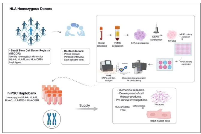
Figure 4: Graphical summary of the undergoing HLA-based banking in Saudi Arabia.
320
Conclusions
321
Our study lays the foundation for the roadmap towards HLA-based banking of human induced 322
pluripotent stem cells (iPSCs) in Saudi Arabia (Figure 4). By interrogating the Saudi Stem Cell 323
Donor Registry, we identified a subset of homozygous donors that could offer considerable 324
coverage for the Saudi populace. Our analysis revealed that achieving 30% and 50% coverage 325
necessitate the generation of 13 and 39 iPS lines, respectively. As a proof of principle, we 326
successfully generated the first HLA-iPS line (2 clones), that offer 6.1% coverage of the Saudi 327
population. By employing clinically-relevant methodologies, we ensured the safety and quality of 328
these iPSCs. Notably, whole-genome sequencing confirmed the genomic stability of the 329
generated lines, hence alleviating concerns of high-risk mutations during reprogramming and 330
expansion processes. Our study substantiates the feasibility of HLA-based iPSC banking in Saudi 331
Arabia and paves the way for a resilient infrastructure in regenerative medicine and personalized 332
therapeutics.
333
List of abbreviations
334
SSCDR: Saudi Stem Cell Donor Registry; HLA: Human leukocyte antigen; EPCs: Erythroid 335
progenitor cells (EPCs); hESC: Human embryonic stem cell; iPSC: Induced pluripotent stem cells;
336
hPSCreg: Human Pluripotent Stem Cell Registry; WGS: Whole Genome Sequencing; SNPs: Single 337
nucleotide polymorphisms; SVs: Structural variants; AMD: Age-related macular degeneration;
338
GMP: good manufacturing practice; RPE: Retinal pigmented epithelial cells.
339
Supplementary Information
340
Additional file 1: Supplementary Figure 1. Alfraih et al haplotype frequency.
341
Additional file 2: Supplementary Figure 2. HLA-iPSC lines authentication.
342
Additional file 2: Supplementary Figure 3. Tandem duplication on chromosome 16 in cell line 343
iPSC#1 visualized using IGV.
344
Additional file 3: Supplementary table 1. Comparison of 5-locus based haplotype frequencies 345
based on the SSCDR database and KFSH&RC dataset (Alfraih et al. 2021).
346
Additional file 4. Supplementary table 2. Single nucleotide polymorphisms for donor blood 347
sample and cell lines iPSC#1 and iPSC#2. The fraction of genotype calls matching with donor 348
was calculated for biallelic SNPs.
349
Additional file 6. Supplementary Table 3: Subtractive SNP calls for cell lines iPSC#1 and iPSC#2 350
vs. donor.
351
Acknowledgements
352
We thank King Abdullah International Medical Research Center (KAIMRC) and the Saudi Stem Cell 353
Donor Registry (SSCDR) for facilitating the initiation of HLA-based iPS banking. We thank current, 354
and future donors for their valuable donations.
355
Authors’ contributions
356
KA contributed through the conception of the idea, the design of the work, interpretation of data, 357
and wrote the manuscript. MA contributed through sample processing, iPS generation, validation 358
assays and differentiation and writing the manuscript. RL designed the iterative algorithm and 359
analyzed the SSCDR HLA database to identify haplotypes and coverage, WGS data analysis and 360
writing the manuscript. MA, MB, HA, DA, AZ, DM, have all contributed in iPS derivation and 361
validation tests. MD performed karyotype analysis. Khaled Alghamdi performed the STR tests. DJ 362
supervised the data curation of the SSCDR. FH assessed the clinical significance of identified 363
variants revealed by WGS. DGC, JT and AH have participated in the design of the work and 364
revision of the document.
365
Funding
366
This work is funded by KAIMRC grant RJ20/134/J. Additional funding was provided through 367
KAUST’s Smart Health Initiative.
368
Availability of data and materials
369
All the presented data is available for consultation.
370
Declarations
371
Ethics approval and consent to participate.
372
The interrogation of the HLA database of the SSCDR, informed consents and donor sample 373
collection were approved by the institutional review board (IRB) and research ethics committee 374
of KAIMRC (RJ20/134/J).
375
Competing interests 376
The authors declare no competing interests.
377
Figure legends
378
Supplementary Figure 1: Comparison of cumulative 5-locus haplotype frequency of the SSCDR 379
HLA database and the Alfraih et al.
380
Supplementary Figure 2: Cell lines authentication. (A) Short Tandem Repeat (STR) profiling 381
guaranteed the genetic identity between the established iPSC lines and the donor EPCs. (B) PCR 382
analysis detected the absence of the episomal plasmids in the indicated lines at passage 12. (C) 383
RT-qPCR showing negative mycoplasma test in HLA-iPSC lines.
384
Supplementary Figure 3: Tandem duplication on chromosome 16 in cell line iPSC#1 visualized 385
using IGV.
386
Materials and Methods
387
Haplotype frequency analysis 388
HLA haplotype frequencies in the Saudi Arabian population were estimated based on haplotype 389
information stored in the Saudi Stem Cell Donor Registry (https://kaimrc.ksau-hs.edu.sa/?page_id=1481) 390
database. This database, comprising 64,315 individuals at the time of analysis, was analyzed using the EM 391
algorithm as implemented in Hapl-o-Mat v 1.1 (DOI: 10.1007/978-1-4939-8546-3_19) to estimate 392
population level haplotype frequencies using two digit resolution. The haplotype coverage was estimated 393
as detailed in Álvarez-Palomo (https://doi.org/10.1186/s13287-021-02301-0) using an iterative algorithm.
394
In each iteration, the most frequent haplotype was identified and all matching individuals were counted 395
and removed from the dataset before the next iteration on the remaining dataset. Importantly, the 396
haplotype matching procedure was modified by considering each locus independently and allowing 397
matches on either of the two possible alleles per locus. Matching was performed based on the loci A, B, 398
and DRB1.
399
Cellular reprogramming and iPS generation.
400
Ethical Approval:
401
This study was approved by the Institutional Review Board of King Abdullah International Medical 402
Research Center (KAIMRC) (Protocol# RJ20/134/J). Initial donor recruitment was done by the 403
Saudi Stem Cell Donor Registry staff. Personal interview was conducted, and informed consents 404
were obtained by the research team.
405
PBMCs isolation and enrichment of erythroid progenitors:
406
Peripheral blood was collected from Saudi patients into an EDTA-containing blood collection tube and 407
treated with RosetteSepTM Human Progenitor Cell Basic Pre-Enrichment antibody cocktail according to the 408
manufacturer’s instructions (Stem Cell Technologies Catalog#15226). After PBMCs separation and 409
isolation, 1 million cells were cultured for 8 days in StemSpanTM SFEM II medium (Stem Cell Technologies 410
Catalog #09605) supplemented with 1X StemSpanTM Erythroid Expansion Supplement (Stem Cell 411
Technologies Catalog #02692).
412
Transfection of Erythroid Progenitor Cells:
413
Expanded erythroid cells were reprogrammed with Episomal iPSC Reprogramming Kit (Thermofisher 414
Catalog#A15960). Around 3 x 10⁵ cells were electroporated with 1 μg of each episomal vector using Neon 415
Transfection System (Thermofisher). The emerging ESC-like colonies were manually picked and 416
transferred into 96-well plates coated with rhLaminin-521 (Thermofisher Catalog#A29248) in mTeSR™
417
Plus medium (Stem Cell Technologies Catalog #100-0276). iPSCs were dissociated ReLeSR (Thermofisher 418
Catalog # 100-0484) using 1:10-1:30 splitting ratio and incubated at 37 ̊C, 5% CO2, 20% O2 incubator.
419
Molecular characterization of pluripotency and genomic integrity.
420
Immunocytochemistry:
421
Cells were fixed in 4% (w/v) paraformaldehyde for 15 min, permeabilized in PBS containing 0.1% (v/v) 422
Triton X-100 for 10 min, and subsequently blocked in PBS containing 1% Gelatin for 45 min. Cells were 423
incubated with primary antibodies overnight at 4 °C and probed with the appropriate secondary 424
antibodies for 1 hour at room temperature (ThermoFisher Scientific). Primary and secondary antibodies 425
were resuspended in 0.2% Gelatin in PBS. The nuclei were counterstained with 1 μg /mL DAPI nuclear 426
staining (Thermo Fisher Scientific).
427
Quantitative Reverse Transcription PCR (RT-qPCR):
428
Total RNA was extracted using RNeasy Kit (Qiagen Catalog# 74104) and reverse transcribed using the High- 429
Capacity cDNA Reverse Transcription Kit (Applied BiosystemsTM Catalog# 4374966). The RT-qPCR assay 430
was carried out using FastStart SYBR Green Master Mix (ROCHE) as described previously (Alsayegh et al., 431
2018).
432
In-vitro Differentiation:
433
The generated iPSCs were differentiated into three germ layers using the STEMdiffTM Trilineage 434
Differentiation Kit (Stem Cell Technologies Catalog #05230).
435
Flow Cytometry Analyses:
436
Cells were stained with OCT4, NANOG, SOX2, and cTnI antibodies diluted in 2% FBS in PBS for 30 mins on 437
ice in the dark with occasional vortexing. It was then washed with PBS and analyzed on BD FACS ARIA cell 438
sorter. FITC-positive cells were measured in stained vs unstained cells.
439
Karyotyping:
440
For G banding karyotyping, iPSC lines were treated with 0.3 μg/mL KaryoMAXTM ColcemidTM (1 μg) for 15 441
min, dissociated by TrypLE, and incubated in hypotonic solution (75 mM potassium chloride) at 37 °C for 442
20 min. iPSCs were then fixed in methanol/glacial acetic acid 3:1 and stored at 4 °C. At least 50 metaphases 443
were karyotyped at the department of pathology and laboratory medicine (Ministry of the National Guard 444
- Health Affairs).
445
Neural Progenitor Cells (NPCs) differentiation:
446
The generation of central nervous system (CNS)-type neural progenitor cells (NPCs) from HLA-iPSCs was 447
performed according to Monolayer Culture Protocol (STEMdiffTM SMADi Neural Induction Kit Catalog 448
#08581).
449
Cardiomyocyte differentiation:
450
The differentiation of hESCs toward beating cardiomyocyte was performed following STEMdiffTM 451
Ventricular Cardiomyocyte Differentiation Kit (Stem Cell Technologies Catalog #05010) in accordance with 452
the manufacturer instructions. In brief, iPSCs were detached using gentle cell dissociation reagent and 453
seeded at 1.2 x 10⁶ cells/well on Matrigel-coated 12 well plates in presence of mTeSRTM Plus medium and 454
10 μM Y-27632. Subsequently, the differentiation was initiated by replacing culture medium with 455
Cardiomyocyte Differentiation Medium A for 48 hrs. at 37 ◦C, 5%CO2. Cardiomyocyte Differentiation 456
Medium B was added for another 48 hrs. Then, Cardiomyocyte Differentiation Medium C was replaced on 457
day 4 and 6. We perform a full-medium change with Cardiomyocyte Maintenance Medium every other 458
day up to 20 days.
459
Episomal Plasmids Screening:
460
DNA was extracted using AllPrep DNA/RNA/ Mini Kit (Qiagen Catalog# 80204). PCR was performed using 461
EBNA-1 primers that detect all five episomal plasmids (expected size 666 bp) according to manufacture 462
guidelines (Thermo Fisher Scientific Catalog # A15960).
463
Mycoplasma Detection:
464
Mycoplasma contamination was assessed using LookOut Mycoplasma qPCR Detection (SIGMA) 465
(Supplementary Figure 1C).
466
Statistical Analysis:
467
RT-qPCR data are represented as mean ± standard deviation (SD). Statistical significance was determined 468
in Student’s t-test (unpaired; two-tailed). A Bonferroni correction was applied to the p-value from multiple 469
comparisons. *p < 0.05.
470
Short Tandem Repeats (STR) Identity Assay:
471
Extracted genomic DNA from HLA-iPSCs and PBMCs was analyzed for 24 polymorphic STR markers using 472
GenePrint 24 system (Promega, Madison, USA) following manufacturer’s protocol and were amplified 473
using PCR and followed by ABI capillary electrophoresis. In this analysis, a 24 autosomal STR were analysed 474
D8S1179, D21S11, D7S820, CSF1PO, D3S1358, THO1, D13S317, D16S539, D2S1338, D19S433, vWA, TPOX, 475
D18S51, D5S818, D1S1656, D2S441, D10S1248, Penta E, Penta D, DYS391, D12S391, D22S1045 FGA and 476
Amelogenin using ABI 3130/3500 Genetic Analyzer.
477
Whole Genome Sequencing (WGS):
478
The Nextera library prep kit (Illumina) was used to prepare libraries for WGS resequencing on the Novaseq 479
6000 sequencer (Illumina). The short-read sequences obtained from a blood sample as control, as well as 480
the two cell lines iPSC#1 and iPSC#2 were assessed with FastQC (Andrews, n.d.). Adapter and low quality 481
regions were trimmed with Trimmomatic v0.33 (Bolger et al., 2014) using parameters: 2:30:10 LEADING:3 482
TRAILING:3 SLIDINGWINDOW:4:20 MINLEN:40, leaving 280, 262, and 245 million reads for the blood, 483
iPSC#1 and iPSC#2 dataset respectively. Trimmed reads were mapped to the human reference genome 484
assembly UCSC hg38 analysis set using BWA mem (Li & Durbin, 2010), which yielded a median coverage 485
between 23X and 26X. Duplicates were marked and read groups added with Picard tools (Broad Institute, 486
2018). The donor sample as well as both generated cell lines were HLA genotyped using xHLA 487
(https://www.pnas.org/doi/10.1073/pnas.1707945114). Single nucleotide polymorphisms were jointly 488
called in all samples with GATK HaplotypeCaller (McKenna et al., 2010) following GATK best practices 489
recommendations as well as with GATK Mutect2. In case of Mutect, mapped reads from HLA-iPSC#1 and 490
HLA-iPSC#2 were run separately as treatment while the blood sample was provided as normal reference.
491
Single nucleotide polymorphism calls were then filtered requiring a minimal allele frequency of 20%. The 492
obtained polymorphisms were then annotated using the Ensembl Variant Effect Predictor VEP (McLaren, 493
Genome Biol. 2016). Detected SNVs were tested for overlap with genes listed in the Catalogie of Somatic 494
Mutations In Cancer (COSMIC) Cancer Gene Census (https://cancer.sanger.ac.uk/census) and the Shibata 495
cancer gene panel (https://www.pmda.go.jp/files/000152599.pdf). Variants which were predicted to 496
have a high impact on the aforementioned gene set were manually examined.
497
Structural variants were called with Delly v1.1.6 and Manta v1.6 in subtractive mode specifying the blood 498
sample as control and both cell lines separately as treatments. Structural variants passing the quality filter 499
for each caller and being classified as “precise” were retained. Variants calls for each cell line from Delly 500
and Manta were then compared using SURVIVOR (Jeffares et al. 2017, Nat. Comm.) and only overlapping 501
calls were retained for further analysis, allowing for at most 500bp distance between break points. This 502
procedure revealed no structural variants in HLA-iPSC#2 and one tandem duplication HLA-iPSC#1.
503
504
References
505
Takahashi K, Yamanaka S. Induction of pluripotent stem cells from mouse embryonic and adult fibroblast 506
cultures by defined factors. cell. 2006 Aug 25;126(4):663-76.
507
Yu J, Vodyanik MA, Smuga-Otto K, Antosiewicz-Bourget J, Frane JL, Tian S, Nie J, Jonsdottir GA, Ruotti V, 508
Stewart R, Slukvin II. Induced pluripotent stem cell lines derived from human somatic cells. science. 2007 509
Dec 21;318(5858):1917-20.
510
Singh VK, Kalsan M, Kumar N, Saini A, Chandra R. Induced pluripotent stem cells: applications in 511
regenerative medicine, disease modeling, and drug discovery. Frontiers in cell and developmental biology.
512
2015 Feb 2;3:2.
513
Mandai M, Watanabe A, Kurimoto Y, Hirami Y, Morinaga C, Daimon T, Fujihara M, Akimaru H, Sakai N, 514
Shibata Y, Terada M. Autologous induced stem-cell–derived retinal cells for macular degeneration. New 515
England Journal of Medicine. 2017 Mar 16;376(11):1038-46.
516
Taylor CJ, Peacock S, Chaudhry AN, Bradley JA, Bolton EM. Generating an iPSC bank for HLA-matched 517
tissue transplantation based on known donor and recipient HLA types. Cell stem cell. 2012 Aug 518
3;11(2):147-52.
519
Taylor CJ, Bolton EM, Bradley JA. Immunological considerations for embryonic and induced pluripotent 520
stem cell banking. Philosophical Transactions of the Royal Society B: Biological Sciences. 2011 Aug 521
12;366(1575):2312-22.
522
Nakatsuji N, Nakajima F, Tokunaga K. HLA-haplotype banking and iPS cells. Nature biotechnology. 2008 523
Jul;26(7):739-40.
524
Habka D, Mann D, Landes R, Soto-Gutierrez A. Future economics of liver transplantation: a 20-year cost 525
modeling forecast and the prospect of bioengineering autologous liver grafts. PloS one. 2015 Jul 526
15;10(7):e0131764.
527
Huang CY, Liu CL, Ting CY, Chiu YT, Cheng YC, Nicholson MW, Hsieh PC. Human iPSC banking: barriers and 528
opportunities. Journal of biomedical science. 2019 Dec;26:1-4.
529
Bravery CA. Do human leukocyte antigen-typed cellular therapeutics based on induced pluripotent stem 530
cells make commercial sense?. Stem cells and development. 2015 Jan 1;24(1):1-0.
531
Taylor CJ, Bolton EM, Pocock S, Sharples LD, Pedersen RA, Bradley JA. Banking on human embryonic stem 532
cells: estimating the number of donor cell lines needed for HLA matching. The Lancet. 2005 Dec 533
10;366(9502):2019-25.
534
Gragert L, Eapen M, Williams E, Freeman J, Spellman S, Baitty R, Hartzman R, Rizzo JD, Horowitz M, Confer 535
D, Maiers M. HLA match likelihoods for hematopoietic stem-cell grafts in the US registry. New England 536
Journal of Medicine. 2014 Jul 24;371(4):339-48.
537
Park M, Seo JJ. Role of HLA in hematopoietic stem cell transplantation. Bone marrow research. 2012;2012.
538
Opelz G, Döhler B. Effect of human leukocyte antigen compatibility on kidney graft survival: comparative 539
analysis of two decades. Transplantation. 2007 Jul 27;84(2):137-43.
540
Johnson RJ, Fuggle SV, O’Neill J, Start S, Bradley JA, Forsythe JL, Rudge CJ. Kidney Advisory Group of NHS 541
Blood and Transplant Factors influencing outcome after deceased heart beating donor kidney 542
transplantation in the United Kingdom: an evidence base for a new national kidney allocation policy.
543
Transplantation. 2010;89:379-86.
544
Lee S, Huh JY, Turner DM, Lee S, Robinson J, Stein JE, Shim SH, Hong CP, Kang MS, Nakagawa M, Kaneko 545
S. Repurposing the cord blood bank for haplobanking of HLA-homozygous iPSCs and their usefulness to 546
multiple populations. Stem Cells. 2018 Oct 1;36(10):1552-66.
547
Álvarez-Palomo B, García-Martinez I, Gayoso J, Raya A, Veiga A, Abad ML, Eiras A, Guzmán-Fulgencio M, 548
Luis-Hidalgo M, Eguizabal C, Santos S. Evaluation of the Spanish population coverage of a prospective HLA 549
haplobank of induced pluripotent stem cells. Stem cell research & therapy. 2021 Dec;12:1-8.
550
Opelz G, Döhler B. Pediatric kidney transplantation: analysis of donor age, HLA match, and posttransplant 551
non-Hodgkin lymphoma: a collaborative transplant study report. Transplantation. 2010 Aug 15;90(3):292- 552
7.
553
Araki R, Hoki Y, Suga T, Obara C, Sunayama M, Imadome K, Fujita M, Kamimura S, Nakamura M, Wakayama 554
S, Nagy A. Genetic aberrations in iPSCs are introduced by a transient G1/S cell cycle checkpoint deficiency.
555
Nature communications. 2020 Jan 10;11(1):197.
556
Chou BK, Mali P, Huang X, Ye Z, Dowey SN, Resar L, Zou C, Zhang YA, Tong J, Cheng L. Efficient human iPS 557
cell derivation by a non-integrating plasmid from blood cells with unique epigenetic and gene expression 558
signatures. Cell research. 2011 Mar;21(3):518-29.
559
Perriot S, Canales M, Mathias A, Du Pasquier R. Generation of transgene-free human induced pluripotent 560
stem cells from erythroblasts in feeder-free conditions. STAR protocols. 2022 Sep 16;3(3):101620.
561
Xu H, Wang BO, Ono M, Kagita A, Fujii K, Sasakawa N, Ueda T, Gee P, Nishikawa M, Nomura M, Kitaoka F.
562
Targeted disruption of HLA genes via CRISPR-Cas9 generates iPSCs with enhanced immune compatibility.
563
Cell stem cell. 2019 Apr 4;24(4):566-78.
564
Kitano Y, Nishimura S, Kato TM, Ueda A, Takigawa K, Umekage M, Nomura M, Kawakami A, Ogawa H, Xu 565
H, Hotta A. Generation of hypoimmunogenic induced pluripotent stem cells by CRISPR-Cas9 system and 566
detailed evaluation for clinical application. Molecular Therapy-Methods & Clinical Development. 2022 Sep 567
8;26:15-25.
568
Fu Y, Foden JA, Khayter C, Maeder ML, Reyon D, Joung JK, Sander JD. High-frequency off-target 569
mutagenesis induced by CRISPR-Cas nucleases in human cells. Nature biotechnology. 2013 Sep;31(9):822- 570
6.
571
Cradick TJ, Fine EJ, Antico CJ, Bao G. CRISPR/Cas9 systems targeting β-globin and CCR5 genes have 572
substantial off-target activity. Nucleic acids research. 2013 Nov 1;41(20):9584-92.
573
Yoshida S, Kato TM, Sato Y, Umekage M, Ichisaka T, Tsukahara M, Takasu N, Yamanaka S. A clinical-grade 574
HLA haplobank of human induced pluripotent stem cells matching approximately 40% of the Japanese 575
population. Med. 2023 Jan 13;4(1):51-66.
576
Alfraih F, Alawwami M, Aljurf M, Alhumaidan H, Alsaedi H, El Fakih R, Alotaibi B, Rasheed W, Bernas SN, 577
Massalski C, Heidl A. High-resolution HLA allele and haplotype frequencies of the Saudi Arabian population 578
based on 45,457 individuals and corresponding stem cell donor matching probabilities. Human 579
Immunology. 2021 Feb 1;82(2):97-102.
580
Gourraud PA, Gilson L, Girard M, Peschanski M. The role of human leukocyte antigen matching in the 581
development of multiethnic “haplobank” of induced pluripotent stem cell lines. Stem cells. 2012 Feb 582
1;30(2):180-6.
583
Panther L, Ornelas L, Jones MR, Gross AR, Gomez E, Liu C, Berman B, Svendsen CN, Sareen D. Generation 584
of iPSC lines with high cytogenetic stability from peripheral blood mononuclear cells (PBMCs). bioRxiv.
585
2021 Sep 28:2021-09.
586
Kamimura S, Suga T, Hoki Y, Sunayama M, Imadome K, Fujita M, Nakamura M, Araki R, Abe M.
587
Insertion/deletion and microsatellite alteration profiles in induced pluripotent stem cells. Stem Cell 588
Reports. 2021 Oct 12;16(10):2503-19.
589
Bang JS, Choi NY, Lee M, Ko K, Lee HJ, Park YS, Jeong D, Chung HM, Ko K. Optimization of episomal 590
reprogramming for generation of human induced pluripotent stem cells from fibroblasts. Animal Cells and 591
Systems. 2018 Mar 4;22(2):132-9.
592
Yu J, Hu K, Smuga-Otto K, Tian S, Stewart R, Slukvin II, Thomson JA. Human induced pluripotent stem cells 593
free of vector and transgene sequences. Science. 2009 May 8;324(5928):797-801.
594
Okita K, Matsumura Y, Sato Y, Okada A, Morizane A, Okamoto S, Hong H, Nakagawa M, Tanabe K, Tezuka 595
KI, Shibata T. A more efficient method to generate integration-free human iPS cells. Nature methods. 2011 596
May;8(5):409-12.
597