This thesis is submitted in partial fulfillment of the requirements for the degree of Master of Science in Molecular Biology and Biotechnology. The cells showed a decreased survival during treatment with Safranal which was associated with induced autophagy. This was confirmed by SDS-PAGE and western blot showing an increase in the expression of the main autophagic protein markers such as Beclin-1 and Atg 12.
Furthermore, the combination of Safranal and Sorafenib showed antagonistic effect on Safranal-inhibiting cell viability. There are many people to thank and acknowledge for their guidance and support during my master's journey. All members of the committee have dealt with patience and support during my dissertation and I could not be less grateful.
Wafaa Ramadan of the University of Sharjah for guiding them through combinatorial experiments at an early stage of the research. Finally, I would like to thank my parents, Mr. and Mrs. from the bottom of my heart. Special thanks to my brothers and sisters, who each in their own way encouraged, supported and lifted me up.
Introduction
Overview
Research Problem
Literature Review
Natural compounds have recently gained momentum and are being investigated as therapeutics for various diseases, including HCC. Many natural compounds such as; Solamargin (Chinese herb derivative), capsaicin (spice), curcumin (spice), resveratrol (plant polyphenol), silibinin (flavonoid), etc. Asians have long used saffron as a spice and food coloring. nations of the Middle East.
It has also been used as a traditional medicine, mostly acting as an anti-inflammatory and pain reliever. Lately, Saffron and its derivatives; Safranal, Crocin and Crocetin have shown anticancer, antioxidant and anti-inflammatory properties in vitro and in vivo. Safranal is one of the main components of Saffron's essential volatile oil, which gives it its characteristic smell and aroma.
The results of the study, which was performed on HepG2 cells, showed that Safranal inhibits growth, survival and cellular proliferation in vitro. The toxicity is much less with combination therapy than with single drug treatment, the chance of developing drug resistance is less, and in fact the ability to introduce a cancer stem cell inhibitor has been shown to reduce the incidence of relapse. [26]. 27] using Safranal alone or in combination with crocin, morphine, diclofenac and naloxone to investigate the effect on orofacial pain in rats.
28], there was also an in vivo study in which rats were examined for the effects of safranal and metformin on learning and memory. Sorafenib has been tested on various cancer cell lines in combination with other drugs, natural compounds and specific inhibitors [29–32]. The use of sorafenib in combination with other anticancer drugs, preferably natural compounds, may change the course of the HCC treatment regimen.
Recent advances have helped to understand the molecular complexity of HCC, oncogenes and cell signaling. These findings serve as an opportunity for the development of therapeutic agents that can successfully target these pathways and their critical checkpoints [ 5 , 6 ].
Materials and Methods
- Cell Culture
- Drugs Preparation
- Cell Viability Assay
- Autophagic Vacuoles Detection
- Detecting Lysosomes using Lysosomal Staining Reagent
- Protein Extraction and Quantification
- SDS-PAGE and Western Blot
Briefly, the cells were seeded at a density of 5000 cells/well in a 96-well clear bottom plate in 100 µl of complete growth medium. The cells were allowed to adhere before being treated with Safranal IC25 and IC50 for 24 hours. The cells were then washed again with 1X assay buffer and then visualized using an IX53 microscope (Olympus).
Briefly, cells were seeded at a density of 20,000 cells/well in 2 mL of 6-well plate media and incubated. Cells were then treated with Safranal IC25 and Safranal IC50 concentrations and incubated for 24 hours. Cells were then washed, twice, and staining processing solution was then added for 30 min.
The proteins were then extracted using and according to the protocol of RIPA Lysis and Extraction Buffer (Sigma). Protein quantitation was performed using Bradford Reagent (Sigma) and absorbance was measured using GloMax Microplate Reader (Promega). Proteins were transferred to PVDF membranes prior to incubation with various primary antibodies; LC3B, p.AKT, AKT, pERK1/2, Beclin1 and Atg12.
Results
- Safranal and Sorafenib Inhibit HepG2 cell proliferation
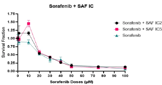
- Safranal and Sorafenib combination showed an antagonistic effect
- Autophagic vacuoles are formed upon treatment with Safranal
- Treatment with Safranal results in formation of autolysosomes
- Effect of Safranal on autophagic proteins expression
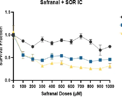
First, we used constant doses of Sorafenib at IC25 and/or IC50 in combination with increasing doses of Safranal µM) as shown below in Figure 3. Here, the combined treatment did not show any significant difference compared to the single treatment with Sorafenib. This may be due to Sorafenib's high potency and effect on the inhibition of HepG2 cell viability.
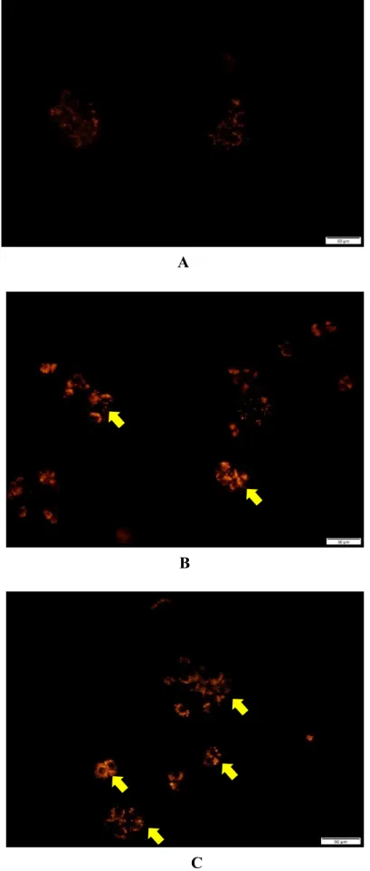
To determine the effect of Safranal on autophagy, cells were treated with IC25 and IC50 doses of Safranal, and autophagic vacuoles were visualized using an autophagy assay kit, as shown in Figure 5. The data showed the formation of autophagic vacuoles, which were more visible with treatment of HepG2 cells with Safranal at its IC50 value (Figure 5C). The cells were treated with Safranal IC25 and IC50 and autolysosomes were visualized using a fluorescence microscope (Figure 6).
The data showed the formation of autolysosomes upon treatment of HepG2 cells with Safranal (Figure 6B and C). To validate the previous findings on autophagy, the expression of the main proteins involved in autophagy was analyzed in HepG2 cells. For this, cells were treated with Safranal at IC25 or IC50 and proteins were extracted and analyzed by SDS-PAGE followed by western blot using specific antibodies (Figure 7 and Figure 8).
All the pro-autophagic markers (LC3B, Beclin 1, Atg 12) tested showed an increased expression in a dose-dependent manner with the treatment with Safranal compared to control. This is very similar to the observations on cell viability and autophagy induced by Safranal treatment. Interestingly, Safranal treatment significantly reduced the phosphorylation level of the kinase AKT (pAKT), which is known to control the survival pathway (Figure 7).
This is consistent with the previous data showing that Safranal inhibits cell viability and induces autophagy. All the variations can be considered specific as no change was observed on GAPDH, used as a loading control (Figure 7).

Discussion
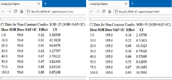
Combination index is used to quantitatively represent the synergistic (CI<1), additive (CI=1) and antagonist (CI >1) effect of the drugs in combination. In other words, the mechanism of action of autophagy is the transport of damaged or unwanted substances from various parts of the cell to the lysosome for degradation or recycling. Treatment with Safranal IC25 and IC50 of HepG2 cells leads to the increase of these autophagic vacuoles in the cell.
There was an increased formation of vacuoles with Safranal IC50 treatment compared to IC25 (Figure 5B and Figure 5C). Inflammation is one of the hallmarks of HCC and autophagy has functioned as a tumor suppressor mechanism by suppressing this inflammation. At the molecular level and to confirm the previous analyzes of induced autophagy, the expression of key autophagic protein markers was analyzed by Western blot.
LC3 is one of the most abundant proteins present in autophagic vacuoles (autophagosome), therefore it is also one of the most used markers for autophagy and autophagic activities. As seen in Figure 7, with treatment with Safranal, the expression of LC3 increased compared to control. In this study, the AKT levels after treatment with Safranal remained the same, while the phosphorylated form of AKT (p.AKT) decreased, suggesting inhibition of cell survival and thereby an induction of autophagy.
The binding of ATP molecule to both of these dimers then leads to phosphorylation of MEK, thus leading to the activation of the ERK pathway. For ERK pathway to be activated, ATP must bind to both the catalytic domains of the dimer. In the case of mutant cell lines, binding of ATP to either domain does not cause a response as RAS is inhibited.
But in wild-type cell lines, binding of ATP to one of the catalytic domains causes transactivation of the second catalytic domain. The response of the ERK pathway to treatment with the Safranal-Sorafenib combination depends on drug concentrations, as shown by the varying response in Figure 11.
Conclusion
Yang JD, Henegouwen P, Gores GJ, et al (2019) A global view of hepatocellular carcinoma: trends, risk, prevention and management. Tang W, Chen Z, Zhang W, et al (2020) The mechanisms of sorafenib resistance in hepatocellular carcinoma: theoretical basis and therapeutic aspects. Baig B, Halim SA, Farrukh A, et al (2019) Current status of nanomaterial-based treatment for hepatocellular carcinoma.
Zhang S, Ni Y, Zhao C, et al (2018) Capsaicin enhances the antitumor activity of sorafenib in hepatocellular carcinoma cells and mouse xenograft tumors. Amin A, Hamza AA, Bajbouj K, et al (2011) Saffron: A potential candidate for a new anticancer drug against hepatocellular carcinoma. Al-Hrout A, Chaiboonchoe A, Khraiwesh B, et al (2018) Safranal induces DNA double-strand breaks and ER stress-mediated cell death in hepatocellular carcinoma cells.
Bajbouj K, Schulze-Luehrmann J, Diermeier S, et al (2012) The anticancer effect of saffron in two p53 isogenic colorectal cancer cell lines. Yao X, Zhao C, Yin H, et al (2020) Synergistic antitumor activity of sorafenib and artesunate in hepatocellular carcinoma cells. Lin Y, Lin L, Jin Y, et al (2014) Combination of Matrine and Sorafenib reduces the aggressive phenotypes of hepatocellular carcinoma cells.
Liu J, Liu Y, Meng L, et al (2017) Synergistic antitumor effect of Sorafenib in combination with ATM inhibitor in hepatocellular carcinoma cells. Huang F, Wang BR, Wang YG (2018) The role of autophagy in tumorigenesis, metastasis, targeted therapy and drug resistance of hepatocellular carcinoma. Yang S, Yang L, Li X, et al (2019) New insights into autophagy in hepatocellular carcinoma: mechanisms and therapeutic strategies.
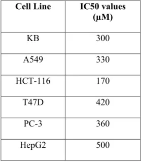
Jabini R, Ehtesham-Gharaee M, Dalirsani Z, et al (2017) Evaluation of the cytotoxic activity of Crocin and Safranal, constituents of saffron, in oral squamous cell carcinoma (KB cell line). Morisaki T, Umebayashi M, Kiyota A, et al (2013) The combination of Celecoxib with Sorafenib synergistically inhibits hepatocellular carcinoma cells in vitro.