Lung cancer is the second most common cancer with the highest mortality worldwide in 2020, despite advances in targeted and immunotherapies. Based on the already published data, this preclinical study aims to explore the anticancer potential of DCA in lung cancer alone and in combination with chemo and targeted therapies using two non-small cell lung cancer (NSCLC) cell lines, namely A549 and LNM35. This project was addressed through the investigation of the impact of DCA on lung cancer cell viability, migration, invasion and colony growth in vitro and on tumor growth and metastasis using the chick embryo-chorioallantoic membrane (CAM) and nude mouse models in vivo.
On the other hand, DCA did not inhibit the in-vitro migration and invasion and the in-vivo appearance and growth of lymph node metastases in nude mice with the highly metastatic lung cancer cells LNM35. In summary, these findings demonstrate that DCA is a safe and promising therapeutic agent for lung cancer and pave the way for further pre-clinical studies evaluating the impact of DCA in combination with not only the first generation, but also the second and third generation of EGFR confirm. -Tki in-vivo.
Introduction
- Lung Cancer Risk Factors
- Lung Cancer Types
- Lung Cancer Stages
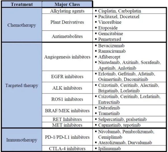
- NSCLC Treatment
- Cancer Hallmarks
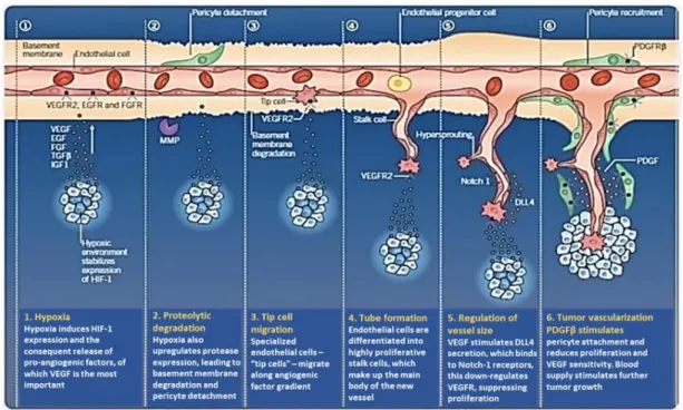
- Angiogenesis in Cancer
- Invasion & Metastasis in Cancer
- Metabolic Reprogramming
- Dichloroacetate (DCA)
- Aim and Objectives
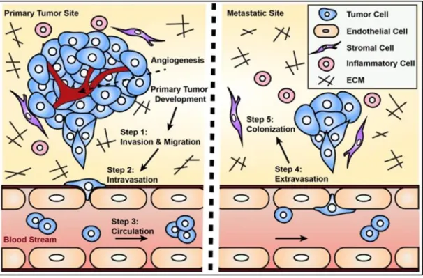
Metastasis has been reported to be a leading cause of cancer-related mortality (Meirson et al., 2020). In addition, lymphatic endothelial cells secrete CCL21 or CXCL12 to enable the chemotaxis of the tumor cells expressing CCR7 or CXCR4 receptors ( Perlikos et al., 2013 ). The potential role of DCA in the treatment of cancer is due to its ability to reverse the Warburg effect (James et al., 2017).
Materials and Methods
- Cell Culture and Reagents
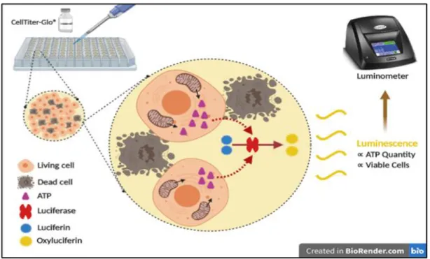
- Cellular Viability
- Clonogenic Assay
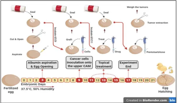
- In Ovo Tumor Growth Assay
- Vascular Tube Formation Assay
- HUVEC Spheroids Sprouting Assay
- Wound Healing Motility Assay
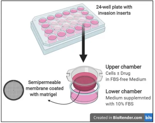
- Matrigel Invasion Assay
- Statistical Analysis
In the second set of experiments, cells were treated for 48 h with increasing concentration of Gefitinib and Erlotinib HCl (5-80 µM). In the second set of experiments, formed colonies were treated for 7 days with a combination of DCA and Frondoside A Hydrate, DCA and Gefitinib, or DCA and Erlotinib HCl. Data were presented by comparing the mean weight of tumors in the control group and the DCA-treated group.
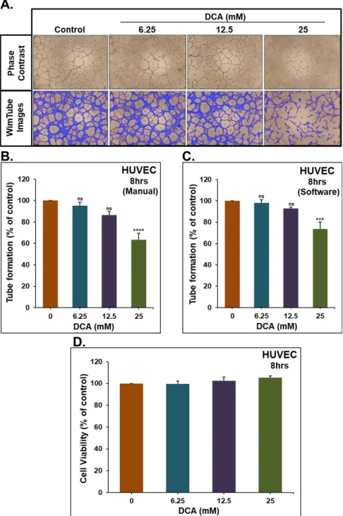
Drug toxicity was assessed by comparing the percentage of live embryos in control and DCA-treated groups at the end of the experiment. Effect of DCA on tumor growth was presented by comparing the mean tumor weight at the end of the experiment between the control group and DCA-treated group. The impact of DCA on the ability of HUVECs to form capillary-like structures was assessed by measuring the total lengths of the tubes formed in control and DCA-treated wells.
The effect of the different concentrations of DCA on the viability of HUVECs was determined using CellTiter-Glo® Luminescent Cell Viability assay (Promega Corporation, Madison, USA) as previously described in the section on cellular viability. Cancer cells were then seeded in the upper chambers at a density of 1x10⁵ cells/0.5 ml in medium without fetal bovine serum, in the presence and absence of DCA. The plate was kept in a humidified incubator at 37°C and 5% CO 2 for 24 hours, after which the non-penetrating cells in the upper chambers were removed by gently rubbing the area with a cotton swab.
The effect of DCA on cell invasion was presented as a percentage (%) comparing invading cells in the presence of DCA with the control.
Results
Effect of DCA on the Growth of NSCLC Tumor Xenografts
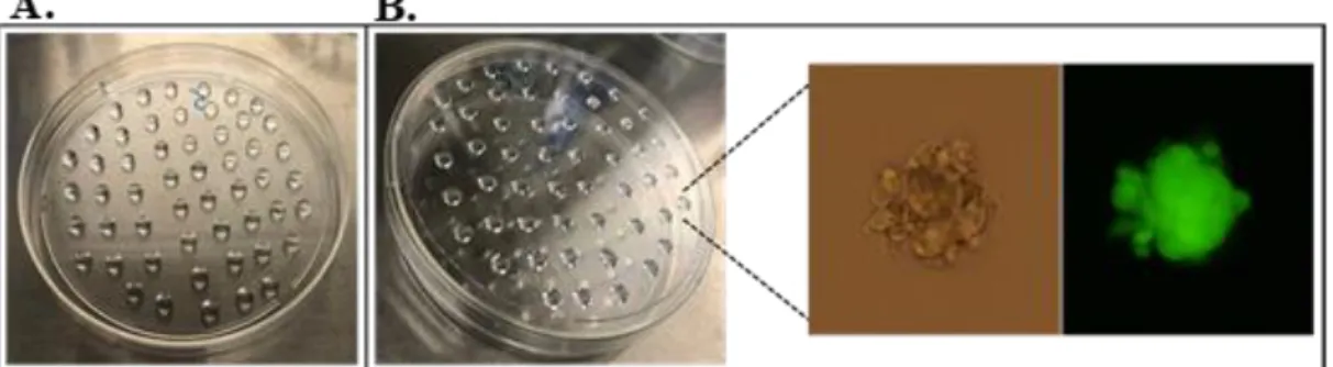
To confirm the pharmacological relevance of the in vitro results, the anticancer effect of DCA was evaluated in vivo using the chicken embryo CAM assay. As observed in Figure 11, 50 mM DCA significantly reduced the growth of A549 tumor xenografts (Figure 11A) by approximately 40%, while it did not show a significant reduction in the growth of LNM35 tumor xenografts (Figure 11B). Therefore, 100 mM DCA was studied on LNM35 tumor xenografts and it significantly reduced growth by approx. 40% (Figure 11C).
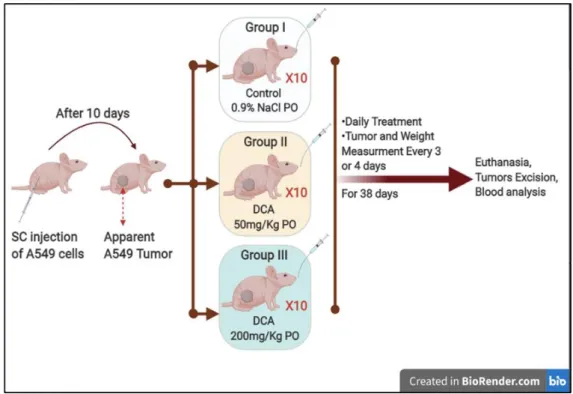
Toxicity was also assessed by comparing the percentage of live embryos in control and DCA-treated groups. The impact of DCA on tumor xenografts was also evaluated in vivo using athymic mice inoculated with A549 and LNM35 cells. Therefore, the mice with A549 tumor xenografts were treated orally every day (5 days/week) with 50 mg/kg and 200 mg/kg DCA for 38 consecutive days.
Treatment with DCA (50 mg/kg) did not significantly decrease the volume of A549 tumor xenografts, whereas DCA (200 mg/kg) significantly decreased the volume by about 45% (Figure 12A). On the other hand, the growth of LNM35 tumor xenografts was monitored and mice were orally treated with 200 mg/kg and 500 mg/kg DCA every day (5 days/week) for 14 and 24 consecutive days, respectively. Treatment with DCA (200 mg/kg) showed no reduction in the volume of LNM35 tumor xenografts (Figure 13A), whereas DCA (500 mg/kg) significantly reduced tumor volume by nearly 75% (Figure 13B).
36 Figure 13: Effect of DCA on the growth of LNM35 xenographed in nude mice in vivo.

Effect of DCA on the Formation of Capillary-Like Structures
An inverted microscope was used for contrast pictures and Wimasis software was used to clarify the pictures. HUVEC cell viability was determined as described in the material and methods under similar conditions of embedded spheroids.
Effect of DCA on NSCLC Metastasis In-Vivo and Invasion and
Cells that invaded the Matrigel and passed the 8 μm pores were determined as described in Materials and Methods. Photographs of scratches induced in confluent monolayers of LNM35 (E) and A549 (F) cells in the presence and absence of different concentrations of DCA at 0, 2, 6, and 24 h.

Effect of DCA in Combination with Chemotherapeutic
44 Figure 18: Effect of DCA in combination with chemotherapeutic agents on the viability of NSCLC cells. Cellular viability was determined using the CellTiter Glo luminescent assay as described in Materials and Methods.
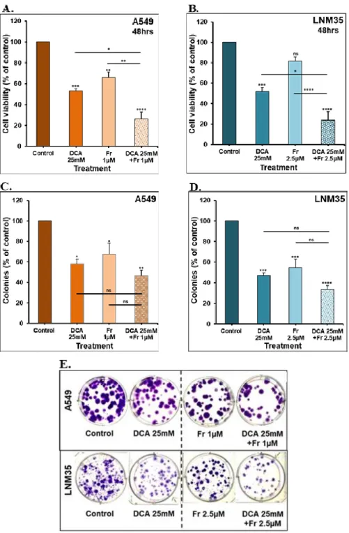
Effect of DCA in Combination with Frondoside A on NSCLC
A549 (C) and LNM35 (D) colonies were treated with the indicated concentrations, fixed, stained and counted as described in Materials and Methods. E) Representative images of control and combination colonies for A549 and LNM35 cancer cells are shown.
Effect of DCA in Combination with EGFR-TKi on NSCLC
Exponentially growing A549 (A, C) and LNM35 (B, D) cells were treated with vehicle, Gefitinib or Erlotinib (5 - 80 μM) for 48 h. Cell viability was determined using the CellTiter-Glo luminescence assay as described in materials and methods. Treatment of cells for 48 hours with 25 mM DCA significantly increased the effect of Gefitinib on cell viability of A549 (Figure 21A) and LNM35 (Figure 21B). Then, the clonogenic assay was performed to assess the effect of the combination on the growth of pre-formed colonies of both cell lines.
Gefitinib leads to a significant reduction in the percentage of total colonies of both cell lines (Figure 21C,D). In addition, this combination shows significant decrease in the cell density of the individual colonies of both cell lines (Figure 21E,F). Similarly, DCA enhances the inhibitory effect of Erlotinib on the cellular viability of A549 and LNM35 (Figure 22A,B).
The percentage of total A549 and LNM35 colonies was significantly reduced by 30-40% with Erlotinib (Figure 22C, D). Despite the insignificant reduction in the percentage of A549 colonies with the combination, the cell density of each colony was significantly reduced compared to the individual treatments (Figure 22E). Similarly, the cell density of the LNM35 colonies was reduced in the combination-treated group (Figure 22F).

Discussion
On the other hand, higher IC50 was reported in cervical cancer cells Hela and SiHa cells (Li et al., 2017), while DCA (20 mM) failed to inhibit the cellular viability of breast cancer MCF-7 cell line (Woo et al. , 2016). It was reported that 10 mM sodium DCA was effective in reducing PBT24 tumor growth but not U87 tumor growth, reflecting some differences in the biology of the two cell lines (Stakišaitis et al., 2021). On the other hand, a significant growth delay was also observed in HT-29 xenografts treated with oral DCA (200 mg/kg) daily for four days (Lin et al., 2014).
The anticancer effect of DCA was reported to be partially due to induction of apoptosis, which was observed in colorectal cancer cells (Madhok et al., 2010) and NSCLC cells (Lu et al., 2018) or due to inhibition of angiogenesis . On the other hand, Zhao and colleagues recently reported that DCA stimulates angiogenesis in vascular dementia rats by improving endothelial precursor cell function (Zhao et al., 2019). It was previously reported that LNM35 cell line is the first human lung cancer cell line with lymphogenic metastatic properties with 100% incidence after a subcutaneous inoculation (Kozaki et al., 2000).
It has been reported that DCA showed promising anticancer effects when combined with some natural compounds such as: Curcumin (Kan et al., 2018) and Betulin derivatives (Mihoub et al., 2018). Despite the remarkable benefits, many patients acquired therapeutic resistance after 10-14 months of treatment due to secondary mutation in EGFR gene (Yuan et al., 2019). In addition, patients with some other mutations such as KRAS and PIK3CA showed primary resistance to the treatment of EGFR-TKi (Tetsu et al., 2016).
Furthermore, inhibition of autophagy was reported to alleviate the antitumor effects of EGFR-TKi (Han et al., 2011; Meng et al., 2019).
Conclusion
Dichloroacetate should be considered in platinum-based chemotherapy in hypoxic tumours, rather than as monotherapy in advanced non-small cell lung cancer. Lovastatin overcomes resistance to gefitinib in human non-small cell lung cancer cells with K-Ras mutations. Programmed cell death ligand-1-mediated enhancement of hexokinase 2 expression is inversely related to T cell effector gene expression in non-small cell lung cancer.
Establishment and characterization of the human lung cancer cell line NCI-H460-LNM35 with consistent lymphogenic metastases via subcutaneous and orthotopic propagation. Trends in the incidence, treatment and survival of patients with lung cancer over the past four decades. Dichloroacetate enhances the antitumor efficacy of chemotherapeutics by inhibiting autophagy in non-small cell lung cancer.
Bidesmoside betulin saponin containing L-rhamnopyranoside moieties induces apoptosis and inhibits the growth of lung cancer cells in vitro and in vivo. Hypersensitivity of EGFR wild-type non-small cell lung cancer cells to the EGFR-tyrosine kinase inhibitor erlotinib. Suppression of pyruvate dehydrogenase kinase-2 re-sensitizes paclitaxel-resistant human lung cancer cells to paclitaxel.
Dichloroacetate potentiates tamoxifen-induced cell death in breast cancer cells via epidermal growth factor receptor downregulation.