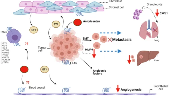
This was associated with a significant decrease in the extent of tumor metastases to the liver and lungs. Furthermore, histological staining of liver tissue revealed a 43% decrease in the number of tumor foci in the Ambrisentan-treated group compared to the control group.
Introduction
Endothelin axis
- Discovery of the endothelin receptors
- Endothelin biosynthesis
- Endothelin receptors (ETRs)
In humans, ETAR is found in the vasculature and is expressed primarily in vascular smooth muscle cells, whereas ETBR is expressed primarily in smooth muscle cells and endothelial cells. In addition, ligand binding to ETBR can mediate vasoconstriction in certain vascular beds as well as bronchoconstriction in the respiratory tract (Aubert & Jeanneret, 2016).
ET Signaling pathway
- G-Protein mediated GPCR signaling
- Cellular contraction
- Endothelin-1 Receptor/β-Arrestin-1 Axis
- Crosstalk with other pathways
In smooth muscle, binding of ET1 to ETAR induces activation of phosphatidyl inositol-specific phospholipase C (PI-PLC), leading to the formation of inositol 1,4,5 triphosphate (IP3) and diacylglycerol (DAG). It induces actin assembly activities and several upstream regulators, leading to local invasion and metastasis (Semprucci et al., 2016).
Physiological role of endothelin
- Receptors distribution
- Blood flow and vasoconstriction
- Other physiological function
Endothelin signaling is extremely complex and activation of the ET axis can stimulate a number of different effector systems, such as phospholipases D and A2, protein kinase A and C (PKA and PKC), as well as the protein tyrosine kinase and MAP kinase (MAPK). /ECR) pathways (Bremnes et al., 2000; Sasaki et al., 1998). ET-mediated activation of NF-κB, β-catenin, hypoxia-inducible factor 1-alpha (HIF1-α), and Rho factor (ρ) has also been described (Nelson et al., 2003).
Endothelin axis expression in cancer
In most of these cells, ET is thought to act as a co-mitogen, enhancing the action of other growth factors such as platelet-derived growth factor (PDGF) (Kohno et al., 1994).
Endothelin receptor antagonists
- Bosentan
- Ambrisentan
- Macitentan
- ERA and pregnancy
Ambrisentan has advantages over other ETAR blockers, such as lower reported rates of liver toxicity (Newman et al., 2007). ET-1/ETAR signaling pathway has been shown to be involved in early induction, migration and maintenance of neural crest specification (Bonano et al., 2008).
Properties of tumors
- Regulation of tumor metastasis
- Tumors and the immune system
Also, the recruitment of myeloid-derived suppressor cells (MDSC) to metastatic sites is induced by the inflammatory chemokines CCL2 and chemokine ligand 5 (CCL5) (Borsig et al., 2014). It has been found that in response to the expression of (CXC) chemokine ligand 12 (CXCL12) (CXC), cancer cells expressing chemokine receptor 4 (CXCR4) metastasize to the lymph nodes and lungs (Müller et al., 2001) .
Breast cancer
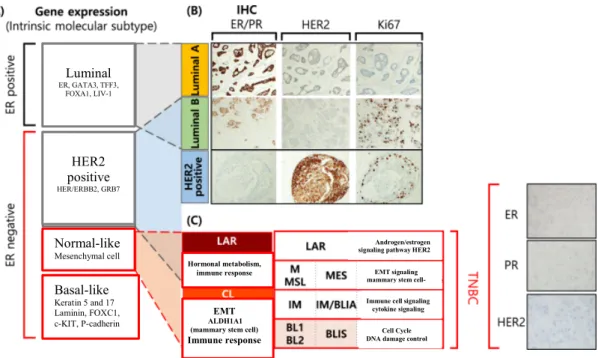
- Triple negative breast cancer
The 4T1 tumor, transplanted into BALB/c mice, is highly invasive and can spontaneously metastasize from the primary tumor in the mammary pad to several distant organs, including blood, lungs, liver, lymph nodes, brain, and bones, making it a great model is to study metastatic breast cancer. ER estrogen receptor, PR progesterone receptor, HER2 human epidermal growth factor receptor 2, IHC immunohistochemistry, LAR luminal androgen receptor, CL claudin low, M mesenchymal, MSL mesenchymal stem-like, IM immunomodulatory, BL basal-like, MES mesenchymal, BLIA basal-like immunoactivated, BLIS basal-like immune-suppressed, EMT epithelial-mesenchymal transition, TNBC triple-negative breast cancer (Lee, Oh, Go, Han, &. Choi, 2020). It has been discovered that these neutrophils collect at the premetastatic site in the lung and produce leukotrienes which in turn will recruit highly tumorigenic breast cancer cells to the lung and thereby promote tumor cell seeding and colonization (Xie et al., 2017). .
ET- axis and tumor growth
Different tumor cells escape apoptosis by modulating known cell survival pathways, such as dependent protein kinase B (Akt), Phosphoinositide 3-kinases (PI3K) and NF-κB (Banerjee et al., 2007; Nelson et al., 2005). In prostate cancer cells, the use of Atrasentan, selective ETAR antagonist, inhibited the NF-κB DNA binding activity, correlated with a decrease in the levels of the anti-apoptotic proteins Bcl-2, Bcl-xl and survivin, which apoptotic cell cause death (Banerjee et al., 2007). Furthermore, when the specific ETAR antagonist BQ123 was used, it restored the apoptotic response (Del Bufalo et al., 2002).
ET1 role in tumor cell invasion and metastasis
- MMPS and integrins
- RHO-GDI2
It also increases the MMP/TIMP ratio by activating membrane type-1-MMP (MT1-MMP) and decreasing the release of tissue inhibitor of MMP1 (TIMP1) and TIMP2, which promotes rapid ECM degradation (Rosanò et al. , 2001; Wu et al., 2012). In cultured glioblastoma (GBM) cells, exogenous ET-1 induced tumor cell migration that was associated with an increase in the expression of MMP-9 and MMP-13 (Hsieh et al., 2014). When DNA microarrays were performed to identify genes affected by RhoGDI2 reconstitution, suppression of ET1 was observed ( Titus et al., 2005 ).
Effect of ET-1 on angiogenesis
Decreased expression of GDP dissociation inhibitor (RhoGDI2), a metastasis inhibitor, was found to decrease survival in patients with advanced lung metastatic bladder cancer. In the same model, treatment with atrasentan and zibotentan inhibited lung metastasis, suggesting the importance of ET1 in the bladder cancer microenvironment for promoting lung metastasis, thereby identifying ET1 as a potential biomarker for lung metastasis (Said et al., 2011; Titus et al. ., 2005).
ET-1 and immune cells
- Inflammation and macrophages
- Dendritic cells
- Lymphocytes
In ovarian cancer cells, ETBR signaling reduced tumor-infiltrating lymphocytes, and the use of ETBR antagonists abrogated this effect (Buckanovich et al., 2008; Kandalaft et al., 2009; Kandalaft et al., 2011). Furthermore, ETBR has been shown to inhibit the expression of endothelial intercellular adhesion molecule 1 (ICAM1), preventing the adhesion of T cells to the endothelium.
ET-1 antagonists in clinical trials
Hypothesis
Aim and Objectives of the study
Materials and Methods
Materials
- Mice
- Standard solutions
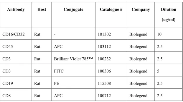
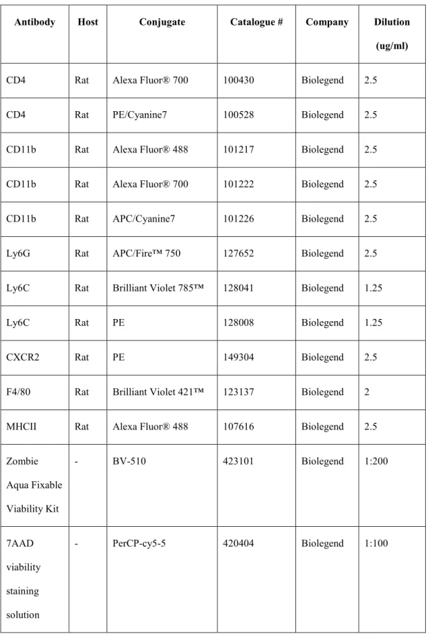
- List of antibodies used for FACS staining
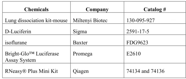
- List of materials, kits used and suppliers
- List of antibodies used for immunocytochemistry
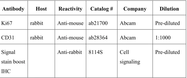
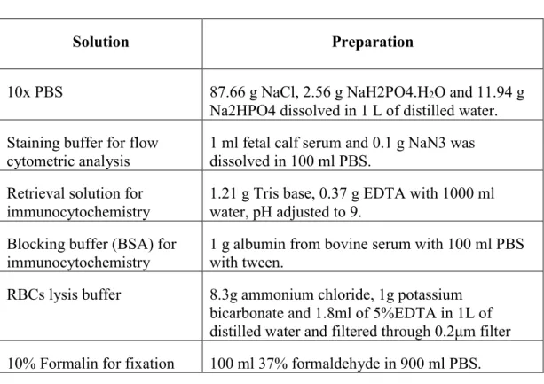
RBCs lysis buffer 8.3g ammonium chloride, 1g potassium bicarbonate and 1.8ml 5%EDTA in 1L distilled water and filtered through 0.2μm filter 10% Formalin for fixation 100ml 37% formaldehyde in 900ml PBS. Antibody Host Reactivity Catalog # Company Dilution Ki67 Rabbit Anti-Mouse ab21700 Abcam Prediluted CD31 Rabbit Anti-Mouse ab28364 Abcam 1:1000 Signal.
Methods
- Breast cancer cell line
- Ambrisentan preparation
- Experimental protocol
- Spleen tissue processing
- Lung tissue processing
- Blood processing for single-cell suspension
- Flow cytometry
- Liver tissue fixation and H and E staining
- Assessment of liver metastasis
- CD31 immunohistochemistry staining and tumor
- Lung metastasis assessment
- In vivo bioluminescence imaging (IVIS)
- Quantitative RT‑ PCR analysis
- In ovo tumor growth assay
- Statistical analysis
The lungs were excised at the end of the experiment, washed with PBS, cut into small pieces which were transferred to GentleMACS C tubes (Miltenyi Biotec, Germany), for processing using a lung dissociation kit and a GentleMACS dissociator (Miltenyi), according to the manufacturer's instructions. At the end of the experiment, the lungs were collected and the right lungs, and the left lungs were processed separately and their weights were recorded. Cancer cells were trypsinized, washed with complete medium and suspended in PBS. to obtain enough surviving embryos at the end of the experiments).
Results
Myelopoiesis is a hallmark of the 4T1 breast cancer model
No gross adverse effects observed following Ambrisentan
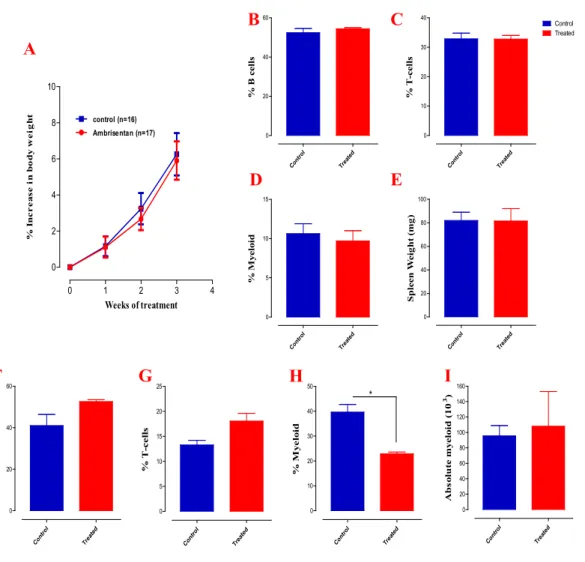
In another experiment, mice were treated with Ambrisentan daily for two weeks, after which spleen and blood were collected and 6-color flow cytometry was used to examine the effect of Ambrisentan on immune cell populations. Mice were implanted with 4T1 cells, then sacrificed at various time points as indicated, whereupon blood and spleens were collected and analyzed by flow cytometry. Mice were implanted with 4T1 cells, then sacrificed at various time points as indicated, whereupon blood and spleens were collected and analyzed by flow cytometry to determine the distribution of myeloid cell populations (continued).
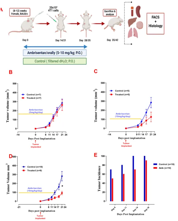
Ambrisentan retards tumor growth in a dose and
First, given that ambrisentan has been approved for use in PAH patients at a dose of 5-10 mg/kg (Galiè et al., 2008; Kingman et al., 2009), this study evaluated the oral administration of ambrisentan at a dose 5-10 mg. /kg dose range in mice. Daily administration of ambrisentan at a dose of 5 mg/kg/day had no obvious effect on the growth of 4T1 tumors (Figure 8B). However, increasing the dose to 10 mg/kg/day resulted in an approximately 55% reduction in primary tumor volume compared to untreated mice 3 weeks after implantation (Figure 8C).
Ambrisentan improves host survival in orthotopically‑implanted
Oral treatment with ambrisentan was started 2 or 3 weeks before the implantation of the 4T1 cells and continued for an additional 2 weeks after implantation. Effect of ambrisentan administration at a dose of 5 mg/kg/day (B) or 10 mg/kg/day (C) on tumor growth. Effect of pretreatment with 10 mg/kg ambrisentan for 3 weeks on tumor volume (D) and incidence (E).
Ambrisentan retards tumor growth and metastasis, - in vivo
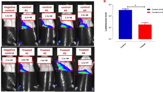
Primary tumor growth can be monitored and measured by quantifying tumor photon flux, which is proportional to the number of light-emitting cancer cells. The bioluminescence signal at the primary tumor implantation site was measured for each animal and used as an indicator of tumor growth. Data collected by IVIS showed a 5.3-fold reduction in tumor growth in Ambrisentan-treated mice (Figure 10C).
Inhibition of liver tumor metastasis in Ambrisentan treated
These foci clearly appeared as small aggregates of tumor cells (Figure 12A, C) and strongly stained with anti-Ki67 antibody (Figure 12B, E), indicating a high proliferative state. Images were acquired after masking the signal from the implantation site and focusing on the metastatic trunk regions.
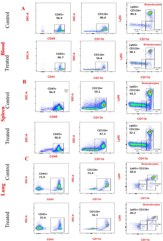
Reduced tumor growth correlates with decreased myelopoiesis
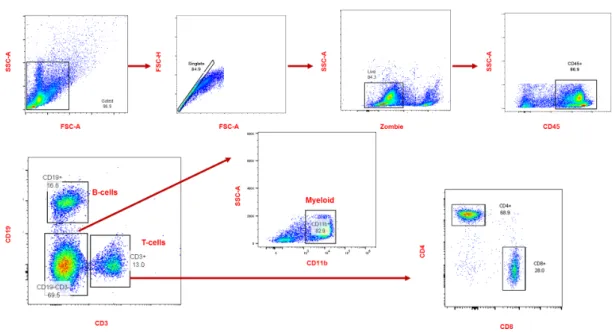
After exclusion of doublets, debris and dead cells, immune cells were identified using the panhematopoietic marker CD45+ cells and further analyzed for B cells and T cells using their CD19+ and CD3+ markers, respectively. Ambrisentan treatment resulted in a significant reduction of myeloid cells in the blood, spleen, and lung, averaging ~35%, 18%, and 63%, respectively ( Fig. 15 A–F ). After elimination of duplicates, debris and dead cells, immune cells were identified using the panhematopoietic marker CD45. CD45+ cells were further analyzed for Ly6G+CD11b+.
Ambrisentan impairs metastasis to the lung
Furthermore, as a result of a decrease in the myeloid cell population in the spleen, Ambrisentan-treated mice have a significant ~26%. In sharp contrast, the extent of metastasis was reduced by 97.8% to 14 cells/mg in Ambrisentan-treated mice (Figure 16C). The graph shows ~97% decrease in the number of metastatic tumor cells/mg lung in treated mice.
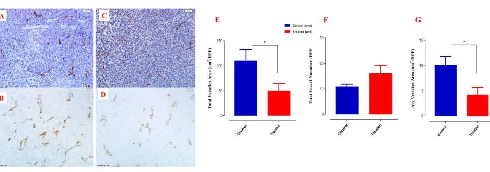
Ambrisentan treatment reduces tumor vascularity
Ambrisentan decreased the expression level of CXCL1 and
Representative tumor sections from control (A, B) or ambrisentan-treated (C, D) mice are shown, with (A, C) or no (B, D) counterstaining. E-G) The number of blood vessels was calculated as the average of the number in 10 fields per section randomly selected from non-necrotic areas of the tumors. The area of each blood vessel in the field was determined and tumor vascularity was calculated as the average of the total vessel area in µm2/HPF.
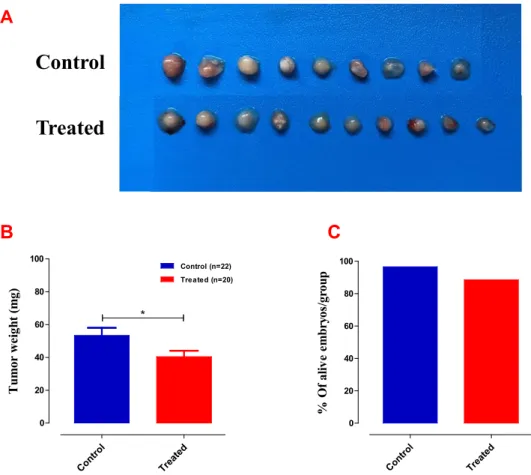
Treatment with Ambrisentan retards tumor growth in ovo
To prepare the 10 mg/kg Ambrisentan dose, the day 11 fertilized egg was weighed and the embryo weight was used to prepare a working solution of Ambrisentan in 1 ml saline, then 100 µl was added/egg. Toxicity was also evaluated by comparing the number of dead embryos in control and treated groups. 4T1-Luc2 cells were inoculated at E9 and after two days when tumors were detectable they were treated with 10 mg/kg Ambrisentan every other day, E11, E13, E15 and E17.
Discussion
The chemokine CXCL1 has a crucial role in the host immune response and mainly acts to recruit neutrophils along the chemokine gradient ( Sawant et al., 2016 ). Furthermore, CXCL1 was shown to be upregulated in LM2-4175 (LM2) breast cancer cells, which grow aggressively and metastasize to the lung (Acharyya et al., 2012). Ly6G+ granulocytes and Ly6C+ monocytes (Ly6C+) in the lungs of animals bearing CXCL1-negative tumors (Acharyya et al., 2012).
Conclusion
Ambrisentan, an endothelin receptor type A-selective endothelin receptor antagonist, for the treatment of pulmonary arterial hypertension. Endothelin A receptor/β-arrestin signaling to the Wnt pathway renders ovarian cancer cells resistant to chemotherapy. Endothelin A receptor drives invadopodia function and cell motility through the β-arrestin/PDZ-RhoGEF pathway in ovarian carcinoma.