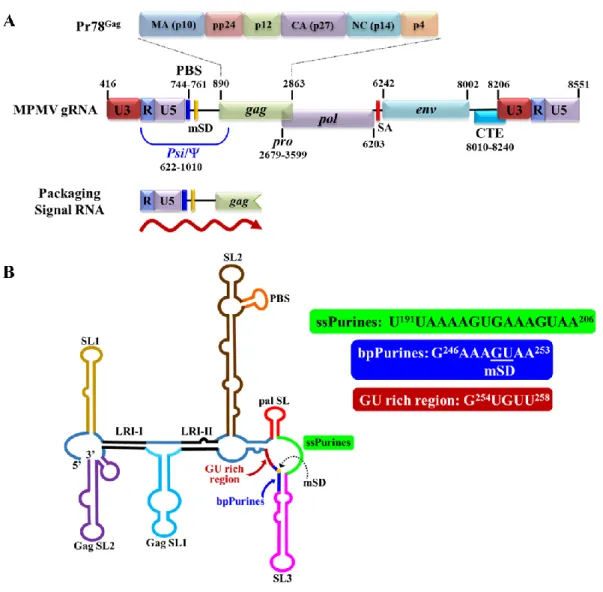
This involves binding of the retroviral Gag polyprotein to sequences at the 5' end of the viral genome, the packaging signal. These revealed that Pr78Gag binds to ssPurines and the A252AGUGUU258 loop, corresponding to two unpaired adenosine residues of bpPurines and the adjacent region called.
Introduction
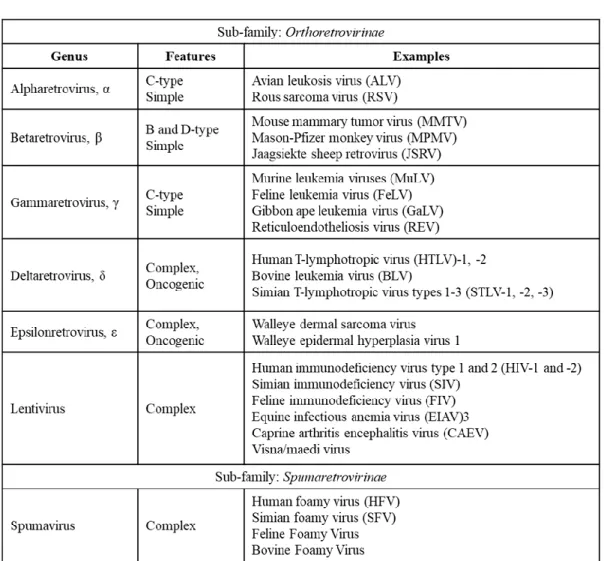
Retroviruses
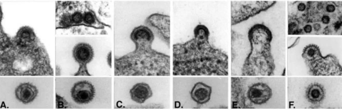
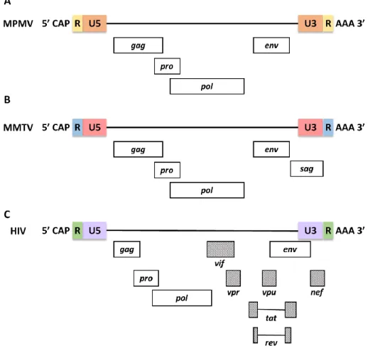
In addition, retroviruses have also been categorized by their morphogenic or assembly pathways identified by electron microscopy (Coffin et al., 1997; Goff, 2001). In contrast, members of the Beta retrovirus and Spumavirus pre-assemble their immature nuclei in the cytoplasm, adopting a B-type (mouse mammary tumor virus, MMTV) or D-type (Mason-Pfizer monkey virus, MPMV) morphology (Figure 2). .
Virion Structure
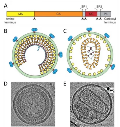
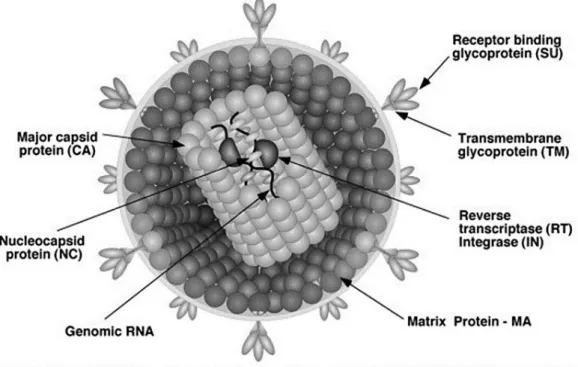
Apart from the RNA genome of the virus, the mature viral core also consists of the enzymes reverse transcriptase (RT) and integrase (IN) which are required for active viral replication and integration during infection (Figure 3) (Coffin et al ., 1997). The apparently spherical nucleocapsid is eccentric for members of the genus Betaretrovirus (Figure 2B), concentric for members of the genera Alpharetrovirus, Gammaretrovirus, Deltaretrovirus and Spumavirus (Figure 2A, C, D and F), and rod or truncated cone-shaped for members of the genus Lentivirus (Figure 2E; Figure 4B, C, D and E).
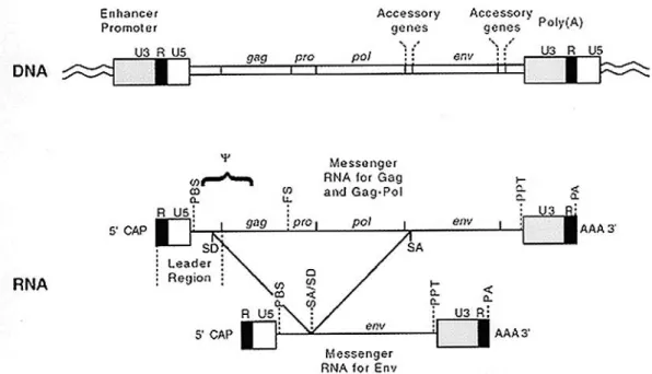
The Retroviral Genome and its Organization
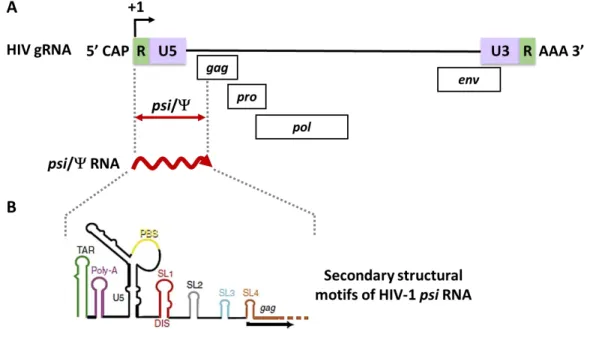
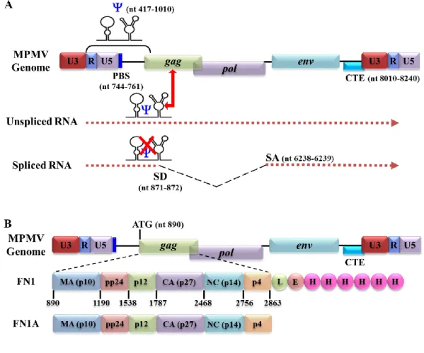
The 5' LTR of the retroviral genome also consists of sequences necessary for the packaging of retroviral genomic RNA (gRNA). Gag binding sites that are involved in the specific binding of the Gag polyprotein to packaging sequences are responsible for increasing the selective packaging of retroviral RNA (Ali et al., 2016; Dubois et al., 2018a; Kuzembayeva et al. , 2014; Rizvi et al., 2010; Webb et al., 2013).
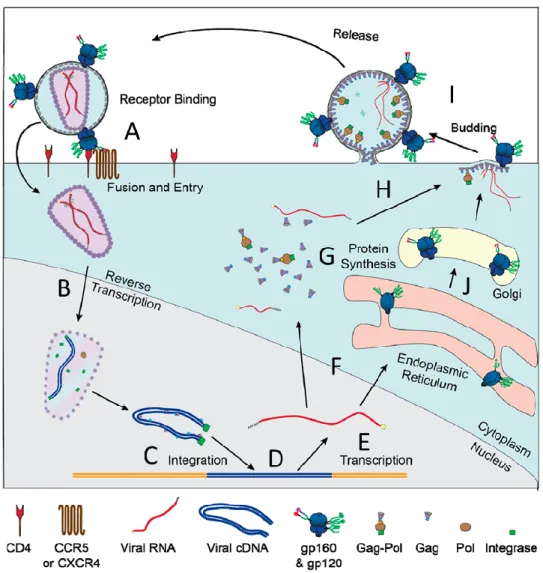
Retroviral Life Cycle
- Fusion and Entry
- Uncoating, Reverse Transcription, and Integration
- Transcription and Nuclear Export
- Translation
- Unspliced Full Length RNA: Packaging versus Translation
- Assembly, Budding, and Release
- Maturation
Once the viral transcripts are exported from the nucleus and into the cytoplasm, they are translated into the appropriate proteins using the cellular translation machinery (Figure 7G). After Gag translation in the cytoplasm, a small amount of RSV Gag translocates back to the nucleus, binds to uncleaved gRNA, and exports the gRNA from the nucleus for packaging (Maldonado et al., 2020).
Retroviral Gag-gRNA Interactions
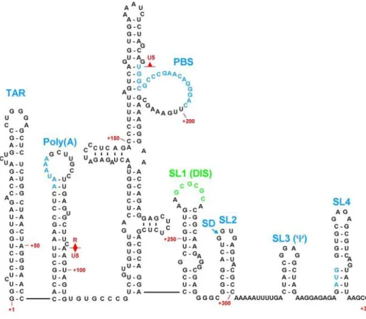
Recent in vitro biochemical studies performed using the full HIV-1 Pr55Gag have shown that a unique high affinity binding is located within an internal loop of the SL1 (Figure 11; Abd El-Wahab et al., 2014). It is likely that these two regulatory regions may function through a putative LRI that has yet to be identified (Figure 12; Abd El-Wahab et al., 2014).
Mason-Pfizer Monkey Virus
The CTE of MPMV has also been successfully used to significantly increase the titer of MLV-based vectors (Zhao et al., 2000). However, it is crucial that the relevant aspects of MPMV replication are fully understood before MPMV-based vectors can be used for human gene therapy.
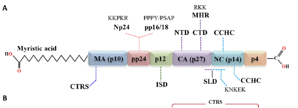
MPMV Gag Polyprotein: Pr78 Gag
- Matrix (MA)
- Capsid (CA)
- Nucleocapsid (NC)
- Phosphoprotein 24 (pp24)
- p12
The structure of the CA-CTD is known to be conserved among many retroviruses, while the structure of the NTD varies significantly (Bharat et al., 2012; Schur et al. This region was followed by a nonspecific stretch of basic residues (similar to the canonical interaction domain (I) of the retrovirus NC), which was found to be essential for the assembly function of the gap-like domain (Bohmová et al., 2010).
The MPMV Packaging Signal RNA
Deletion analysis of this region in MPMV revealed a significant reduction in in vitro RNA dimerization capacity (Aktar et al., 2013. A more detailed analysis of the secondary structural motifs of MPMV packaging sequences identified two important purine-rich regions.
Objectives
The background, the method used and the results obtained regarding this specific purpose have been discussed in Chapter 3 of this thesis. Specific objective III: Establish biological correlation between the Pr78Gag binding site(s) and MPMV gRNA packaging and propagation. This was achieved by comparing the results obtained in Specific Objective 2 with those recently published using a biologically relevant gRNA packaging and propagation assay (Ali et al., 2020).
The background and results obtained in relation to this specific objective were discussed in Chapter 3 of this dissertation.
Abstract
Introduction
Together, these observations suggest that specific selection of gRNA from cellular and spliced RNAs is a complex phenomenon that takes place in the context of the entire Gag polyprotein, as recently shown for HIV-1 (Abd El-Wahab et al., 2014; Bernacchi et al., 2017;Smyth et al. In addition, the sequences downstream of this region enhance the packaging of HIV-1 RNA, while the upstream sequences inhibit the packaging efficiency (Abd El-Wahab et al., 2014) achieved by a protease ( PR) encoded by the virally encoded progene (Bradac & Hunter, 1984; Henderson et al., 1985; Hrusková-Heidingsfeldová et al., 1995).
This was followed by immobilized metal affinity chromatography (IMAC) to purify the protein to homogeneity using high pressure liquid chromatography (HPLC) (Bewley et al., 2017; McKinstry et al., 2014; Tanwar et al., 2017).
Materials and Methods
- Nucleotide Numbering System
- Construction of Prokaryotic MPMV Gag Expression Plasmids
- Construction of Eukaryotic MPMV Gag Expression Plasmids
- Bacterial Strains and Media
- Large Scale Expression of Recombinant Pr78 Gag -His 6 -
- IMAC Protein Purification and Size Exclusion
- SDS-polyacrylamide Gel Electrophoresis and Western
- Eukaryotic Expression of Recombinant Pr78 Gag -His 6 -tag
- Reverse Transcriptase Polymerase Chain Reaction (RT-PCR)
- VLP Production in Prokaryotic Cells and Transmission
- In Vitro Assembly of Recombinant Pr78 Gag -His 6 -tag
After HisTRAPTM column elution, Pr78Gag-His6-tagged protein was concentrated using Amicon® Ultra molecular weight cut-off membrane) and was further fractionated by gel filtration/size exclusion chromatography using a Superdex GL column ( GE Healthcare) previously equilibrated with 50 mM Tris. -HCl (pH 8.0) and 1.0 M NaCl. Expression and purification of recombinant Pr78Gag-His6-tagged protein was monitored by SDS-PAGE and western blot. To monitor the formation of VLPs by recombinant Pr78Gag-His6 tag protein in bacterial cells (after induction with IPTG), cells were pelleted, washed with 0.1 M PBS and fixed in Karnovsky's fixative.
To observe the in vitro assembly of VLPs, purified recombinant Pr78Gag-His6 protein was mixed (in 20 mM Tris (pH 7.4) containing 1.0 M NaCl and 10 mM DTT.

Results and Discussion
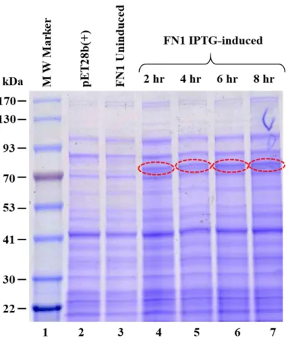
- Bacterial Expression of Recombinant MPMV Pr78 Gag -His 6 -tag
- The Bacterially-expressed MPMV Pr78 Gag -His 6 -tag Protein
- MPMV Pr78 Gag -His 6 -tag Protein is Expressed in the Soluble
- Further Purification of the Soluble Fraction Containing
- Concentration and Further Purification of the IMAC-Purified
- The Recombinant Pr78 Gag -His 6 -tag Protein Can Assemble
- Recombinant Pr78 Gag -His 6 -tag Protein Expressed in Eukaryotic
The IMAC-purified protein was then monitored for purity of recombinant MPMV Pr78Gag-His6-tag protein by SDS-PAGE and immunoblotting. To establish the purity of the full-length recombinant MPMV Pr78Gag-His6-tag Gag fusion, the A260/A280 ratio of the protein was measured spectrophotometrically. Therefore, the ability of purified full-length MPMV Pr78Gag-His6-tagged fusion protein to assemble in vitro to form VLPs was tested.
-F) Transmission electron micrographs showing in vitro assembled virus-like particles from purified recombinant full-length MPMV Pr78Gag-His6 tag fusion protein expressed from FN1 in the presence of yeast tRNA.
Funding
Amplification of the SJ2-specific cDNAs isolated from Gag VLPs formed by either the MPMV Pr78Gag-His6 tag fusion protein (FN7) or without His6 tag (FN9) revealed that transfer vector RNA was efficiently packaged by both types of VLPs (Figure 24C panels V and VI). Interestingly, the VLPs formed by the C-terminal His6-tagged version of the Gag protein appeared slightly more efficient in encapsulating the transfer vector RNA than its untagged version (Figure 24C; panels V and VI). Since the Gag NC is thought to be the main domain involved in the interaction with the genomic RNA using its positively charged zinc finger domains, addition of the His6 tag may have increased the basic nature of the polyprotein and stabilized its interaction with the RNA. further.
Therefore, the slight increase in packaging may be attributed to the increased positive charge added to the full-length MPMV Pr78Gag due to the presence of positively charged His6 tag as recently suggested for HIV-1 Pr55Gag nucleic acid interaction in vitro (Bewley et al. , 2017).
Identification of Pr78 Gag Binding Sites on the Mason-Pfizer Monkey
Abstract
Introduction
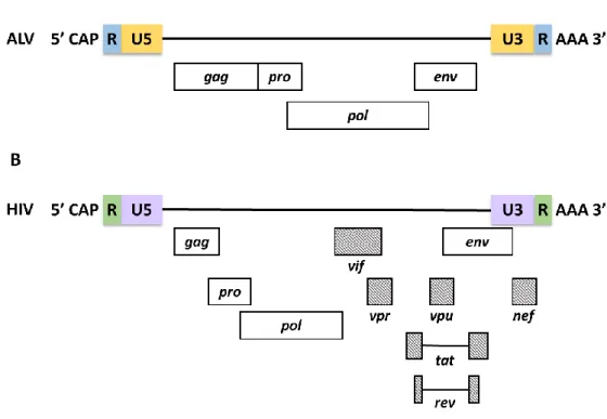
However, the use of full-length HIV-1 Pr55Gag has revealed the presence of a high affinity binding site on the internal (G//AGG) loop of SL1 of the HIV Psi RNA (Abd El-Wahab et al., 2014) . These sequences extend from the 5' UTR to the gag open reading frame, similar to most other retroviruses (Ali et al., 2016). This observation is consistent with HIV-1 where the primary Gag binding site (G//AGG) is located on the internal loop of SL1, the apical loop of which also functions as DIS (Abd El-Wahab et al., 2014).
Interestingly, the last half of the ssPurin sequence repeats downstream in a base-pair fashion to form bpPurins (Figure 25B; Jaballah et al., 2010).
Materials & Methods
- Nucleotide Numbering System
- Expression and Purification of Pr78 Gag
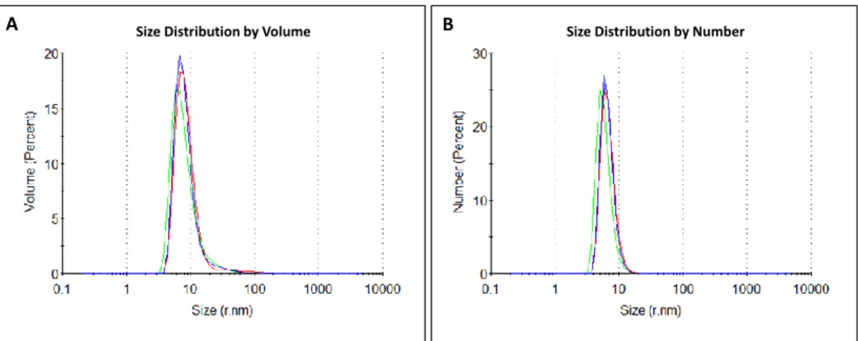
- Physical Characterization of Pr78 Gag by Dynamic Light
- Plasmid Construction for Spliced env, ss- and bp-Purines
- In vitro Transcription and Purification of Unlabeled and
- Band-Shift and Competitive Band-Shift Assays
- RNA Footprinting and High-throughput Selective 2’-Hydroxyl
Briefly, reverse transcription of modified and unmodified RNAs was performed using two arrays (OTR 18/19 and 22/23). To extend both the modified and unmodified sample, 1 µl of each of the VIC-labeled primers OTR 18 (1 µM) and 22 (2 µM) was added to the resuspended RNA and incubated for 2 min at 90 °C, then cooled on ice for 2 min. 2 µl of 5X RT buffer was added to each of the samples and incubated at room temperature for 10 minutes.
The statistical significance between the SHAPE reactivities of each of the nucleotides obtained in the presence and absence of Pr78Gag was calculated using a paired two-tailed t-test and a significant decrease or increase in hSHAPE reactivity with ' A p-value less than <0.05 was considered significant (Appendix C and D).
Results
- Characterization of Recombinant Full-length Purified MPMV
- Pr78 Gag Discriminates between Full-length, Unspliced
- Pr78 Gag Binds Redundantly to both the ssPurines and
- MPMV Pr78 Gag Binds to the ssPurines, bpPurines, and
- Pr78 Gag Footprints in the Absence of the ssPurines indicate
- The ssPurines is Partially Base Paired in the Spliced env RNA
Average reactivity data from triplicate experiments with protein was applied to the secondary structure of the MPMV packaging signal obtained in the absence of Pr78Gag. In the case of bpPurines, only 2 out of 8 of the nucleotides indicated significant attenuation in reactivities and thus potential Gag binding. Given the extended binding pattern of Pr78Gag observed at the ssPurines region, we wanted to determine the binding pattern of Pr78Gag to the packaging signal RNA in the absence of the ssPurines.
The ssPurines achieve a partial base-paired conformation in the secondary structure of the spliced env RNA.
Discussion
The latter is located downstream of the mSD; thus, it is present only in genomic and unspliced RNA. Regardless, these data suggest that the ssPurinas and the single-stranded GU region (A252AGUGU257) function redundantly, leading to the provocative question of how the presence of Pr78Gag binding sites upstream of the mSD in the spliced env RNA overcomes binding selective with Gag precursor proteins. The answer probably lies in the effect of splicing on the structure of Gag binding sites.
The presence of specific Gag binding sites upstream of the mSD has been observed for a number of retroviruses, including HIV-1 (Abd El-Wahab et al., 2014) and MMTV (Chameetachal et al., 2021).
Funding
Based on structural differences, results here also show that Pr78Gag can effectively distinguish unspliced MPMV Psi RNA from spliced env RNA, revealing how MPMV differentiates between the two RNA substrates. Thus, identification of structural elements crucial in MPMV gRNA packaging should help understand not only the mechanism of virion assembly by retroviruses, but also facilitate the development of safe and effective retroviral vectors for human gene therapy.
Conclusions & Future Directions
Conclusions
The results of such assays, performed using various deletion mutants of the purine-rich motifs, revealed that both regions bind to Pr78Gag in a redundant manner. Finally, footprinting experiments followed by hSHAPE mapped Pr78Gag binding to two single-stranded loops: 1) ssPurins and 2) the A252AGUGUU258 loop, which corresponds to two 3' unpaired adenosine nucleotides of bpPurins and GU-rich regions. In addition, imprinting experiments performed on FN26, a mutant containing a deletion of the major ssPurin imprinting region, revealed binding of Pr78Gag to bpPurin nucleotides and a GU-rich region, recapitulating the redundant ability of Pr78Gag to bind to this region in the absence of ssPurins.
Notably, ssPurines are located upstream of the mSD and are therefore present in both unspliced and spliced RNAs, and the A252AGUGUU258 loop is located downstream of the mSD; thus, present only in unspliced RNA.
Future Directions
Mutations in the Basic Region of the Mason-Pfizer Monkey Virus Nucleocapsid Protein Affect Inversely. Membrane interactions of the Mason-Pfizer simian virus matrix protein and its budding-defective mutants. NMR structure of the N-terminal domain of capsid protein of the mason-pfizer simian virus.
Mutational analysis of the predicted RNA secondary structure of the Mason-Pfizer simian virus packaging signal. Mutational analysis of the bipartite dimer binding structure of human immunodeficiency virus type 1 genomic RNA. Identification of the primary site of human immunodeficiency virus type 1 RNA dimerization in vitro.