ANTIMICROBIAL PROPERTIES AND PHYTOCHEMICAL ANALYSIS OF MEDICINAL PLANTS USED FOR THE TREATMENT OF EAR INFECTIONS
By
SINORITA CHAUKE
DISSERTATION
Submitted in fulfilment of the requirements for the degree of MASTER OF SCIENCE
In
BOTANY
in the
FACULTY OF SCIENCE AND AGRICULTURE (School of Molecular and Life Sciences)
at the
UNIVERSITY OF LIMPOPO
SUPERVISOR: Prof. SM Mahlo
2023
i DECLARATION
I declare that the dissertation hereby submitted to the University of Limpopo, for the degree of Master of Science in Botany has not previously been submitted by me for a degree at this or any other university; that it is my work in design and in execution, and that all material contained herein has been duly acknowledged.
Chauke, S (Ms) Date
ii DEDICATION
I dedicate this work to my loving and supportive parents Mr. D.M and Mrs. L. Chauke.
iii ACKNOWLEDGEMENTS
I thank God almighty for his undying love and endless mercies, for giving me wisdom and the strength needed to initiate, plan, implement, and establish this research project. I am grateful for his full support, as well as the people he has blessed me with, who have helped me in every way possible. In addition, I acknowledge the protection and preservation that has been bestowed upon me by God so far.
My heartfelt thanks go to my supervisor, Professor S.M Mahlo for her guidance, patience, support, and humility. It is truly an honour to have worked with a woman of her stature on this project.
My sincere gratitude to my father, Mr. D.M Chauke for always being a champion for education, and always encouraging me to go as far as I can in my studies. I also thank my father for supporting and helping me with plant collections.
I am grateful to Mr. P. Makukule, Mr. D. and Mrs. L. Sibuyi for allowing me to collect some plant species in their homes.
I thank my mother, Mrs. L. Chauke for her endless encouragement, love, and care always and throughout the period of study.
To my close friend E.M Kgoedi, I am grateful for always praying for me and praying with me.
I appreciate Miss T.C Machaba and Miss T.T Ramavhale for assisting me with laboratory experiments.
I am grateful to my father, the University of Limpopo, and the Council for Scientific and Industrial Research Inter-Bursary Support (CSIR-IBS) for providing substantial financial support toward my studies.
iv ABSTRACT
Ear infections are a major health concern that negatively affects the health and welfare of individuals across the globe. The infection is caused by a wide spectrum of bacterial, fungal, and viral pathogens. Treatments of ear infections involve the use of antimicrobials such as antibiotics, antifungals, and antivirals. However, most microbial pathogens have developed resistance to the available antimicrobial drugs. Hence, the study aimed to identify plant species used in traditional medicine as a remedy for ear infections and investigate their antifungal activities against the selected fungal pathogens (Aspergillus fumigatus and Candida albicans). These fungal pathogens cause ear infections in humans. Eight plant species including Carpobrotus edulis L., Cotyledon orbiculata L., Dichrostachys cinerea (L.) Wight & Arn., Erythrina lysistemon Hutch., Flacourtia indica (Burm. f.) Merr., Psidium guajava L., Ricinus communis L., and Sansevieria hyacinthoides (L.) Druce were selected from the ethnomedicinal plant's database of over 300 medicinal plants used for therapeutic purposes in humans.
Fresh and dried leaves of selected plants were extracted with solvents of various polarities such as acetone, hexane, methanol, and water. In the current study, methanol extracted a larger quantity (30.75%) of plant materials followed by acetone (6.5%) from dried leaf extract of C. edulis and P. guajava. Acetone extracted more plant material (8.05%) from fresh leaf extract of C. orbiculata. Acetone was the second-best solvent for extracting a larger quantity of dried leaf materials as compared to other solvents.
Thin layer chromatography (TLC) was used to analyse the phyto-constituents of different plant extracts. The TLC plates were developed using different eluent solvent systems such as Benzene: ethanol: ammonia hydroxide (BEA), Chloroform: ethyl acetate: formic acid (CEF) and Ethyl acetate: methanol: water (EMW). The TLC chromatograms were visualized under UV radiation at 360 nm. In TLC chromatograms separated with BEA, chemical components with a similar Rf value of 0.88 were observed in acetone, hexane, and methanol-dried leaf extracts of R. communis and S. hyacinthoides. Surprisingly, TLC chromatograms separated in BEA, dried leaf extracts contained the highest number of phyto-constituents with a total of 73 followed by 30 in CEF and EMW (29). However, in chromatograms of fresh leaf extracts a total
v
of 12 compounds were visible in BEA, followed by 5 compounds in EMW, and 1 in CEF. Therefore, the BEA solvent system was the best eluent for separating compounds. In addition, different bands were observed after spraying the TLC plates with vanillin reagent.
Antifungal activities of plant extracts were determined using serial microdilution assay against the selected fungal pathogens (Aspergillus fumigatus and Candida albicans).
Noteworthy activities (0.02 mg/ml) against C. albicans were observed from P. guajava acetone extract and S. hyacinthoides acetone fresh leaf extracts. The methanol-dried leaf extract of C. edulis was active against A. fumigatus with MIC of 0.02 mg/ml while fresh leaf extract was active with MIC of 0.31–2.5 mg/ml. The dried leaf water extracts of C. edulis and D. cinerea had an excellent activity of 0.02 mg/ml against A. fumigatus.
The bioautography assay was used to determine the number of active components in different plant extracts. Antifungal compounds were visible in dried leaf extracts of P.
guajava, R. communis, and S. hyacinthoides. A total of 19 antifungal compounds were observed against A. fumigatus. Dried leaf extracts of P. guajava, R. communis, and S.
hyacinthoides had an active component with an Rf value of 0.88 against A. fumigatus.
In TLC bioautograms developed in BEA, two active compounds with similar Rf values of 0.20 were visible in acetone and methanol extract of P. guajava against C. albicans.
The results of this study support the traditional use of the selected plant species to combat ear infections and related ailments in humans. The crude extracts have the potential to serve as an ototopic. The antifungal compounds also have the potential to be isolated and used in the formulation of ototopical drugs that may help lift the health burden caused by ear infections across the globe.
vi ABBREVIATIONS
A Acetone
A.F Aspergillus fumigatus
AIDS Acquired Immunodeficiency Syndrome AOE Acute Otitis Externa
AOM Acute Otitis Media
BEA Benzene: Ethyl acetate: Acetone C.A Candida albicans
CEF Chloroform: Ethyl acetate: Formic acid COE Chronic Otitis Externa
COM Chronic Otitis Media
CSOM Chronic Suppurative Otitis Media EMW Ethyl acetate: Methanol: Water
H Hexane
HIV Human Immunodeficiency Virus INT p-Iodonitrotetrazolium Violet
M Methanol
MIC Minimum Inhibitory Concentration NOE Necrotizing Otitis Externa
OE Otitis Externa OI Otitis Interna
OM Otitis Media
Rf Retention Factor
SD Sabouraud Dextrose
TLC Thin-Layer Chromatography URTI Upper Respiratory Tract Infection URVI Upper Respiratory Viral Infection W Distilled Water
vii Table of Contents
CHAPTER 1 ... 1
INTRODUCTION ... 1
1.1 Medicinal plants ... 1
1.2 Rationale... 2
1.3 Aim ... 4
1.4 Objectives ... 4
1.5 Outline of study ... 5
CHAPTER 2 ... 6
LITERATURE REVIEW... 6
2.1 Introduction ... 6
2.2 Indigenous knowledge ... 6
2.3 Importance of traditional medicine ... 6
2.4 Conservation of medicinal plants ... 7
2.5 Use of medicinal plants in drug production ... 7
2.6 Traditional treatments for ear infections ... 9
2.7 Ear infections ... 9
2.7.1 Types of ear infections ... 9
2.7.2 Causes or risk factors of ear infections ... 12
2.7.3 Microbial pathogens that cause ear infections ... 13
2.8 Extracranial complications of ear infections ... 14
2.8.1 Labyrinthitis ... 14
2.8.2 Facial nerve paralysis ... 14
2.8.3 Mastoiditis ... 14
2.9 Ear infections and URTIs ... 14
2.10 Ear infections and HIV/AIDS ... 15
2.11 Antibiotic treatment of ear infections caused by bacteria ... 15
2.11.1 Oral antibiotics ... 15
viii
2.11.2 Topical antibiotics ... 16
2.12 Antifungal treatment of ear infections caused by fungi ... 16
2.13 Antimicrobial drug resistance ... 17
2.14 Fungal pathogens ... 17
2.14.1 Aspergillus fumigatus ... 17
2.14.2 Candida albicans ... 18
2.15 Bacterial pathogens ... 18
2.15.1 Escherichia coli... 18
2.15.2 Pseudomonas aeruginosa ... 19
2.15.3 Staphylococcus aureus ... 19
2.16 Botanical descriptions of the plant species used in the study ... 20
2.16.1 Carpobrotus edulis L. ... 20
2.16.1.1 Ethnomedicinal uses ... 21
2.16.2 Cotyledon orbiculata L. ... 22
2.16.2.1 Ethnomedicinal uses ... 22
2.16.3 Dichrostachys cinerea (L.) Wight & Arn. ... 23
2.16.3.1 Ethnomedicinal uses ... 23
2.16.4 Erythrina lysistemon Hutch. ... 24
2.16.4.1 Ethnomedicinal uses ... 24
2.16.5 Flacourtia indica (Burm. f.) Merr. ... 25
2.16.5.1 Ethnomedicinal uses ... 26
2.16.6 Psidium guajava L. ... 26
2.16.6.1 Ethnomedicinal uses ... 27
2.16.7 Ricinus communis L. ... 28
2.16.7.1 Ethnomedicinal uses ... 29
2.16.8 Sansevieria hyacinthoides (L.) Druce ... 30
2.16.8.1 Ethnomedicinal uses ... 30
ix
2.17 Conclusion ... 32
CHAPTER 3 ... 33
PLANT EXTRACTION AND PHYTOCHEMICAL ANALYSIS OF SELECTED PLANT SPECIES ... 33
3.1 Introduction ... 33
3.2 Materials and methods ... 34
3.2.1 Plant selection and collection ... 34
3.2.2 Extraction of dried plant material ... 35
3.2.3 Extraction of fresh plant material. ... 36
3.2.4 Phytochemical analysis ... 38
3.3 Results and discussion ... 38
3.3.1 Extraction of dried and fresh plant material ... 38
3.3.2 Phytochemical analysis ... 41
3.4 Conclusion ... 43
CHAPTER 4 ... 44
ANTIFUNGAL ACTIVITIES OF SELECTED PLANT SPECIES AGAINST FUNGAL PATHOGENS THAT CAUSE EAR INFECTIONS ... 44
4.1 Introduction ... 44
4.2 Methods and materials ... 45
4.2.1 Fungal strains and inoculum quantification ... 45
4.2.2 Serial microdilution assay ... 45
4.2.3 Bioautography assay ... 46
4.3 Results and discussion ... 46
4.3.1 Serial microdilution assay ... 46
4.3.2 Plant extracts with excellent antifungal activity ... 47
4.3.3 Total activity of plant extracts ... 47
4.3.4 Bioautography assay ... 52
4.4 Conclusion ... 57
x
CHAPTER 5 ... 58 CONCLUSION ... 58 REFERENCES ... 61
xi List of Figures
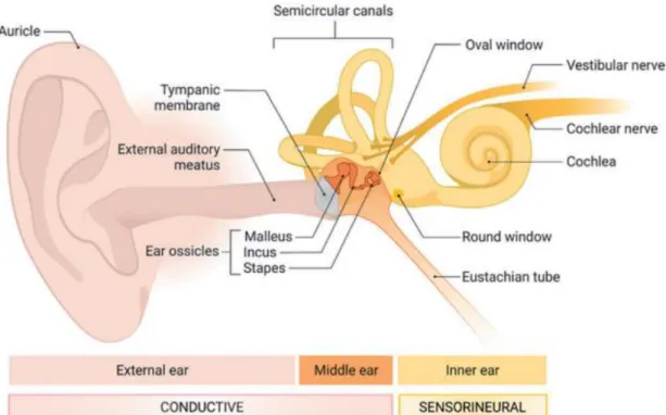
Figure 2. 1 The structure of the ear with indications of the main compartments of the ear, namely: Outer ear, middle ear, and inner ear (Payne and Wong, 2022).
... 10
Figure 2. 2 Flow chart indicating the three parts of the ear and the type of infection. Images from left to right: Otoscopic images of OE (Osguthorpe and Nielsen, 2011), OM (Bhutta et al., 2017), and 3D T2 TSE MRI scan of OI (Wu et al., 2014). ... 12
Figure 2. 3 Microscopic image of Aspergillus fumigatus (Adhavan, 2020). ... 17
Figure 2. 4 Microscopic image of Candida albicans (Tyavambiza, 2018)... 18
Figure 2. 5 Microscopic image of Escherichia coli (Singh et al., 2015)... 19
Figure 2. 6 Microscopic image of Pseudomonas aeruginosa (Tyavambiza, 2018). . 19
Figure 2. 7 Microscopic image of Staphylococcus aureus (Tyavambiza, 2018). ... 20
Figure 2. 8 Images of Carpobrotus edulis L. (Bilomu ra ku nava) leaves (A) and flower (B). ... 21
Figure 2. 9 Cotyledon orbiculata L. (Bilomu ra tindleve) whole plant (A), leaves (B) and flowers (C). ... 23
Figure 2. 10 Dichrostachys cinerea (L.) Wight & Arn. (Ndzhenga) whole plant (A), leaves (B), and flowers (C). ... Figure 2. 11 Erythrina lysistemon Hutch. (Muvale/ Nsisimbana) whole plant (A), leaves (B), flowers (C), and pods (D). ... 25
Figure 2. 12 Flacourtia indica (Burm. f.) Merr. (Xivambula) whole plant (A), leaves (B), and flowers (C). ... 26
Figure 2. 13 Psidium guajava L. (Mugwava) whole plant (A), leaves (B), flowers (C), and fruit (D). ... 28
Figure 2. 14 Ricinus communis L. (Nhlampfurha) whole plant (A), leaves (B), flowers and fruit (C). ... 30
Figure 2. 15 Sansevieria hyacinthoides (L.) Druce (Mhangane) whole plant (A). ... 31
Figure 3. 1 A map showing study areas in Bushbuckridge Local Municipality. 35 Figure 3. 2 Schematic representation of the extraction procedure for dried and fresh leaf materials. ... 37
Figure 3. 3 The percentage of crude extracts from dried leaves of different plants extracted using acetone, hexane, and methanol. ... 40
xii
Figure 3. 4 Percentage of crude extracts from fresh leaves of succulent plants extracted using acetone, hexane, and methanol. ... 40 Figure 3. 5 TLC chromatograms of dried leaf extracts of C. edulis, C. orbiculata, D.
cinerea, E. lysistemon, F. indica, P. guajava, R. communis, and S.
hyacinthoides in BEA, CEF, and EMW. Lanes from left to right: A= Acetone, H= Hexane, M= Methanol, and W= water. ... 42 Figure 4. 1 Bioautograms of F. indica, P. guajava, R. communis, and S. hyacinthoides
leaf extracts developed in BEA, CEF and EMW solvent systems with their corresponding chromatograms. White bands indicating compounds with antifungal activity against A. fumigatus. Lanes from left to right: A= Acetone, H= Hexane, M= Methanol and W= Distilled water. ... 53 Figure 4. 2 Bioautograms of R. communis and S. hyacinthoides leaf extracts developed in CEF and EMW solvent systems with their corresponding chromatograms. White bands indicating compounds with antifungal activity against A. fumigatus. Lanes from left to right: A= Acetone, H= Hexane, M=
Methanol and W= Distilled water. ... 54 Figure 4. 3 Bioautogram and corresponding chromatogram of F. indica and P. guajava in BEA with white bands indicating compounds with antifungal activity against C. albicans. Lanes from left to right: A=Acetone, H=Hexane, M=Methanol and W= Distilled water. ... 55
xiii List of tables
Table 2. 1 Drugs derived from plants or plant products ... 8 Table 4. 1 Minimum inhibitory concentrations of fresh and dried leaves extracts of eight
different plant species tested against two fungal pathogens………..49 Table 4. 2 Plant extracts with excellent antifungal activity (0.02 mg/ml) against the tested fungal pathogens at 24 and 48 hours. ... 50 Table 4. 3 Total activity of plant extracts against A. fumigatus and C. albicans. ... 51 Table 4. 4 Rf values of compounds inhibiting fungal growth separated with BEA and
EMW using dried leaf extracts of P. guajava. ... 55 Table 4. 5 Rf values of compounds inhibiting fungal growth separated with BEA, CEF
and EMW using dried leaf extracts of R. communis. ... 55 Table 4. 6 Rf values of compounds inhibiting fungal growth separated with CEF and
EMW using dried leaf extracts of S. hyacinthoides. ... 56
1 CHAPTER 1
INTRODUCTION 1.1 Medicinal plants
Medicinal plants continue to play an important role in securing the health of people in African developing countries (Agidew, 2022). In South Africa, local people in rural and urban areas rely on herbal medicines for their primary healthcare since it is cheap, easily accessible, and safe (Fennel et al., 2004). Hence, medicinal plants are receiving global recognition due to their importance in healthcare (Aburigal et al., 2022). In Limpopo province, local people and traditional health practitioners use medicinal plants to combat various ailments in humans (Shikwambana and Mahlo, 2020). The local community prefers medicinal plants for medicinal purposes since they are effective, easily accessible, and low-cost. Hence, there is a growing interest in the study of medicinal plants (Larida et al., 2022). Herbal medicine is a major component of South African cultures and traditions. This medicine is also known as phytomedicine which uses plants for medicinal purposes. More so, herbal medicines are used to vitalise the immune system to prevent and cure diseases (Taylor et al., 2001).
Medicinal plants are an essential source of medicine with effectual properties to treat various ailments in humans. More importantly, these plants contain active components that are used to cure diseases and ease pain (Madhu et al., 2016). The chemical compounds present in the different plant parts may have physiological properties in the human body. Plant parts such as flowers, fruits, leaves, resins, rhizomes, roots, seeds, and stems are used in traditional medicine (Alqethami and Aldhebiani, 2021).
Medicinal plants provide a great source of information for a wide diversity of chemical compounds that can be used to derive the drugs that will be effective against various ailments in humans (Yadav et al., 2014). Furthermore, medicinal plant drugs are divided into two categories. Firstly, they are incorporated in mixtures containing different compounds, like infusions, essential oils, tinctures, or extracts. Secondly, they are used as pure, chemically defined active principles (Taylor et al., 2001).
Plants undergo a metabolic process that results in primary and secondary metabolites.
Primary metabolites such as amino acids, simple sugars (glucosides), proteins, and lipids are involved in cellular processes. Secondary metabolites are chemically active
2
compounds produced in response to stress with complex structures and more limited distribution than primary metabolites (Kumari et al., 2017). Secondary metabolites are classified into different groups based on their structure. The most important of these secondary metabolites with medicinal properties are alkaloids, glycosides, flavonoids, tannins, saponins, and resins. These bioactive compounds vary depending on the plant in which they are produced (Alqethami and Aldhebiani, 2021).
The chemical compounds that are naturally found in plants are called phytochemicals.
They are responsible for the colour and organoleptic properties of the plant.
Phytochemicals could be available as dietary supplements, but the potential health benefits of phytochemicals are derived from the consumption of the whole plant.
Several phytochemicals have a wide range of activities that helps give immunity against long-term diseases (Yadav et al., 2017). It has been discovered that phytochemical compounds have antibacterial and antifungal activities (Rangasamy et al., 2007). In this dissertation, the study investigates medicinal plants used for the treatment of ear infections by traditional health practitioners and local people residing in Limpopo province (Capricorn District) and Mpumalanga province (Ehlanzeni District). Based on the literature, the study has never been conducted previously in the two Districts.
1.2 Rationale
An ear infection is caused by bacteria, fungi, and viruses in the ear (Wolk, 2016).
These microorganisms accumulate in the ear discharge and cause inflammation, especially in the middle and external ear (Appiah-Korang et al., 2014). Furthermore, the infections may lead to ear tumours, metastatic tumours, or primary tumours in the surrounding organs (Mahmoudian-Sani et al., 2017). The overuse and misuse of antibiotics have caused microbial pathogens to be more resistant to methicillin and vancomycin (Aneja et al., 2012). Recently, drugs such as quinolones, amoxicillin- clavulanate, levofloxacin, moxifloxacin, and ciprofloxacin are used to combat ear infections in humans (Szmuilowicz and Young, 2019). However, some of these drugs may be toxic and ineffective due to antimicrobial drug resistance developed by pathogens (Chong et al., 2021). Screening of medicinal plants with potential antimicrobial properties can lead to the discovery of novel antifungal agents that could resolve fungal ear infections. Therefore, the study focused on investigating medicinal
3
plants that are used by traditional health practitioners for the treatment of ear infections.
South Africa is rich in the knowledge of plants used in traditional medicines which need to be justified by scientific evidence (Dyubeni and Buwa, 2012). Plants are a good source of bioactive compounds with antimicrobial activity (Gunatilaka, 2006).
Furthermore, these plants contain phytochemicals that have therapeutic properties that are affordable and less toxic (Otimanam et al., 2022). The World Health Organization (WHO) also regards medicinal plants as the best source of a variety of drugs (Yadav and Agarwala, 2011). About 78% of new chemical compounds derived from medicinal plants are used as a potential alternative to treat various infections in humans (Mustafa et al., 2017). Aspergillus fumigatus and Candida albicans are the most common fungal pathogens responsible for ear infections (Prasad et al., 2014).
The use of plant-based natural products in drug development may lead to the discovery of new bioactive compounds that can provide effective curative agents for microbial infections. According to Dewatisari et al. (2022), medicinal plants are the solution to the rising concern of antimicrobial drug resistance. In previous studies, notable antimicrobial activities of some plant extracts have been observed (Kebede et al., 2021). Ear infections in children are a major health concern and may be associated with hearing impairment, and delayed speech, and language development (Karunanayake et al., 2016). These infections affect over 90% of children with 20% of the infections being chronic (Ryan et al., 2020). It is estimated that 60% of the cases of ear infections in humans may lead to disabling hearing loss or death in extreme conditions (Hailu et al., 2016). Hence, these infections account for a large proportion of the health concerns in developing countries (Sahu et al., 2014). Without the discovery of new compounds through various methods such as the screening of medicinal plants, this major health burden may continue to rise.
The invention of a new antimicrobial drug or eardrop solution from plant-based natural products could be a solution. Most treatments for ear infections are western, however, some of these drugs are less effective because of resistance to antimicrobial drugs (Yang et al., 2022) and may have adverse side effects on humans (Magdy et al., 2022) hence, there is a need to investigate a different approach that is used in traditional
4
medicine. Local people and traditional health practitioners use different medicinal plants and approaches to treat ear infections and other diseases. However, there is a lack of scientific evidence to support the ethnomedicinal use of these plants to combat ear infections and related ailments. Therefore, there is a need to focus research on traditional medicine and phytotherapy since they can provide novel drugs that are effective, cheap, and non-toxic (Mahmoudian-Sani et al., 2017).
1.3 Aim
The study aimed to select medicinal plants used to treat ear infections from a database of ethnomedicinal plants and to determine the antimicrobial activity of the selected plants against fungal pathogens.
1.4 Objectives
The objectives were to:
i. select eight plant species used to treat ear infections from a database of ethnomedicinal plant species for further phytochemical analysis and biological assays.
ii. investigate the chemical components of various plant extracts.
iii. determine the antifungal activity of acetone, hexane, methanol, and water extracts of selected plants against Aspergillus fumigatus and Candida albicans.
iv. investigate the effectiveness of the fresh and dried leaf materials against the tested fungal pathogens.
v. determine the number of antifungal compounds in different plant extracts.
5 1.5 Outline of study
Chapter 1 entails the general background of the importance of medicinal plants, the rationale, aim, and objectives of the study.
Chapter 2 entails a more detailed literature review of the use of medicinal plants in traditional medicine and drug production as well as a literature review of the different types of ear infections, causes, complications, and treatments. The botanical descriptions and literature review of ethnomedicinal uses of selected plants are discussed, and the conclusion is also given.
Chapter 3 deals with the methods employed during plant extraction and phytochemical analysis and their respective results, discussions, and conclusions.
Chapter 4 focuses on the antifungal activities of different plant extracts. The methodologies for serial microdilution and bioautography assays are outlined followed by the results, discussion, and conclusions.
Chapter 5 focuses on the summary and overall conclusion of the study.
Recommendations for future work have been given and the references that were used for the study are listed.
6 CHAPTER 2
LITERATURE REVIEW 2.1 Introduction
This chapter deals with a literature review on medicinal plants, traditional medicine, treatment of ear infections, antimicrobials used for ear infections, indigenous knowledge, and conservation of medicinal plants. Thorough literature reviews were conducted and compared with the previous and current findings on the uses of different plant species.
2.2 Indigenous knowledge
Indigenous knowledge is the set of knowledge, rules, standards, skills, and mentalities generated by native people of an area (Kebebew, 2016). The native people are responsible for protecting their indigenous knowledge (Emmanuel and Didier, 2012).
This indigenous knowledge is transferred from one generation to the next through oral communication (Shikwambana and Mahlo, 2020). However, it is quite difficult to obtain traditional knowledge from indigenous people due to the lack of trust (Zerabruk and Yirga, 2012). There is a lack of documentation on the use of medicinal plants to combat various diseases in humans, in Limpopo and Mpumalanga provinces. Hence, it necessitated the research to focus on the medicinal usage of plant species for the treatment of ear infections and related ailments in humans.
2.3 Importance of traditional medicine
Traditional medicine refers to the knowledge, beliefs, and practices that are applied in the diagnosis, prevention, and elimination of diseases (Kebebew, 2016). Traditional health practitioners provide healthcare needs to the community based on cultural, religious, spiritual, and social beliefs (Zuma et al., 2016). Additionally, South African traditional health practitioners are divided into diviners, faith healers, and herbalists (De Andrade and Ross, 2005). Many people in urban and rural areas across Africa prefer traditional medicine than conventional medicine (Rainatou et al., 2021). South Africans believe that traditional medicine is a form of healthcare practice that different communities use to treat various ailments. It was found that nearly 80% of the South African population utilises traditional medicine for primary healthcare (Mahwasane et
7
al., 2013). The use of plants in traditional medicine is regarded as a form of cultural practice (Zizka et al., 2015). Hence, traditional knowledge of medicinal plants is best sourced from traditional health practitioners. Moreover, traditional health practitioners also believe that medicinal plants must be respected so that they can be effective in treating ailments (Shinwari, 2010). Furthermore, many significant modern drugs have been discovered based on traditional knowledge of medicinal plants (Fabricant and Farnsworth, 2001).
2.4 Conservation of medicinal plants
The increasing population growth is a factor that may lead to the loss of medicinal plants in the wild due to agricultural practices and deforestation (Zerabruk and Yirga, 2012). Overharvesting of medicinal plants is on the rise since communities depend on medicinal plants for their healthcare needs (Chen et al., 2016; Bukuluki et al., 2014).
According to Heywood (2017), the loss of medicinal plants decreases the chances of novel drug discovery. It is, therefore, important to preserve or document the ethnomedicinal knowledge of plants since it is at risk of diminishing (Khan et al., 2018).
Hence, local people and traditional health practitioners are encouraged to grow medicinal plants in their homes. More importantly, they should be taught the conservation measures that need to be taken when collecting endangered plant species.
Traditional health practitioners and local people have methods of conserving medicinal plants. They preserve medicinal plants through selective harvesting, domesticating medicinal plants, and growing medicinal plants at burial sites and sacred forests. In addition, traditional health practitioners may hide the names, uses, and locations of some medicinal plants (Kibonde, 2020). Medicinal plants can also be conserved in situ and ex situ. In situ conservation includes bio reserves, natural parks, and wild nurseries. Ex situ methods of conservation include botanic gardens, field gene banks, and seed banks (Kadam and Pawar, 2020).
2.5 Use of medicinal plants in drug production
Medicinal plants are used to complement conventional medicine due to their manifold uses (De Oliveira Melo et al., 2022). They have affordable and less toxic phytochemicals with potential therapeutic properties (Otimanam et al., 2022). These
8
phytochemicals have good molecular properties such as greater rigidity, lower mass, fewer heavy metals, and structural diversity (Mathur and Hoskins, 2017). They are in demand since they can also produce physiological and pharmacological effects in living cells (Mathur and Hoskins, 2017). Medicinal plants increase the chances of new drug discovery since they are rich in chemically diverse components. The new drugs can be in the form of pure compounds or homogenous extracts (Mustafa et al., 2017).
Hence, plants are used as raw materials for the extraction of active compounds that can be used in drug development (Singh, 2015). These active compounds can be found in different plant parts such as the bark, flowers, leaves, roots, seeds, and stems. Medicinal plants form the basis of many modern medicines (Khumalo et al., 2022). Hence, medicinal plants are regarded as the main source of novel drugs (Srinivas et al., 2013). Several drugs have been developed or isolated from medicinal plants as recorded in table 2.1 (Anand et al., 2019).
Table 2. 1 Drugs derived from plants or plant products
Plant species Plant-derived drugs/molecules Allium sativum L. Allicin (diallylthiosulfnate)
Artemisia annua L. Artemisinin
Atropa belladonna L. Tiotropium bromide (Spiriva®) Camptotheca acuminate Decne. Camptothecin
Cannabis sativa L. Cannabidiol and tetrahydrocannabinol Catharanthus roseus (L.) G. Don Vinblastine and vincristine
Colchicum autumnale L. Colchicine
Digitalis purpurea L. Digoxin and digitoxin Galanthus woronowii Losinsk. Galantamine (Reminyl®) Filipendula ulmaria (L.) Maxim Aspirin
Papaver somniferum L. Codeine, Papaverine and Apomorphine hydrochloride (Apokyn®)
Taxus brevifolia Nutt. Paclitaxel (Taxol®) Taxus brevifolia Nutt.
Taxus chinensis (Pilg.) Rehder
Paclitaxel
9 2.6 Traditional treatments for ear infections
Traditional health practitioners are more knowledgeable about various traditional methods used for the treatment of different ailments in humans, including ear infections. There are different ways used by traditional healers to treat ear infections.
Predominantly, the majority of traditional health practitioners use a variety of plants to treat ear infections. However, some traditional health practitioners use chicken fat, snake fat, millipede, lizard fat, water buffalo fat, sardine fat, powdered tortoise, powdered owl, and powdered monkey brain to treat ear infections (De Andrade and Ross, 2005).
2.7 Ear infections
2.7.1 Types of ear infections
Infections of the ear are clinically called otitis (Neves et al., 2018). Otitis is an inflammation of the ear (Aldhaher et al., 2018). Hence, an ear infection is a term denoted to the inflammation of the ear caused by infectious organisms such as bacteria, yeasts, and viruses (Hegde et al., 2021). Otitis is divided into otitis externa (OE), otitis media (OM), and otitis interna (OI) for the outer, middle, and inner ear respectively (Figure 2.1) (Szmuilowicz and Young, 2019). Symptoms of ear infections include clumsiness, fever, fluid draining from the ear, fussiness, loss of appetite, ringing sounds in the ear, and vomiting (Ayub et al., 2015).
10
Figure 2. 1 The structure of the ear with indications of the main compartments of the ear, namely: Outer ear, middle ear, and inner ear (Payne and Wong, 2022).
OE is an inflammation of the outer ear canal that may extend to the soft tissues surrounding the outer ear (Harris and Viljoen, 2021). OE can be acute or chronic (Bulut et al., 2021). Acute otitis externa (AOE) is usually caused by bacteria while chronic otitis externa (COE) is caused by fungi, allergies, or dermatitides (Osguthorpe and Nielsen, 2021). Factors that contribute to OE include excessive sweating, stress, wearing a hearing aid, and removal of ear wax (Bhat et al., 2015). Symptoms of OE include severe ear pain, blood-stained otorrhea, swelling (Bhat et al., 2015), severe headache, a feeling of fullness in the ear, and hearing loss (Alnawaiseh et al., 2011).
OM refers to infections of the middle ear which are usually related to upper respiratory tract infections (URTIs) (Aldhaher et al., 2018). The main causes of OM are bacteria and viruses (Cho et al., 2015). However, in very rare cases fungal pathogens are also responsible for OM (Bennett et al., 2016). These microorganisms can enter the middle ear from the external ear through a perforated tympanic membrane (Gaur and Khan, 2019). OM can be acute or chronic (Olive-Busom et al., 2021). Chronic Suppurative Otitis Media (CSOM) is the continual discharge through a chronic perforation of the tympanic membrane. Acute otitis media (AOM) results from a viral infection in the
11
respiratory tract leading to damage to the eustachian tube (Siyad and Venkataramanan, 2021). The presence of dense or watery fluid that can cause temporary hearing loss is considered a symptom of both acute and chronic OM (Sabir et al., 2021; Won et al., 2021).
OI is the inflammation of the inner ear which involves the sensory organs (Ayub et al., 2015). OI is mostly regarded as one of the complications following middle ear infections (Madsen et al., 2001). It is also believed that OI can extend from the brain in the presence of meningitis (Madsen et al., 2001). The types of microbial pathogens responsible for OI are of bacterial, fungal, and viral origin (Gheorghe et al., 2021).
Symptoms of OI include labyrinthitis also known as vertigo (Ayub et al., 2015).
12
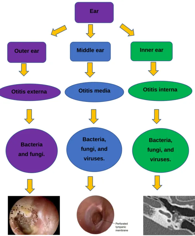
Figure 2. 2 Flow chart indicating the three parts of the ear and the type of infection.
Images from left to right: Otoscopic images of OE (Osguthorpe and Nielsen, 2011), OM (Bhutta et al., 2017), and 3D T2 TSE MRI scan of OI (Wu et al., 2014).
2.7.2 Causes or risk factors of ear infections
Ear infections are caused by genetic and environmental factors with the most significant environmental factor being air pollution (Deng et al., 2017). Some immune
Outer ear
Otitis externa
Middle ear
Otitis media
Inner ear
Otitis interna Ear
Bacteria and fungi.
Bacteria, fungi, and
viruses.
Bacteria, fungi, and
viruses.
13
responses are associated with the genetic makeup of an individual. Therefore, genetic makeup can increase the chances of ear infections in an individual (Hammaren-Malmi et al., 2005). High levels of ear infections can be caused by a lack of breastfeeding, poor hygiene and nutrition, passive smoking, and inadequate healthcare (Karkos et al., 2004). Breastfeeding protects children from ear infections, while formula feeding may increase the chances of developing ear infections (Curry et al., 2002). Pacifier usage also increases the chances of middle ear infections (Curry et al., 2002).
However, the use of ear cleaners (89.1%), ear manipulation (95.6%), and swimming in the sea (9.1%), and in pools (5%) are the most important factors causing ear infections (Curry et al., 2002). Rates of OM are high in winter and low during the summer season, corresponding to rates of URTIs. Kiakojuri et al. (2019) found that ear infections were more frequent in autumn (57.3%) than in other seasons and lowest during spring.
2.7.3 Microbial pathogens that cause ear infections
Viruses are one of the causative pathogens of ear infections, especially middle ear infections. Viruses cause inflammation in the middle ear, extending to the outer ear.
They include adenovirus, coronavirus, enterovirus, types A and B influenza virus, rhinovirus, and parainfluenza virus (Ayub et al., 2015). In other types of infection, such as AOM, these viruses interact with bacteria. The bacteria include Haemophilus influenzae, Moraxella catarrhalis, and Streptococcus pneumoniae (Pettigrew et al., 2011). Martin et al. (2005) found that fungal pathogens associated with ear infections include Aspergillus flavus, Aspergillus fumigatus, Aspergillus niger, Candida albicans, and Candida tropicalis. It was found that some species of bacteria such as Klebsiella, Pseudomonas, Staphylococcus, and Streptococcus were responsible for outer ear infections (Enoz et al., 2009). Additionally, inner ear infections can be caused by bacteria such as (Neisseria meningitidis), fungi (Aspergillus niger), and viruses (cytomegalovirus and zika virus) (Gheorghe et al., 2021).
14
2.8 Extracranial complications of ear infections 2.8.1 Labyrinthitis
Labyrinthitis is one of the most common complications of OM (Trinidad et al., 2005). It is caused by the spread of infection through the oval or round window of the middle ear into the inner ear, which often leads to vertigo and sensorineural hearing loss (Bennett et al., 2016).
2.8.2 Facial nerve paralysis
Facial nerve paralysis can be caused by acute or chronic OM (Ciorba et al., 2015).
The facial nerve is responsible for the movement and sensation of the face (Gao et al., 2022). It is prone to a tumour, infection, trauma, surgery, and other injuries. The recovery of the facial nerve takes time thereby affecting the psychological and social activities of people (Gao et al., 2022). Facial nerve paralysis has adverse mental and physical effects such as dysarthria, impaired mastication, poor peripheral vision, keratopathy, nasal obstruction, and oral incompetence (Wang et al., 2022).
2.8.3 Mastoiditis
Mastoiditis is regarded as an inflammation of the mastoid air cells, a portion of the temporal bone. The majority of the time it occurs as a consequence of AOM.
Mastoiditis forms a cascade of events leading to fever, headache, lethargy, and pain behind the ear (Patel and Olympia, 2022).
2.9 Ear infections and URTIs
URTIs are infections involving the ear, nose, and throat (Njoroge and Bussmann, 2006). The ear is connected to the nose and throat through the eustachian tube which extends to the nasopharynx (Alberti, 2001). URTIs include ear infections, epiglottitis, laryngitis, pharyngitis, rhinitis, sinusitis, and tonsillitis (Bhuvaneshwari et al., 2020).
Ear infections, mainly OM are regarded as a complication of Covid-19 (Raad et al., 2021). AOM is also a complication of upper respiratory viral infections (URVIs). It occurs in about 50% of children with URVIs caused by an adenovirus, respiratory syncytial virus, or coronavirus. In addition, about one-third occurs in those associated with the influenza virus, parainfluenza virus, enterovirus, or rhinovirus. Co-colonization of bacteria in the nasopharynx also increases the risk of AOM in children (Sawada et al., 2019). Human rhinovirus is also involved in AOM in children, and it has been
15
reported to be associated with antibiotic failure in mixed bacterial-viral OM (Pitkaranta et al., 1998).
2.10 Ear infections and HIV/AIDS
Human immunodeficiency virus (HIV) is a retrovirus that infects cells of the immune system impairing their function and resulting in the deterioration of the immune system.
The virus infects and damages helper T-cells, weakening both cell-mediated and humoral immunity. Destruction of both cell-mediated and humoral immunity predisposes an individual to develop acquired immunodeficiency syndrome (AIDS) with ear, nose, and throat manifestations (Shija et al., 2020). It exposes the patients to opportunistic infections such as AOM or CSOM. Furthermore, a low CD4 cell count increases the chances of middle ear fluid build-up paralleling the chances of developing OM (Obasikene et al., 2014). Children that are HIV-positive are more susceptible to middle ear infections. The greater immunosuppression in HIV-positive children is related to both high rates and severity of OM (Torre III et al., 2016). A study revealed that HIV-positive adults may have auditory and otologic disorders such as tinnitus (26%), vertigo (17%), hearing loss (27.5%), and middle ear abnormalities (41%) (Dawood et al., 2020).
2.11 Antibiotic treatment of ear infections caused by bacteria 2.11.1 Oral antibiotics
There are broad differences in the antibiotic prescriptions given to patients suffering from ear infections. Antibiotic prescription for ear infections depends on the aetiology of each specific ear infection. For instance, neomycin is effective only against S.
aureus and Proteus sp. while polymyxin B is effective against the aeruginosa genus and anaerobes (Ayub et al., 2015). In addition, oral antibiotic treatments are recommended for immunosuppressed individuals (Viswanatha and Naseeruddin, 2011). Oral antibiotics used for the treatment of ear infections include azithromycin, amoxicillin, co-amoxiclav, benzylpenicillin, cephalosporins, clindamycin, gentamycin, and vancomycin (Pantagia et al., 2021). However, amongst those that are used amoxicillin is a preferred oral antibiotic against gram-positive bacteria (Ayub et al., 2015). Oral antibiotics have adverse side effects such as diarrhoea, rash, and vomiting (Hullegie et al., 2021). In a study conducted by Zhang and Chen (2019), it was found that the use of oral antibiotics may increase the risk of colon cancer. Amongst the
16
adverse side effects, oral antibiotics also increase the chances of microorganisms developing resistance to the antibiotics (Hoskison et al., 2013).
2.11.2 Topical antibiotics
Topical antibiotics are considered the most effective mode of treatment for ear infections with clinical cure rates of 80% within ten days of treatment (Mughal et al., 2021). Topical antibiotics reach the pathogens on infected tissue at high concentrations than systemic antibiotics (Bhat et al., 2015). Topical fluoroquinolones such as ciprofloxacin and ofloxacin are reported to be more effective as compared to any other kind of antibiotic (Vivero-Lopez et al., 2021). It is also reported that ear drops containing quinolones are safer to use in the middle ear (Bhat et al., 2015). Quinolones are structural derivatives of quinoline from quinine which was obtained from the bark of the Chinchona plant (Heeb et al., 2011). However, about 58% of cultures have developed resistance to fluoroquinolones (Noonan et al., 2018).
In addition, aminoglycosides such as amikacin and gentamicin are preferred to treat gram-negative bacteria through a topical application (Ayub et al., 2015).
Aminoglycoside ear drops are applied with care because they are toxic to the ear (ototoxic) (Bhat et al., 2015). For instance, gentamicin causes adverse side effects including ataxia, imbalance, oscillating vision, hearing loss, and vertigo (Wooltorton, 2002). Examples of aminoglycoside ear drops such as kanamycin, tobramycin, streptomycin, and dihydrostreptomycin are also toxic (Matz et al., 2004).
2.12 Antifungal treatment of ear infections caused by fungi
Ear infections caused by Aspergillus and Candida species are treated using topical antifungals such as clotrimazole cream (Ayub et al., 2015), ketoconazole cream, and cresylate otic drops (Ho et al., 2006). However, ketoconazole and other topicals such as salicylic acid and griseofulvin are less effective (Debta et al., 2020). Examples of effective topicals include amphotericin B, econazole cream, and thiomersal (Debta et al., 2020). Drugs such as antihistamines and decongestants (e.g., acetaminophen and phenylpropanolamine) are used. However, these have side effects including dizziness, dry mouth, headache, high blood pressure, and sedation (Ayub et al., 2015).
Topical formulations such as acetic acid, aluminium acetate, boric acid, silver nitrate, and topical steroids are used for AOE (Bhat et al., 2015).
17 2.13 Antimicrobial drug resistance
Resistance to antibiotics exhibited by microbial pathogens continues to escalate due to the overuse and misuse of antibiotics (Aneja et al., 2012). The use of systemic antimicrobials increases the development and recurrence of resistant organisms (Schaefer and Baugh, 2012). The failure of patients to complete their course of antibiotics also contributes to the development of resistance. A large number of antibiotic eardrops are sold in the market without a doctor’s prescription which condones the development of resistant microbes (Gaur and Khan, 2019). For example, Staphylococcus aureus has developed a methicillin-resistant strain that is difficult to treat (Thapaliya et al., 2017). Additionally, the bacterial pathogen Escherichia coli is resistant to ceftriaxone and amoxiclav antibiotics (Hussein, 2022).
C. albicans is resistant to the antifungal drug fluconazole while A. fumigatus is resistant to triazoles (Gow et al., 2022).
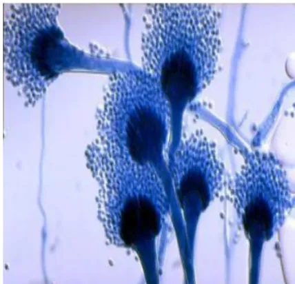
2.14 Fungal pathogens 2.14.1 Aspergillus fumigatus
A. fumigatus is an airborne fungus (Figure 2.3) resulting in high mortality rates in patients suffering from invasive fungal infections (Heinekamp et al., 2015). The fungus commonly targets immunocompromised individuals thereby causing life-threatening invasive diseases (Schrettl et al., 2008). A. fumigatus is the second most common cause of fungal infections causing various diseases such as aspergilloma, invasive aspergillosis, and allergic bronchopulmonary aspergillosis (Kaur and Singh, 2013). A.
fumigatus is also a common pathogen isolated from OE (Prasad et al., 2014).
Figure 2. 3 Microscopic image of Aspergillus fumigatus (Adhavan, 2020).
18 2.14.2 Candida albicans
C. albicans is a dimorphic fungus (Figure 2.4) that can grow as yeast and in other cases as hyphae in response to external stimuli. However, there are other morphological forms of C. albicans including the opaque, the pseudohyphal cell, and the chlamydospore (Whiteway and Bachewich, 2007). It is part of the human microbiome inhabiting the gastrointestinal tract, reproductive tract, oral cavity, and skin of most humans. In healthy individuals, it is often harmless but alterations in the host microbiota can enable C. albicans to overgrow and cause an infection (Nobile and Johnson, 2015). C. albicans causes two major types of infections in humans known as superficial infections (oral or vaginal candidiasis), and life-threatening systemic infections (Mayer et al., 2013). In OE, C. albicans is the most common fungal pathogen (Prasad et al., 2014).
Figure 2. 4 Microscopic image of Candida albicans (Tyavambiza, 2018)
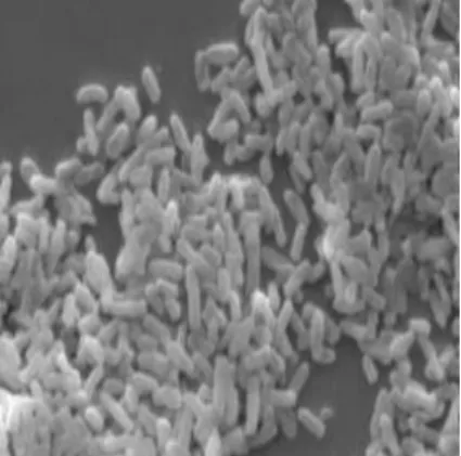
2.15 Bacterial pathogens 2.15.1 Escherichia coli
E. coli is a normal inhabitant of the animal and human gut (Figure 2.5). This bacterium can be found in soil, vegetation, and water. It is one of the common causes of bloodstream infections, OM, urinary tract infections, and wounds. The prevalence and vulnerability of E. coli show geographic variations in various environments and populations (Kibret and Aber, 2011).
19
Figure 2. 5 Microscopic image of Escherichia coli (Singh et al., 2015).
2.15.2 Pseudomonas aeruginosa
P. aeruginosa is a gram-negative rod-shaped bacterium (Figure 2.6) with a wide distribution in diverse environments, including water, soil, plants, animals, and a tendency to be present in locations associated with human activity (Spernovasilis et al., 2021). P. aeruginosa causes opportunistic infections in both animals and humans.
According to the World Health Organization (WHO), P. aeruginosa is resistant to the currently available drugs such as fluoroquinolones and penicillins (Langendonk et al., 2021). Infections of the ear skin caused by P. aeruginosa range from mild to life- threatening which include OE, necrotizing otitis externa (NOE), and perichondritis (Spernovasilis et al., 2021).
Figure 2. 6 Microscopic image of Pseudomonas aeruginosa (Tyavambiza, 2018).
2.15.3 Staphylococcus aureus
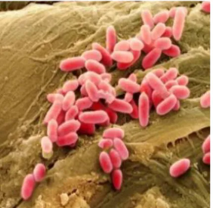
S. aureus (Figure 2.7) is both a commensal bacterium and a human pathogen (Tong et al., 2015). S. aureus, a gram-positive bacterium, asymptomatically colonizes approximately 30% of the population and can infect nearly every tissue in the body. S.
20
aureus readily adapts its metabolic and virulence responses in different tissues, causing superficial (e.g., folliculitis) and invasive infections (e.g., osteomyelitis) (Ford et al., 2021). S. aureus is the most invasive species and aetiological agent of human and animal maladies, including skin infections, abscesses, food poisoning, toxic shock syndrome, septicemia, endocarditis, and pneumonia. S. aureus is one of the most prominent causes of nosocomial- and community-acquired bacterial infections worldwide (Malachowa and DeLeo, 2010). Methicillin-resistant S. aureus (MRSA) constitutes the major threat among antibiotic-resistant agents that cause deaths (Sharaf et al., 2021).
Figure 2. 7 Microscopic image of Staphylococcus aureus (Tyavambiza, 2018).
2.16 Botanical descriptions of the plant species used in the study
Eight plant species used for the treatment of ear infections in Capricorn District, Limpopo province and Ehlanzeni District, Mpumalanga province were selected from a database on ethnomedicinal plants of over 300 plant species used for therapeutic purposes obtained from four Districts of Limpopo province (Capricorn, Mopani, Vhembe, and Waterberg) and Mpumalanga province (Ehlanzeni District). The pictures of the selected plant species were taken from five villages: Gottenburg, Hlalakahle, Hlavekisa, Hluvukani, and Thorndale (Figure 2.8–Figure 2.14).
2.16.1 Carpobrotus edulis L.
Carpobrotus edulis L. with the synonym Mesembryanthemum edule L. belongs to the Aizoaceae family (Figure 2.8). It has several common names including sour fig (English), suurvy (Afrikaans), umgongozi (Zulu), and igcukuma (Xhosa) (Rocha et al., 2017). The herb is native to South Africa and commonly found in the Eastern Cape,
21
Northern Cape, KwaZulu-Natal, Free State, and Western Cape provinces (Omoruyi et al., 2020). The plant thrives on sandy soils and dunes but can be found in all soil types in all provinces of South Africa (van der Watt and Pretorius, 2001). It is a ground-cover succulent plant that occupies coastal ecosystems with Mediterranean climatic conditions (Hafsa et al., 2016). The plant has green-coloured leaves that grow up to 10.8cm in length (Omoruyi et al., 2012) that are thin, blade-like, and succulent (Chokoe et al., 2008). The leaves can also have red, orange, or purple-coloured margins (Akinyede et al., 2020). This perennial herb is resistant to drought and wind conditions (Omoruyi et al., 2020).
2.16.1.1 Ethnomedicinal uses
C. edulis has been used as a form of traditional medicine in South Africa. The leaves, flowers, or fruits of the plant are used raw, or boiled in water and administered orally to treat various bacterial and fungal infections (Mudimba and Nguta, 2019). The Khoi- Khoi and San use the leaf juice to treat diarrhoea and tuberculosis, as a mouthwash for gum infections and sore throat, or applied topically to burn wounds. Traditional health practitioners in the Eastern Cape province also use the plant to treat constipation, diabetes mellitus, high blood pressure, and intestinal worms (Rocha et al., 2017). The leaf juice of C. edulis has been recorded for its use to treat sinusitis, infantile eczema, and internal chest conditions, as well as in soothing bites caused by spiders, ticks, and blue bottle stings (Van der Watt and Pretorius, 2001). Other authors have documented that C. edulis is used in traditional medicine to treat blood pressure, chilblains, earache, toothache, and vaginal and oral thrush (Akinyede et al., 2020).
Figure 2. 8 Images of Carpobrotus edulis L. (Bilomu ra ku nava) leaves (A) and flower (B).
A B
22 2.16.2 Cotyledon orbiculata L.
Cotyledon orbiculata L. belongs to the family of Crassulaceae (Amabeoku et al., 2007).
It is a succulent shrub with woody branches and thick and fleshy leaves that may vary from green to grey, often with a red line around the leaf margins and covered with a waxy layer on the surface (Figure 2.9). The leaves are obovate to narrowly ovoid- shaped. The flowers are yellow to orange-red coloured, usually hanging, tubular and bell-shaped, carried in clusters on the ends of an elongated flower stalk. It is found in Angola, Lesotho, Mozambique, Namibia, South Africa, and Swaziland in sandy or rocky soils, riverbanks, grassland, scrub, fynbos, and karoo biomes at an altitude ranging from 50 m to 3000 m above sea level (Maroyi, 2019). It has been listed in the Red Data List of South Africa due to its over-use (Kumari et al., 2016).
2.16.2.1 Ethnomedicinal uses
The leaves are used to treat corn and warts while the juice from the leaves is used to treat ear and toothaches, and as a hot poultice to treat boils and inflammation (Amabeoku et al., 2007). The traditional use of C. orbiculata includes the treatment of epilepsy using juice from the leaves (Stafford et al., 2005). In Lesotho, the plant is listed as one of the medicinal plants used to treat skin infections as well as boils and mouth ulcers (Moteetee and Kose, 2017). The leaves, leaf sap, and roots are used to treat sexually transmitted diseases such as gonorrhoea, syphilis, and venereal diseases (Maroyi, 2019). The plant is sun-dried and ground into fine powder by the Bapedi people of South Africa to wake fainted individuals whereas fresh leaves are crushed, and the juice is used to relieve aching feet or sniffed to induce sneezing as a way to treat various ailments (Mogale et al., 2019). The leaves and roots are also used to treat chronic diarrhoea (Masafu et al., 2016). It is used to treat haemorrhoids and skin rashes (Maroyi, 2019).
23
Figure 2. 9 Cotyledon orbiculata L. (Bilomu ra tindleve) whole plant (A), leaves (B) and flowers (C).
2.16.3 Dichrostachys cinerea (L.) Wight & Arn.
Dichrostachys cinerea (L.) Wight & Arn. belongs to the Fabaceae plant family (Figure 2.10). It is commonly referred to as a sickle bush, Christmas tree, or Chinese lantern tree (Abdullahi and Yusha’u, 2021). The plant is indigenous to South Africa (Abou Zeid et al., 2009) and found in tropical and subtropical areas (Mishra et al., 2009). D.
cinerea is a thorny shrub that can grow up to 8 metres with thorns that can also bear leaves. The inflorescence consists of a large amount of pink, white, or mauve filaments, which derive from the leaf axes, on approximately 4 cm stems. The flowers may produce a light fragrance; however, the sterile flowers are located under the fertile ones and each bearing a yellow stamen (Saez and Alfayate, 2020).
2.16.3.1 Ethnomedicinal uses
Fresh leaves of the plant are chewed to relieve diarrhoea, earache, as well as toothache. The leaves and bark are used to heal wounds while an infusion of the roots is used to treat abdominal pains, cough, and pneumonia. Powdered roots are also sniffed to stop nose bleeds, while leaves and roots are smoked to relieve head colds, epilepsy, and tuberculosis (Abdullahi and Yusha’u, 2021). The bark of the plant is used to treat dysentery, elephantiasis, and toothache while the leaves are used to treat boils, gonorrhoea, and as a laxative. The plant has also been used as an aphrodisiac and as an astringent for scorpion bites (Lavanya and Ambikapathy, 2016). It is also reported that the tender shoots of the plants are applied to the eye to treat eye infections while the roots are used to treat calculi, joint pains, renal troubles, and the uterus (Jayakumari et al., 2012). Mishra et al. (2009) reported that the plant is used
A B C
24
as a vermifuge and to treat headaches, leprosy, and syphilis. A decoction of the plant roots is used to treat snakebite victims (Mishal et al., 2008).
Figure 2. 10 Dichrostachys cinerea (L.) Wight & Arn. (Ndzhenga) whole plant (A), leaves (B), and flowers (C).
2.16.4 Erythrina lysistemon Hutch.
Erythrina is a genus within the family Fabaceae and comprises over 110 species. The species are found in tropical and subtropical regions across the globe (Figure 2.11).
They can be trees, herbs, or shrubs characterized by orange or red-coloured flowers.
E. lysistemon is a deciduous tree that reaches up to 12 metres in height (Juma and Majinda, 2004). E. lysistemon has a spreading crown and red flowers. It is widely distributed from the North of Tanzania to the Eastern Cape of South Africa (Dao et al., 2009). E. lysistemon is one of the species of Erythrina indigenous to South Africa (Pillay et al., 2001).
2.16.4.1 Ethnomedicinal uses
Species of Erythrina are largely used in folk medicine in many parts of the world (Nde et al., 2012) to treat various ailments such as female infertility, stomach pain, and gonorrhoea (El-Masry et al., 2002). In South Africa, the plant is used in traditional medicine to treat abscesses, arthritis, earache, and wounds (Dao et al., 2009), whereas in India the stem bark paste is used to treat asthma, rheumatism, stomach- ache, dysentery, eczema, dermatitis, and other skin infections (Akter et al., 2016). The Xhosa and Zulu people of South Africa use the leaves and bark to treat abscesses, bronchitis, earache, purulent sores, respiratory infections, sprains, strained ligaments, toothache, tuberculosis and to disinfect wounds (Pillay et al., 2001). The plant is also known for the treatment of eye infections using its leaves (Akter et al., 2016). The plant
A B C
25
is used in folk medicine by Asian, African, and South Americans as a tranquilizer and anti-anxiety as well as for the treatment of amenorrhea, dizziness, eye trouble, headache, sterility, asthma, malaria, epilepsy, liver disorder, and inflammation (Son and Elshamy, 2021).
Figure 2. 11 Erythrina lysistemon Hutch. (Muvale/ Nsisimbana) whole plant (A), leaves (B), flowers (C), and pods (D).
2.16.5 Flacourtia indica (Burm. f.) Merr.
Flacourtia indica (Burm. f.) Merr. is a branched, deciduous, and dioecious shrub (Figure 2.12) or small tree usually up to 5 m high belonging to the Salicaceae family, formerly known as Flacourtiaceae (Nguyen et al., 2021). The plant is native to African and Asian countries and is commonly known as the Indian plum, Governor’s plum, and Madagascar plum (Nguyen et al., 2021). It is indigenous to the Indian Peninsula (Nandhini et al., 2019). The plant is characterized by thorny branches and crenate, deciduous, and obovate or ovately shaped leaves (Tiwari, 2017). It also comprises small, yellow-coloured flowers either in a simple or compound raceme. The fruits of the plant are edible dark red or black-coloured indehiscent drupes (Tiwari, 2017).
A B
C D
26 2.16.5.1 Ethnomedicinal uses
All parts of the plant are used globally in traditional medicine to treat a wide variety of ailments. The leaves are used in Ayurvedic medicine to treat asthma, bronchitis, cough, phthisis, and pneumonia whereas, in Bengal, the leaves are known to be effective against snakebites as well as the treatment of schistosomiasis, malaria, and diarrhoea. The juice of the fresh leaves is used to treat fevers, dysentery, diarrhoea, and intestinal worms. Leaf decoctions are used for gynaecological disorders and hydroceles (Nguyen et al., 2021). The fruits of the plant are used to treat jaundice and enlarged spleen, while the seeds combined with turmeric are used to prevent rheumatic pain, the bark is applied to the body to treat intermittent fever and the root is used to treat nephritic colic (Kundu et al., 2013). The leaves of F. indica are used to treat measles whereas the bark is used to treat chicken pox (Kigen et al., 2016).
Figure 2. 12 Flacourtia indica (Burm. f.) Merr. (Xivambula) whole plant (A), leaves (B), and flowers (C).
2.16.6 Psidium guajava L.
Psidium guajava L. belongs to the plant family of Myrtaceae and subfamily Myrtoideae (Morais-Braga et al., 2016) (Figure 2.13). It is commonly referred to as guava (Fernandes et al., 2014). It is native to tropical America (Sanches et al., 2005) and Mexico (Soliman et al., 2016). However, in South Africa P. guajava is regarded as an invasive species (Ruwanza and Thondhlana, 2022). It flourishes in all tropical and subtropical (Khadhri et al., 2014) areas but it can adapt to varying climatic conditions although it prefers dry climates (Soliman et al., 2016). It grows as an evergreen shrub or a small tree that can reach up to 15 m in height (Jaiarj et al., 1999). However, the plant is capable of growing up to 1500 m in height (Ekeleme et al., 2017).
A B C
27
It is characterized by a thin, smooth, copper-coloured bark that flakes off, showing a greenish layer beneath (Kamath et al., 2008). The guava fruit is exotic and can either be round, ovoid, or pear-shaped with an average diameter that ranges from 4-10cm (Ngbolua et al., 2018) and a weight that can be from as little as one ounce to as much as one pound (Rishika and Sharma, 2012). It is a berry-type of fruit (Morais-Braga et al., 2016) that is yellow with pink flesh and tan-coloured seeds (Joseph and Priya, 2011). It produces white flowers that are about 1 inch in diameter, borne singly or in small clusters in axils of leaves of recent growth (Rishika and Sharma, 2012). It has dark green leaves (Kumar et al., 2021) that can grow up to 6 inches in length and 2 inches wide (Biswas et al., 2013).
2.16.6.1 Ethnomedicinal uses
The bark, fruits, leaves, and roots of the plant all possess medicinal value in traditional medicine (Rishika and Sharma, 2012). The plant has been long used in traditional medicine to treat various ailments in many parts of the world. In South Africa, P.
guajava leaves are used to treat flu (Ruwanza and Thodhlan, 2022) and diabetes (Mbara et al., 2022). The decoction of the bark and leaves is used to treat diarrhoea, dysentery, vomiting, and sore throats, and to regulate menstrual cycles in India (Kamath et al., 2008). The bark is used to treat diarrhoea in children whereas the leaves are used to relieve coughs, pulmonary disorders, wounds, and ulcers, while the fruit is used as a tonic, laxative, and anthelmintic (Khadri et al., 2014). The leaves are chewed to relieve toothache while an aqueous extract of the leaves is used to lower blood glucose levels in individuals with diabetes (Soliman et al., 2016). The whole plant is used to treat female-related disorders such as dysmenorrhoea, miscarriages, uterine bleeding, and premature labour (Rishika and Sharma, 2012). In America, plant leaf extracts are used in herbal formulas for bowel health, and to induce weight loss, while in Brazil the fruit and leaves are used to treat anorexia, cholera, diarrhoea, digestive problems, dysentery, gastric insufficiency, skin problems, sore throat, ulcers, and vaginal discharge (Soliman et al., 2016).
28
Figure 2. 13 Psidium guajava L. (Mugwava) whole plant (A), leaves (B), flowers (C), and fruit (D).
2.16.7 Ricinus communis L.
Ricinus communis L. is a member of the Euphorbiaceae family (Inayor and Ibraheem, 2014) (Figure 2.14). The plant is commonly known as the castor plant or palm of Christ (Jena and Gupta, 2012). According to Manoj (2017), the plant is an evergreen herbaceous or semi-woody, large shrub or small tree that reaches 5 m in height. It is a fast-growing, suckering perennial herb oilseed crop (Abdul et al., 2018). The plant is indigenous to Africa (Shobha et al., 2019) but is an occupant of tropical and temperate regions of the world (Sandhu et al., 2014). It is resistant to droughts and frost (Nemudzivhadi and Masoko, 2015). It thrives in clay and sandy soils (Vasco-Leal et al., 2021). According to Jain and Nafis (2011), the plant is known to be highly poisonous all over the world. Its phenotypic diversity is wide, with variations in growth habits, the colour of the leaves, stem, and seeds, and oil content (Riberio et al., 2016).
Flowers are monoecious, large, and arranged on the thick rachis of an oblong panicle.
The fruit is greenish, deeply grooved, tricoccus capsules, dehiscing longitudinally and septically into six valves. It has smooth seeds that are flattened and oval (Jeyaseelan and Jashothan, 2012). When ripe, the capsule, which contains three seeds, becomes
A B
C D
29
hard and cracked (Sbihi et al., 2018). Leaves are alternate, split like a palm, ovate lobes, sharp tips, green or brown on the surface, and sharp saw teeth on the edge (Park, 2018). The colour of leaves changes from light green to dark red in relation to the amount