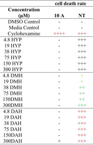
After treatment of normal (MCF10A) mammary epithelial cells and premalignant cHa-ras transfected (MCF10A-NeoT) derived mammary epithelial cells with HYP, DMH and the DAH derivative, the MTS test and the Duncan's multiple series, analysis of variance (ANOVA) post hoc analysis of the MTS results revealed that only the 150 and 300 M DAH derivative had a statistically significant effect on the metabolic activity of the abnormal cell line relative to the dimethyl sulfoxide (DMSO) and no significant effect on the normal MCF- 10A cell line after treatment with any of the test compounds. Supravital PI staining of adherent cells appeared to indicate a much higher rate of induction of cell death in abnormal cells than was evident in the MTS assay and the PI-based flow cytometry or the trypan blue exclusion assays and should be reinvestigated, although result trends were similar.
CANCER
- Causes and prevention of cancer
- Breast cancer
- Treatment of breast cancer
Longer breastfeeding has also been shown to protect women against developing breast cancer (Bray et al., 2004). There are still a number of natural anticancer products in clinical trials (Albrecht et al., 1995a).
TRADITIONAL MEDICINE
- Hypoxis hemerocallidea
- Uses of Hypoxis hemerocallidea
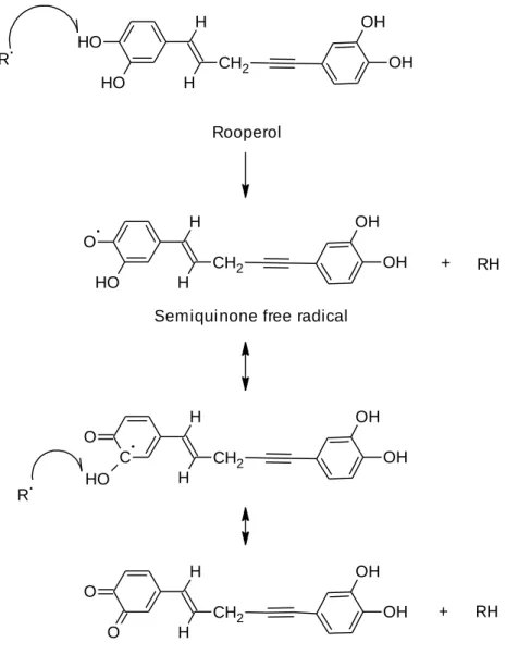
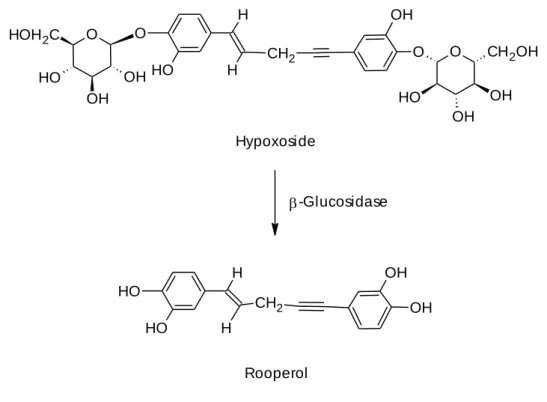
Hypoxoxide in vitro is non-toxic to normal cells, but becomes toxic to cancer cells as they produce the enzyme β-glucosidase that converts hypoxoxide to active rooperol (Albrecht et al., 1995a). Rooperol was found to have strong anti-microbial activity and was able to inhibit cyclooxygenase-1 (COX-1) and cyclooxygenase-2 (COX-2) which contributes to the anti-inflammatory and anti-noceptive activity of this compound. (Laporta et al., 2007a; Ojewole, 2006).
DRUGS EFFECTIVENESS
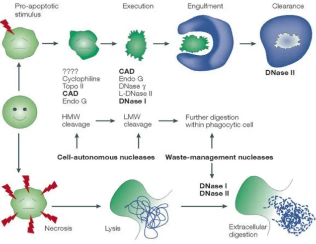
- Cell death
- Apoptosis
- Intrinsic apoptotic pathway
- Extrinsic pathway
- Perforin/granzyme apoptotic pathway
- What do the common executioner caspases do?
- Regulators of apoptotic cell death
- Methods of assessing cell death (apoptosis or necrosis)
These extrinsic pathway death receptors (DR) are characterized by their content of an 80 amino acid long domain referred to as death domain (DD) (Jonnalagadda et al., 2008). Nevertheless, PI appears to represent one of the most important dyes for the detection of cell death (Bradbury et al., 2000).
MCF10A
The MCF10A cells are frequently used as a normal control in breast cancer studies, as they show the morphological characteristic of normal breast epithelial cells (Zientek-Targosz et al., 2008). Cells transformed with such a construct were subsequently named the MCF10A-NeoT cell line (Basolo et al., 1991).
RESEARCH AIMS AND OBJECTIVES
- OVERVIEW
- COLLECTION AND IDENTIFICATION OF THE CONSTITUENTS IN
- EXTRACTION AND ISOLATION
- Materials and equipment
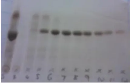
- Extraction and isolation procedures
- Results
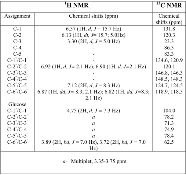
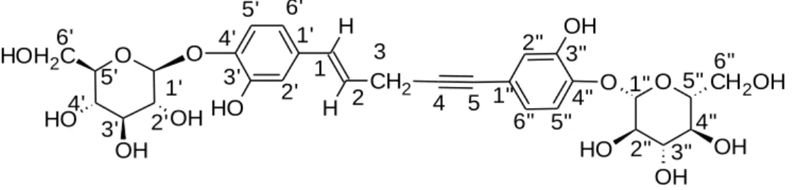
- Elucidation of the hypoxoside structure by 1 H and 13 C NMR spectroscopy
- PREPARATION OF HYPOXOSIDE DERIVATIVES
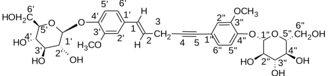
- Methylating hypoxoside
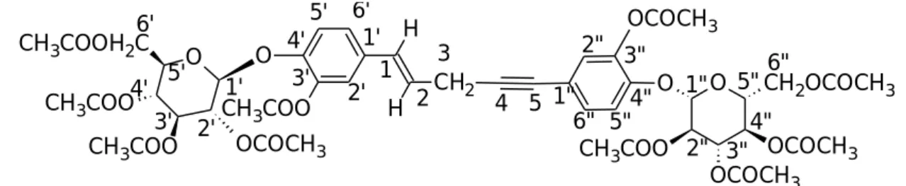
- Acetylation of hypoxoside
The biological activity of the Hypoxis extract was initially associated only with steroid glycosides (Bettolo et al., 1982; Laporta et al., 2007a; Nair and Kanfer, 2006). The analysis of the spectra is shown in Table 2.2 below (all spectra shown in Appendix A).
INTRODUCTION
Therefore, HIF-1 expression alters cancer cell metabolism to alleviate ROS stress and, to compensate for the inefficiency of Warburg metabolism. Cancer cells also attempt to ensure survival by stimulating glutaminolysis to provide compensatory ATP energy.
HYPOXOSIDE AND TWO DERIVATIVES AS POTENTIAL ANTI-
PI methods have been specifically mentioned as suitable for the assessment of early and late apoptosis as well as necrosis (Zamai et al., 2001). Effects of test compounds on metabolism (O'Toole et al., 2003) (i.e. MTS assay) and also cell number were investigated in the present study.
EXPERIMENTAL
- Materials and equipment
- Preparation of the stock and working solutions of hypoxoside and hypoxoside
- Reagents
- Procedure
- Culturing MCF10A and MCF10A-NeoT cell lines
- Reagents
- Procedure
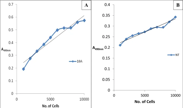
- MTS Assay optimization, compound exposure and data analysis
- Reagents
- Procedure
- Acridine orange/propidium iodide (PI) fluorescence microscopic assay
- Reagents
- Procedure
- Propidium iodide (PI) flow cytometric assay
- Reagents
- Procedure
- Trypan blue hemocytometric assay
- Reagents
- Procedure
44 complex dilution series so that all compounds were dissolved in the same final % DMSO (ie, final concentration of 0.001% DMSO) to allow a more direct comparison of compound effects. The choice of medium is based on experimentation and experience during cell line establishment (Phelan, 1996). The volume of the cell suspension was adjusted so that the number of cells required for the selected area (i.e.
A 1:100 dilution of the stock solution was made to produce a working concentration of 10 µg/ml and the solution was stored at -20oC. The size of the FS signal is approximately proportional to the cell size and is detected by the forward scatter detector ( Shapiro, 2003 ). 2D scatter plot (B) showing the granularity and size of the analyzed cells, and reveals 3 populations, R1, R2 and R3. The 1D-histogram graph (A) shows only 2 stained cell populations, the first region referred to as a negative control region (no staining area) and, subsequently, there is a weakly stained and strongly fluorescent (FL) region.
RESULTS AND DISCUSSION
- The effect of hypoxoside, dimethylhypoxoside and decaacetylhypoxoside
- The influence of hypoxoside and its derivatives on cell death of MCF-10A
- Cell death assessment of MCF10A cell line through AO/PI staining
- Cell death assessment of MCF10A-NeoT cell line through AO/PI staining
- Cell death assessment of the MCF10A and MCF10A-NeoT cell lines by Flow
- Trypan blue hemocytometric cell count
The effect of hypoxoside derivatives on the metabolic activity of the MCF10A cell line (A) and on the MCF10A-NeoT cell line (B), expressed as a fraction of the DMSO control to ensure that the effects were due to drugs and non-diluting solvent were plotted . using a bar graph and a standard error of the mean (error bars, SEM), for n=3 and significance as assessed by ANOVA. It appears that DAH reduced the metabolic activity of the MCF10A cells relative to the control (Figure 3.8A), with HYP and finally DMH having less effects (Figure 3.8A). By visual inspection, the concentration producing the greatest effect on the MCF10A-NeoT cell line appears to be the 300 µM concentration of the DAH compound, i.e.
In the premalignant cell line (i.e., MCF10A-NeoT cells), the DAH compound appears to have the most significant, selective metabolic effect, although the metabolic activity of the MCF10A cells is slightly affected. When the effect of compound type alone is considered, the metabolic activity of the MCF10A-NeoT cells is seen to be not significantly different when treated with the HYP and DMH (Table 3.3, MCF10A-NeoT, Compound Type, both are represented by "b" "). The overall effect of the three compounds to induce cell death does not appear to be significantly different according to the trypan blue staining assay, except that the total cell counts in the MCF10A-NeoT cells appear to be significantly suppressed at 19 µM (and possibly 150 µM which was not tested) and the 300 µM DAH (Figure 3.12).
CONCLUSION
94 The supravital staining method used was useful to confirm the lack of toxicity of the HYP and its derivatives, on the normal breast epithelial cell line, and could indicate the incipient death of the premalignant cell line, even after treatment with all hypoxoside compounds. levels, as indicated by results of supravital adherent cell staining (which is highly sensitive). Such staining results may predict a massive cell death event that may manifest if the 24 hour test exposure time is extended. The exposure time should therefore be extended and another method besides the PI staining method should be investigated to validate induction of cell death.
If the present conclusions are supported by such further investigations and additional cell death induction assays, the trypan blue method together with the MTS assay, total cell count and Duncan's analysis of the MTS results, and 24-hour exposure to the test compounds appear to represent an optimal screening system medicines.
GENERAL DISCUSSION
Supravital staining gave rise to results that could not be reconciled well with the overall interpretation of the results of the present study. In this study, cells were fixed before staining (assessment of cell death) and it was considered that the difference in the result was due to this (Foglieni et al., 2001). These cells can later be incorrectly classified as viable cells if the detection threshold (sensitivity) of the flow cytometer is set too high (Darizynkiewicz et al., 1992).
The concentration of PI and the length of time required for staining may need to be optimized individually for each cell line due to such differences and differences in the cytoplasmic pH of the MCF10A-NeoT cells (unpublished results) that may also influence the intensity of PI coloring. This can be explained by the Warburg metabolism of the cancer cells and the possible reduced conversion of MTS tetrazolium salt by the MCF10A-NeoT. Nevertheless, the results of the MTS assay revealed the obvious selective proliferation of cells in most treatments, but a decrease in metabolic activity in the MCF10A-NeoT with DAH treatment.
CONCLUSION
Isolate and purify hypoxoside from Hypoxis hemerocallidea by ethanol extraction of the tubers and further isolation with a prepacked HPCL reverse phase column - this was achieved. Establish a low-cost, rapid and high-throughput method for preliminary evaluation of the selective effects and cytotoxicity of prepared compounds. A system that appears to work well is the use of Trypan blue and total cell counts along with the MTS assay and Duncan's analysis of the MTS results of 24-hour exposure of the test cell lines.
This suggests that hypoxoside/rooperol activity has a synergistic effect with other unknown compounds such as sterols and sterolins present in impure test extracts (Boukes et al., 2011). This conclusion seems supported by the fact that the methyl derivative (DMH compound) appears to have a similar protective and proliferation-inducing effect as rooperol. This would explain the apparently significant, selective effect of the acetyl derivative, at a concentration of 150 and 300 M, on the premalignant mammary epithelial test cell model.
FUTURE STUDIES
Since rooperol has a strong antioxidant activity, the antioxidant activity of the derivatives (i.e., DMH and DAH) prepared in the present study should be further investigated. The mode of action of the DAH in its selective toxicity to the premalignant cancer cell model used needs to be investigated. Morphological characterization of the cell growth inhibitory activity of rooperol and pharmacokinetic aspects of hypoxide as an oral prodrug for cancer therapy.
Measurement of the ADP:ATP ratio in human leukemic cell lines can be used as an indicator of cell viability, necrosis and apoptosis. The role of membranes in the antibacterial and anti-inflammatory activities of bioactive compounds from the corm extract of Hypoxis rooperi. Isolation, characterization and antioxidant capacity evaluation of bioactive compounds derived from Hypoxis rooperi (African potato) corm extract.
Sensitivity and specificity of the MTS tetrazolium assay for the detection of in vitro cytotoxicity of 20 chemicals using human cell lines. Transcriptional activation of ras oncogenes and implications of BRAC1 in cell cycle regulation through the p53 checkpoint.